

Published Erratum
doi: 10.4103/1673-5374.350234.
Corrigendum: Purinergic signaling systems across comparative models of spinal cord injury
- PMID: 36018196
- PMCID: PMC9727416
- DOI: 10.4103/1673-5374.350234
Item in Clipboard
Published Erratum
Corrigendum: Purinergic signaling systems across comparative models of spinal cord injury
Neural Regen Res.
2023 Mar.
Abstract
[This corrects the article DOI: 10.4103/1673-5374.338993].
Conflict of interest statement
Figures
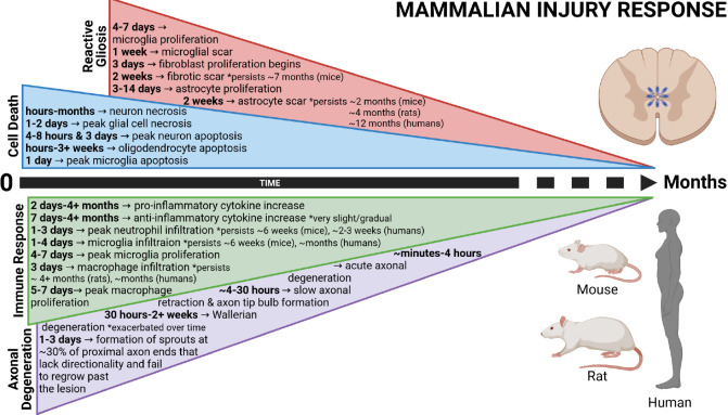
Timeline depicting the secondary cellular injury response following mammalian spinal cord injury. The primary mechanical trauma is exacerbated by prolonged cell death, widespread inflammation, reactive gliosis, and axonal degeneration. These events prevent successful regeneration and limit sensorimotor recovery. Created with BioRender.com with permissions and publication license.
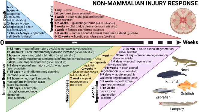
Timeline depicting the secondary cellular injury response following non-mammalian spinal cord injury. The primary mechanical trauma induces transient cell death, controlled inflammation, reactive gliosis, neurogenesis, and axonal regeneration. These events conclude within weeks and facilitate recovery and restoration of locomotor function in non-mammalian vertebrates. Created with BioRender.com with permissions and publication license.
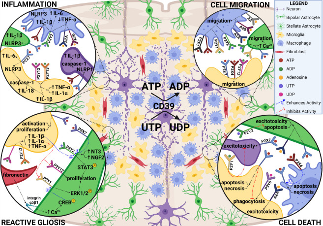
Purinergic signaling within the spinal cord microenvironment during the early injury response. The first several days following mammalian SCI are characterized by widespread cell death, migration of various cell types to the lesion, inflammation, and reactive gliosis. Identified roles for purinergic receptors in these processes is summarized. Created with BioRender.com with permissions and publication license. ADP: adenosine diphosphate; ATP: adenosine triphosphate; CD39: cluster of differentiation 39; Ca2+: calcium; CREB: cAMP response element-binding protein; ERK1/2: extracellular signal-regulated kinases 1/2; IL: interleukin; NGF2: nerve growth factor-2; NLRP1/3: NLR family pyrin domain containing 1/3; NT3: neurotrophin-3; STAT3: signal transducer and activator of transcription 3; TNF-α: tumor necrosis factor alpha; UDP: uridine diphosphate; UTP: uridine triphosphate. Created with BioRender.com with permissions and publication license.
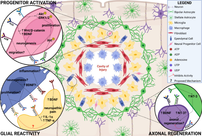
Purinergic signaling within the spinal cord microenvironment during the chronic injury response. After the first week following mammalian SCI, reactive astrocytes become scar forming, ependymal and neural progenitor cells fail to undergo injury-induced proliferation and neuronal differentiation, and axons continue to degenerate. Identified and hypothesized roles for purinergic receptors in these processes is summarized. Created with BioRender.com with publication permissions and publication license. ADP: Adenosine diphosphate; Akt: protein kinase B; ATP: adenosine triphosphate; BDNF: brain derived neurotrophic factor; CD39: cluster of differentiation 39; Ca2+: calcium; CREB: cAMP response element-binding protein; ERK1/2: extracellular signal-regulated kinases 1/2; IL: interleukin; NGF2: nerve growth factor-2; NLRP1/3: NLR family pyrin domain containing 1/3; NT3: neurotrophin-3; STAT3: signal transducer and activator of transcription 3; TNF-α: tumor necrosis factor alpha; UDP: uridine diphosphate; UTP: uridine triphosphate; Wnt: wingless-related intergration site. Created with BioRender.com with permissions and publication license.
Erratum for
-
Purinergic signaling systems across comparative models of spinal cord injury.Neural Regen Res. 2022 Nov;17(11):2391-2398. doi: 10.4103/1673-5374.338993. Neural Regen Res. 2022. PMID: 35535876 Free PMC article. Review.
Similar articles
-
Purinergic signaling systems across comparative models of spinal cord injury.Neural Regen Res. 2022 Nov;17(11):2391-2398. doi: 10.4103/1673-5374.338993. Neural Regen Res. 2022. PMID: 35535876 Free PMC article. Review.
-
Corrigendum: Comparative proteomes change and possible role in different pathways of microRNA-21a-5p in a mouse model of spinal cord injury.Neural Regen Res. 2020 Dec;15(12):2305. doi: 10.4103/1673-5374.285007. Neural Regen Res. 2020. PMID: 32594053 Free PMC article.
-
Corrigendum: TAZ induces migration of microglia and promotes neurological recovery after spinal cord injury.Front Pharmacol. 2022 Sep 9;13:995767. doi: 10.3389/fphar.2022.995767. eCollection 2022. Front Pharmacol. 2022. PMID: 36160380 Free PMC article.
-
Corrigendum: Dissecting the Dual Role of the Glial Scar and Scar-Forming Astrocytes in Spinal Cord Injury.Front Cell Neurosci. 2020 Oct 6;14:270. doi: 10.3389/fncel.2020.00270. eCollection 2020. Front Cell Neurosci. 2020. PMID: 33132841 Free PMC article.
-
The Role of Microglia in Neuroinflammation of the Spinal Cord after Peripheral Nerve Injury.Cells. 2022 Jun 30;11(13):2083. doi: 10.3390/cells11132083. Cells. 2022. PMID: 35805167 Free PMC article. Review.
References
-
- Ali AAH, Abdel-Hafiz L, Tundo-Lavalle F, Hassan SA, von Gall C. P2Y2 deficiency impacts adult neurogenesis and related forebrain functions. FASEB J. 2021;35:e21546. - PubMed
-
- Barry D, McDermott K. Differentiation of radial glia from radial precursor cells and transformation into astrocytes in the developing rat spinal cord. Glia. 2005;50:187–197. - PubMed
-
- Becker CG, Becker T, Hugnot JP. The spinal ependymal zone as a source of endogenous repair cells across vertebrates. Prog Neurobiol. 2018;170:67–80. - PubMed
Publication types
LinkOut - more resources
Full Text Sources
