. 2022 Aug 9:13:986396.
doi: 10.3389/fmicb.2022.986396. eCollection 2022.

Wall teichoic acid-dependent phagocytosis of intact cell walls of Lactiplantibacillus plantarum elicits IL-12 secretion from macrophages

Affiliations

Wall teichoic acid-dependent phagocytosis of intact cell walls of Lactiplantibacillus plantarum elicits IL-12 secretion from macrophages

Naoya Kojima et al. Front Microbiol. .

Abstract

Selected lactic acid bacteria can stimulate macrophages and dendritic cells to secrete IL-12, which plays a key role in activating innate and cellular immunity. In this study, we investigated the roles of cell wall teichoic acids (WTAs) displayed on whole intact cell walls (ICWs) of Lactiplantibacillus plantarum in activation of mouse macrophages. ICWs were prepared from whole bacterial cells of several lactobacilli without physical disruption, and thus retaining the overall shapes of the bacteria. WTA-displaying ICWs of several L. plantarum strains, but not WTA-lacking ICWs of strains of other lactobacilli, elicited IL-12 secretion from mouse bone marrow-derived macrophages (BMMs) and mouse macrophage-like J774.1 cells. The ability of the ICWs of L. plantarum to induce IL-12 secretion was abolished by selective chemical elimination of WTAs from ICWs, but was preserved by selective removal of cell wall glycopolymers other than WTAs. BMMs prepared from TLR2- or TLR4-deficient mouse could secret IL-12 upon stimulation with ICWs of L. plantarum and a MyD88 dimerization inhibitor did not affect ICW-mediated IL-12 secretion. WTA-displaying ICWs, but not WTA-lacking ICWs, were ingested in the cells within 30 min. Treatment with inhibitors of actin polymerization abolished IL-12 secretion in response to ICW stimulation and diminished ingestion of ICWs. When overall shapes of ICWs of L. plantarum were physically disrupted, the disrupted ICWs (DCWs) failed to induce IL-12 secretion. However, DCWs and soluble WTAs inhibited ICW-mediated IL-12 secretion from macrophages. Taken together, these results show that WTA-displaying ICWs of L. plantarum can elicit IL-12 production from macrophages via actin-dependent phagocytosis but TLR2 signaling axis independent pathway. WTAs displayed on ICWs are key molecules in the elicitation of IL-12 secretion, and the sizes and shapes of the ICWs have an impact on actin remodeling and subsequent IL-12 production.

Keywords: IL-12; Lactiplantibacillus plantarum; actin remodeling; cell wall teichoic acid; intact cell wall; macrophage; phogocytosis.


Conflict of interest statement

The authors declare that the research was conducted in the absence of any commercial or financial relationships that could be construed as a potential conflict of interest.

Figures

FIGURE 1
FIGURE 1
Scanning electron microscopy of ICWs of L. plantarum TUA 5099L. (A) Heat-killed cells. (B) Intact cell walls. (C) Acid-treated ICWs. (D) Base-treated ICWs.
FIGURE 2
FIGURE 2
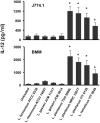
IL-12 secretion from macrophages stimulated with ICWs of lactobacilli. ICWs were prepared from heat-killed cells of several lactobacilli by SDS, pronase, and nuclease treatments. J774.1 cells (5 × 10⁵/ml, upper panel) and BMMs (5 × 10⁴/ml, lower panel) were cultured with 10 μg/ml of ICWs for 24 h and IL-12 secreted in the culture supernatant was determined. Each bar is the mean ± SE for five independent experiments. The results were reproducible for at least three independent ICW preparations for each strain. *P < 0.01 vs. untreated cells.
FIGURE 3
FIGURE 3
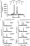
Analysis of cell wall glycopolymers isolated from N-acetylated ICWs of lactobacilli. (A) ICWs of L. plantarum TUA 5099L were N-acetylated and treated with 0.1 M glycine-HCl, pH 2.5 at 100°C for 30 min. After centrifugation at 8,000 × g for 15 min, supernatants were recovered, dialyzed against 5 mM Tris–HCl (pH 7.4), and applied to a DEAE-cellulose column. The column was eluted in a stepwise manner with 5 mM Tris–HCl (pH 7.4) containing 50, 150, 250, 350, and 500 mM NaCl. Fractions (5 ml) were collected and hexoses and phosphorus in each fraction were determined. (B) Chromatography of glycopolymers isolated from N-acetylated ICWs of eight strains of lactobacilli. Glycopolymers isolated from N-acetylated ICWs of each strain were subjected to chromatography on a DEAE-cellulose column, as described above, and hexose or phosphorus was determined. The figure shows representative chromatograms of at least three independent ICW preparations of each strain. Other ICW preparations also gave the same chromatograms. Note that L. plantarum TUA 5099L, NRIC 1067T, and YIT 0139 and L. helveticus YIT 0049 contain glycopolymers eluted at 250 mM NaCl.
FIGURE 4
FIGURE 4
Chemical modification of ICWs of L. plantarum TUA 5099L. (A) Flowchart of treatments of ICWs with mild acid (0.1 M glycine-HCl, pH 2.5, 100°C, 30 min), mild base (0.5 M NaOH, 37°C, 30 min), aqueous hydrogen fluoride (47% HF, 4°C, 16 h), and periodate (0.1 M NaIO4, pH 4.5, 4°C 48 h). (B) Smith degradation of ICWs. Periodate-oxidized ICWs and mock-treated ICWs were hydrolyzed with 0.1 M glycine-HCl, pH 2.5, 100°C for 30 min after N-acetylation, and each resulting supernatant was subjected to chromatography on a DEAE-cellulose column (described in Figure 3). Hexose in each fraction was determined. (C) ICWs were treated with a combination of N-acetylation, mild acid and mild base according to the flowchart in (A). The supernatant obtained by each mild acid treatment (Sup 1 to 5 in A) was subjected to chromatography on a DEAE-cellulose column. Hexose in each fraction was determined. (D) Depictions of chemically modified ICWs prepared as shown in (A). Acid-treated ICWs contain only WTA, base-treated ICWs contain only WPS, and acid-treated NAc-ICWs and HF-treated ICW lack both WTA and WPS.
FIGURE 5
FIGURE 5
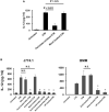
IL-12 secretion from macrophages stimulated with chemically modified ICWs of L. plantarum TUA 5099L. (A) J774.1 cells were cultured with unmodified, periodate-oxidized or mock-treated ICWs (each 10 μg/ml) for 24 h and IL-12 secreted in each supernatant was determined. Each bar is the mean ± SE for three independent experiments. (B) J774.1 cells or BMMs were cultured with chemically modified or unmodified ICWs (10 μg/ml) for 24 h and IL-12 secreted in the culture supernatant was determined. Each bar is the mean ± SE for 4 (for J774.1 cells) and 3 (for BMMs) experiments. Significance was evaluated by one-way ANOVA. N.S., not significant vs. ICW treated cells. *P < 0.01 vs. cells treated with unmodified ICWs.
FIGURE 6
FIGURE 6
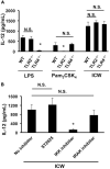
Involvement of TLRs in IL-12 secretion from macrophages stimulated with ICWs. (A) BMMs were prepared from wild-type (WT), TLR2-deficient (TLR2–/–), and TLR4-deficient (TLR4–/–) mice, respectively. BMMs (5 × 10⁴/ml) were cultured with 1 μg/ml of Pam3CSK4, 500 ng/ml of LPS, or 10 μg/ml of ICWs for 24 h and IL-12 secreted in the culture supernatant was determined. Each bar is the mean ± SE for three independent experiments. N.S., not significant vs. BMMs from wild-type mice. *P < 0.01 vs. cells from wild-type mice. (B) BMMs prepared from wild-type mice were pretreated with 10 μM of IKK inhibitor II, 10 μM of ST2825, a MyD88 dimerization inhibitor and 0.5 μM of IRAK1/4 inhibitor I, respectively, for 30 min, and then cells were stimulated with 10 μg/ml of ICWs. After 24 h stimulation, IL-12 secreted in the culture supernatant was determined. Each bar is the mean ± SE for three independent experiments. N.S., not significant vs. cells treated without inhibitors (no inhibitor).
FIGURE 7
FIGURE 7
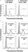
Uptake of ICWs into macrophages. (A) J774.1 cells were incubated with fluorescein-labeled WTA-displaying ICWs of L. plantarum TUA 5099L and WTA-lacking ICWs of L. rhamnosus ATCC 7496T for 30 min at 37°C with gentle rotation. Cells were washed with PBS and fluorescent signals in the cells were then analyzed by flow cytometry. Representative results of two independent experiments are shown. Gray and open histograms indicate untreated and fluorescein-labeled ICWs treated cells, respectively. (B) J774.1 cells were incubated with chemically modified ICWs prepared from fluorescein-labeled ICWs of L. plantarum TUA 5099 for 30 min and fluorescent signals in the cells were analyzed. Representative results of two independent experiments are shown. Gray and open histograms indicate untreated and fluorescein-labeled ICWs treated cells, respectively.
FIGURE 8
FIGURE 8
Effect of actin polymerization inhibitors on uptake of ICWs and IL-12 secretion. (A) J774.1 cells were treated with cytochalasin D (CytD, 1 μM) or chlorpromazine (Chlor, 1 μg/ml) in complete RPMI1640 containing 0.1% DMSO for 30 min at 37°C. Fluorescein-labeled ICWs of L. plantarum TUA 5099L were added and the mixture was incubated for 30 min. Fluorescent signals in the cells were analyzed. Representative histograms (left) and percentages of positive cells among the treated cells (right) are shown. Gray and open histograms indicate untreated and fluorescein-labeled ICWs treated cells, respectively. (B) J774.1 cells and BMMs were treated with CytD (1 μM), latrunculin B (Lat, 1 μM), jasplakinolide (Jas, 0.1 μM), or Chlor (1 μg/ml) in complete RPMI1640 containing 0.1% DMSO for 30 min. Then the cells were stimulated with ICWs of L. plantarum TUA 5099L (10 μg/ml) or LPS (100 ng/ml). Culture supernatants were collected after 24 h and IL-12 in supernatants was determined. Open, gray and black columns indicate unstimulated, LPS-stimulated, and ICW-stimulated cells, respectively. Each bar is the mean ± SE for four independent experiments. *P < 0.01 vs. inhibitor-untreated cells.
FIGURE 9
FIGURE 9
Effects of disruption of overall shapes of ICWs on immunomodulatory abilities of ICWs. (A) Scanning electron microscopy of ICWs and DCWs of L. plantarum TUA 5099L. (B) Uptake of ICWs or DCWs. J774.1 cells were incubated with fluorescein-labeled ICWs or disrupted ICWs (DCWs) of L. plantarum TUA 5099 for 30 min and fluorescent signals in the cells were analyzed. Gray and open histograms indicate untreated and fluorescein-labeled cell walls treated cells, respectively. Representative images under florescent microscopy were also shown. (C) Reduction of uptake of DCWs by HF treatment. J774.1 cells were incubated with fluorescein-labeled DCWs (solid line) or HF-treated DCWs (dashed line) for 30 min and fluorescent signals in the cells were analyzed. Gray histograms indicate cells without DCW treatment. (D) Elicitation of IL-12 secretion from macrophages. J774.1 cells were cultured with ICWs (10 μg/ml) or DCWs (100 μg/ml) for 24 h and IL-12 secreted in culture supernatants was determined. Each bar is the mean ± SE for three independent experiments. (E) Effect of cytochalasin D on uptake of DCWs. J774.1 cells were treated with cytochalasin D in complete RPMI1640 containing 0.1% DMSO for 30 min at 37°C. Fluorescein-labeled DCWs were added and the mixture was incubated for 30 min. Fluorescent signals in the cells were analyzed. Solid and dashed lines indicate untreated and cytochalasin D-treated cells, respectively. Gray histograms indicate cells without DCW treatment.
FIGURE 10
FIGURE 10
Inhibition of immunomodulation by ICWs by pretreatment of cells with DCWs and soluble WTAs. (A) J774.1 cells were treated with various concentrations of DCWs of L. plantarum TUA 5099L (open circles) or HF-treated DCWs (closed circles) for 30 min at 37°C with gentle rotation. Then the cells were cultured with 10 μg/ml of ICWs of L. plantarum TUA 5099L for 24 h, and IL-12 secreted in culture supernatants was determined. Each bar is the mean ± SE for three independent experiments. *P < 0.01 vs. DCW-untreated cells. (B) Cells were treated with 500 μg/ml of purified soluble WTAs or WPS of L. plantarum TUA 5099L for 30 min at 37°C. Then the cells were stimulated with 10 μg/ml of ICWs of L. plantarum TUA 5099L for 24 h, and IL-12 secreted in culture supernatants was determined. Each bar is the mean ± SE for three independent experiments.

Similar articles

Cited by

References

    1. Abdel-Akher M., Hamilton J. K., Montgomery R., Smith F. (1952). A new procedure for the determination of the fine structure of polysaccharides. J. Am. Chem. Soc. 74 4970–4971.
    1. Aderem A., Underhill D. M. (1999). Mechanisms of phagocytosis in macrophage. Annu. Rev. Immunol. 17 593–623. 10.1146/annurev.immunol.17.1.593 - DOI - PubMed
    1. Araki Y., Ito E. (1989). Linkage units in cell walls of gram-positive bacteria. Crit. Rev. Microbiol. 17 121–135. 10.3109/10408418909105745 - DOI - PubMed
    1. Bromley S. K., Burack W. R., Johnson K. G., Somersalo K., Sims T. N., Sumen C., et al. (2001). The immunological synapse. Annu. Rev. Immunol. 19 375–396. 10.1146/annurev.immunol.19.1.375 - DOI - PubMed
    1. Brown G., Willment J. A., Whitehead L. (2018). C-type lectins in immunity and homeostasis. Nat. Rev. Immunol. 18 374–389. 10.1038/s41577-018-0004-8 - DOI - PubMed