. 2022 Aug 12;11(16):2506.
doi: 10.3390/cells11162506.

Simple Detection of Unstained Live Senescent Cells with Imaging Flow Cytometry

Affiliations

Simple Detection of Unstained Live Senescent Cells with Imaging Flow Cytometry

Marco Malavolta et al. Cells. .

Abstract

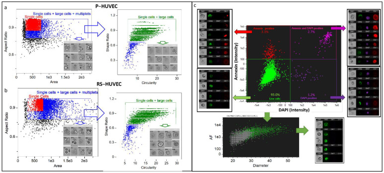
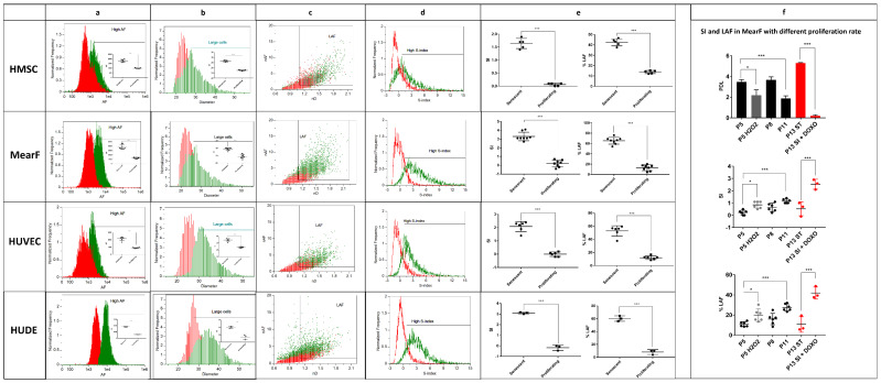
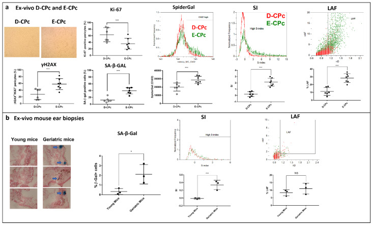
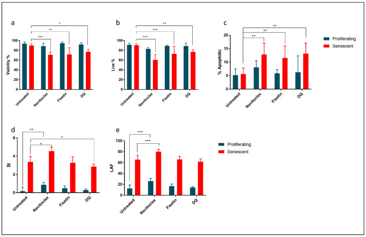
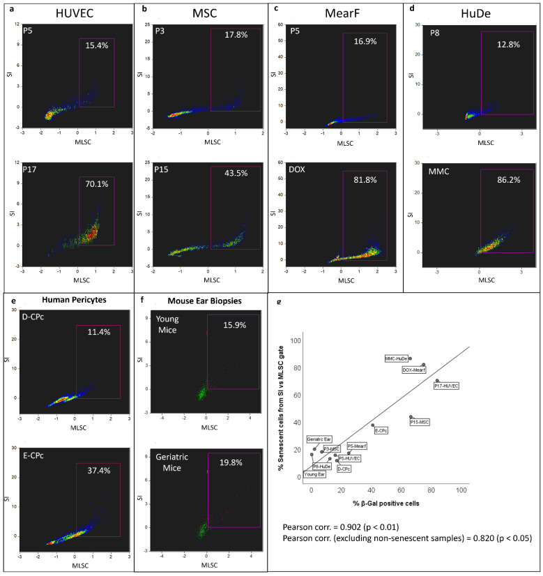
Cellular senescence is a hallmark of aging and a promising target for therapeutic approaches. The identification of senescent cells requires multiple biomarkers and complex experimental procedures, resulting in increased variability and reduced sensitivity. Here, we propose a simple and broadly applicable imaging flow cytometry (IFC) method. This method is based on measuring autofluorescence and morphological parameters and on applying recent artificial intelligence (AI) and machine learning (ML) tools. We show that the results of this method are superior to those obtained measuring the classical senescence marker, senescence-associated beta-galactosidase (SA-β-Gal). We provide evidence that this method has the potential for diagnostic or prognostic applications as it was able to detect senescence in cardiac pericytes isolated from the hearts of patients affected by end-stage heart failure. We additionally demonstrate that it can be used to quantify senescence "in vivo" and can be used to evaluate the effects of senolytic compounds. We conclude that this method can be used as a simple and fast senescence assay independently of the origin of the cells and the procedure to induce senescence.

Keywords: artificial intelligence and machine learning; cellular senescence; imaging flow cytometry; replicative senescence; senolytics.


Conflict of interest statement

Alessandro Serra disclaims that he is affiliate of Luminex, whose portfolio comprises the imaging flow cytometry and software of Amnis used in this study. All other authors declare no conflicts of interest.

Figures

Figure 1
Figure 1
Gating strategy used for the analysis of senescent cells. Representative example from the HUVEC model of the gating strategy applied to proliferating (a) and senescent (b) cells. In the example, the conventional “Single cells” gate used in imaging flow cytometry to select proliferating cells (red color) is not appropriate when senescent cells are present in the sample, as the population is shifted to right in the dot plot “Aspect Ratio” vs. “Area”. However, extending this gate to the right leads to the inclusion of multiplets in the analysis (single cells + large cells + multiplets gate, blue color). A further gate (green gate) set in the high “Circularity” and high “Shape ratio” can quite completely clean the population of multiplets and other artifacts independently by the status of the cells. Cell images below each gate represent the largest events detected in the respective gate. Further selection of live cells (c) can be performed by staining the cells with Annexin-APC and DAPI to remove apoptotic and dead cells. The phenotype of the remaining cells can be monitored based on autofluorescence (intensity of channel 2, band 480–560 nm) and diameter ((width + height) /2)) parameters. In the bottom of panel c, there is an overlayed representative dot plot of a senescent (green events) and proliferating (gray events) sample showing that both autofluorescence and diameter are different between the two samples. P = proliferating; RS = replicative senescent; HUVEC= human umbilical vein endothelial cells; HMSC = human bone marrow mesenchymal stem cells.
Figure 2
Figure 2
Quantitative estimation of the senescent index (SI) and % of large autofluorescent cells (LAF) in various senescence models. (a) Overlayed histograms and quantitative estimation of autofluorescence (AF) in proliferating (red) and senescent cells (green). All senescent samples display increased autofluorescence compared to non-proliferating samples. (b) Overlayed histograms and quantitative estimation of the diameter (D = (width + height)/2)) in proliferating (red) and senescent cells (green). All senescent samples display increased diameter compared to non-proliferating samples. (c) Overlayed representative dot plots of normalized autofluorescence (nAF = “AF”/“mean AF of proliferating samples”) vs. normalized diameter (nD = “D”/“mean D of proliferating samples”) showing that LAF increases in senescent samples (green events) compared to non-senescent samples (red events). The threshold of nAF was set at 1.5 whereas the threshold of D was set at 1.1 for all samples. (d) Overlayed representative histograms of the SI (SI = ((nAF − 1) + 5 × (nD − 1))/2) in proliferating (red) and senescent cells (green). (e) Quantitative estimation of SI and LAF in proliferating and senescent HMSC (n = 5), MearF (n = 8), HUVEC (n = 6) and HuDe (n = 3). (f) Comparison of SI and LAF in MearF with different population doubling level (PDL). SI and LAF significantly increase after stressing conditions that decrease PDL, such as treatment with H2O2 (250µM H2O2 × 2 h + 1 week of resting, n = 6) or at late passages (P11, n = 6) compared to early passages (P5, n = 6). Spontaneous transformation of one of the cultures (P13 ST, n = 3 replicates from the same culture) resulted in a strong increase in PDL and a parallel decrease in SI and LAF. Treatment of P13 ST with doxorubicin 75 nM × 1 week (P13 ST + DOXO, n = 3 replicates from the same culture) strongly increased both SI and LAF. MearF = mouse ear fibroblasts; HUVEC = human umbilical vein endothelial cells; HMSC = human bone marrow mesenchymal stem cells; HuDe = human dermal fibroblasts; * p < 0.05; *** p < 0.001 by Student’s t test.
Figure 3
Figure 3
Estimation of senescence by imaging flow cytometry in “ex vivo” human and mouse samples (a) Estimation of senescence by imaging flow cytometry (IFC) in ex vivo cardiac pericytes (CPcs). On the left of the panel, representative microscope images are shown, and senescence (SA-β-gal and γH2AX positive cells) and proliferative (Ki-67 positive cells) markers showing that CPcs from patients undergoing cardiac transplantation (E-CPc, n = 7) display a high degree of senescence compared to CPcs from healthy donors (D-CPcs, n = 6). A representative histogram and the quantification of senescence performed by flow cytometry Spider-βGal assay (SpiderGal, middle of the panel) also confirmed the higher degree of senescence of E-CPcs. In agreement with these biomarkers, we detected a significant increase in LAF and SI in E-CPcs compared to D-CPcs by IFC (right of the panel). (b) Estimation of senescence by IFC in cells isolated from mouse ear biopsies. Ear biopsies taken from geriatric mice display and increased staining for SA-β-gal (left of the panel). SI also significantly increased in cells isolated form ear biopsies taken from geriatric mice vs. those from young mice. This difference was not detected for LAF. * p < 0.05; *** p < 0.001 by student’s t test.
Figure 4
Figure 4
Treatment of proliferating or senescent mouse ear fibroblasts (MearF) with various senolytics. (a) Viability assay performed by Trypan Blue exclusion showing that exposure (48 h) to navitoclax (10 μM), fisetin (10 μM) or the combination of dasatinib + quercetin (DQ) (100 nM + 10 μM) reduces the viability only in senescent fibroblasts. (b) Live cells estimated by imaging flow cytometry (% of cells negative to DAPI and annexin in the IFC assay) also significantly decrease only in senescent fibroblasts. (c) Apoptotic cells (% of cells positive to annexin and negative to DAPI) significantly increased after treatment with all senolytics. (d) The senescence index (SI) estimated by IFC significantly decreases in senescent samples after treatment with DQ. Conversely, SI increases after treatment with navitoclax. (e) The % of large autofluorescent cells (LAFs) significantly increase in senescent and proliferating samples only after treatment with navitoclax2.5. Quantification of senescent cells estimated by artificial intelligence (AI) and machine learning (ML) * p < 0.05; ** p < 0.01; *** p < 0.001 by ANOVA followed by post hoc.
Figure 5
Figure 5
Estimation of senescence by imaging flow cytometry implemented with artificial intelligence and machine learning in different cellular models. Scatter plots of the super-feature derived from AI and ML elaboration, “ML Senescence Classifier” (MLSC), versus Senescence Index (SI). Panel (a), human umbilical vein endothelial cells (HUVEC) at passage P5 and passage 17 (P17); panel (b), human mesenchymal stem cells (MSC) at passage (P5) and passage (P15); panel (c), murine ear fibroblasts (MearF) at passage 5 (P5) and after treatment with Doxorubin (DOX); panel (d), human dermal fibroblasts (HuDe) at passage 8 (P8) and after treatment with Mitomycin C (MMC), panel (e), “ex-vivo” cardiac pericytes obtained from one healthy donor (D-CPc) and from one patient undergoing cardiac transplantation (E-CPc); (f) “ex-vivo” cells immediately extracted from ear biopsies of young and geriatric mice; panel (g), correlation between the senescent cells gated from the SI vs. MLSC scatter plot versus the % of β-galactosidase (β-Gal) positive cells assessed by light microscopy. Pearson and Spearman correlations (both p < 0.01) are shown in the graph.

Similar articles

Cited by

References

    1. Gorgoulis V., Adams P.D., Alimonti A., Bennett D.C., Bischof O., Bishop C., Campisi J., Collado M., Evangelou K., Ferbeyre G., et al. Cellular Senescence: Defining a Path Forward. Cell. 2019;179:813–827. doi: 10.1016/j.cell.2019.10.005. - DOI - PubMed
    1. Robbins E., Levine E.M., Eagle H. Morphologic changes accompanying senescence of cultured human diploid cells. J. Exp. Med. 1970;131:1211–1222. doi: 10.1084/jem.131.6.1211. - DOI - PMC - PubMed
    1. Rattan S.I.S., Keeler K.D., Buchanan J.H., Holliday R. Autofluorescence as an index of ageing in human fibroblasts in culture. Biosci. Rep. 1982;2:561–567. doi: 10.1007/BF01314216. - DOI - PubMed
    1. Muñoz-Espín D., Serrano M. Cellular senescence: From physiology to pathology. Nat. Rev. Mol. Cell Biol. 2014;15:482–496. doi: 10.1038/nrm3823. - DOI - PubMed
    1. Van Deursen J.M. The role of senescent cells in ageing. Nature. 2014;509:439–446. doi: 10.1038/nature13193. - DOI - PMC - PubMed

Publication types

Grants and funding

This study was supported by Ricerca Corrente funding from Italian Ministry of Health to M.M. and M.P.

LinkOut - more resources