. 2022 Aug 13;12(8):1115.
doi: 10.3390/biom12081115.

Conformational Plasticity of Centrin 1 from Toxoplasma gondii in Binding to the Centrosomal Protein SFI1

Affiliations

Conformational Plasticity of Centrin 1 from Toxoplasma gondii in Binding to the Centrosomal Protein SFI1

Luca Bombardi et al. Biomolecules. .

Abstract

Centrins are calcium (Ca2+)-binding proteins that are involved in many cellular functions including centrosome regulation. A known cellular target of centrins is SFI1, a large centrosomal protein containing multiple repeats that represent centrin-binding motifs. Recently, a protein homologous to yeast and mammalian SFI1, denominated TgSFI1, which shares SFI1-repeat organization, was shown to colocalize at centrosomes with centrin 1 from Toxoplasma gondii (TgCEN1). However, the molecular details of the interaction between TgCEN1 and TgSFI1 remain largely unknown. Herein, combining different biophysical methods, including isothermal titration calorimetry, nuclear magnetic resonance, circular dichroism, and fluorescence spectroscopy, we determined the binding properties of TgCEN1 and its individual N- and C-terminal domains to synthetic peptides derived from distinct repeats of TgSFI1. Overall, our data indicate that the repeats in TgSFI1 constitute binding sites for TgCEN1, but the binding modes of TgCEN1 to the repeats differ appreciably in terms of binding affinity, Ca2+ sensitivity, and lobe-specific interaction. These results suggest that TgCEN1 displays remarkable conformational plasticity, allowing for the distinct repeats in TgSFI1 to possess precise modes of TgCEN1 binding and regulation during Ca2+ sensing, which appears to be crucial for the dynamic association of TgCEN1 with TgSFI1 in the centrosome architecture.

Keywords: SFI1 protein; Toxoplasma gondii; calcium; centrin; protein-peptide interactions.


Conflict of interest statement

The authors declare no conflict of interest.

Figures

Figure 1
Figure 1
Repeat centrin-binding sequences of TgSFI1. (A) Sequence logo of the SFI1 repeats in T. gondii. The total height of all the stacked letters at a single position is proportional to the amount of conservation of the residue [41], and the height of each stacked letter is proportional to the frequency with which the amino acid is observed. (B) The seven peptides used in this work, localized in distinct positions in the TgSFI1 sequence, are aligned with the consensus repeat sequences of the human (HsSFI1) and yeast SFI1 (ScSFI1) proteins. The hydrophobic triad is in bold (reverse orientation).
Figure 2
Figure 2
ITC characterization of the interactions of TgCEN1 and its domains with the R10 peptide in the presence of CaCl2 or EGTA. Representative thermograms (top panels) and the derived binding isotherms (bottom panels) of titration of R10 into TgCEN1, N-TgCEN1, and C-TgCEN1 in the presence of 5 mM CaCl2 or 5 mM EGTA at 25 °C. The ligand dilution blank experiments (peptide titrated into buffer) were subtracted from the binding isotherm obtained in the presence of protein. The first injection of 0.2 μL was made, and then the first data point was removed from data fitting.
Figure 3
Figure 3
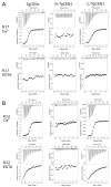
ITC characterization of the interactions of TgCEN1 and its domains with the R17 and R12 peptides in the presence of CaCl2 or EGTA. Representative thermograms (top panels) and the derived binding isotherms (bottom panels) of titration of R17 (A) and R12 (B) into TgCEN1, N-TgCEN1, and C-TgCEN1 in the presence of 5 mM CaCl2 or 5 mM EGTA at 25 °C. The ligand dilution blank experiments (peptide titrated into buffer) were subtracted from the binding isotherm obtained in the presence of protein. A first injection of 0.2 μL was made and then the first data point was removed from data fitting.
Figure 4
Figure 4
Conformational features of Ca2+-TgCEN1-peptide interaction. (A) 1H-15N-HSQC spectra of Ca2+-TgCEN1 in the absence (black) and presence (blue) of 2.5 molar excess of R10. The figure inset box contains the chemical shift changes of the two N-terminal glycines at position 6 of the EF-hand binding loop (G43 and G79) during the titration. Selected protein:peptide molar ratios are 0 (black), 0.5 (yellow), 1 (red), and 2.5 (blue). (B) 1H-15N-HSQC spectra of Ca2+-TgCEN1 in the absence (black) and presence (orange) of 2.5 molar excess of R17. (C) 1H-15N-HSQC spectra of Ca2+-TgCEN1 in the absence (black) and presence (green) of 2.5 molar excess of R12. (D) SEC profiles of Ca2+-TgCEN1 alone (black line) and in complex with R10 (red line), R17 (green line) or R12 (blue line) peptides. All runs were performed on a Superose 12 10/300 GL column in buffer containing 5 mM CaCl2. In all runs, the same concentration of Ca2+-TgCEN1 (2 mg/mL) was used and a 1:2 molar ratio of R10 peptide and 1:1 molar ratio of R17 or R12 peptides was added.
Figure 5
Figure 5
Far-UV CD analysis of R10 binding to intact TgCEN1 and its isolated domains in the presence of CaCl2 or EGTA. (A,B) Far-UV CD spectra of R10 peptide alone (black line), TgCEN1 (red line), and protein–peptide complex (green line) in the presence of (A) 5 mM CaCl2 or (B) EGTA. The CD spectrum resulting from subtraction of the spectrum of protein–peptide complex from that of protein alone was also shown (blue line). (C,D) Far-UV CD spectra of R10 peptide alone (black line), N-TgCEN1 (violet line), or C-TgCEN1 (red line), N-TgCEN1–peptide complex (green line) or C-TgCEN1–peptide complex (blue line) in the presence of (C) 5 mM CaCl2 or (D) EGTA. In all spectra, the same concentration of TgCEN1 (0.2 mg/mL) was used, and a 1:2 molar ratio of R10 peptide was added. For the single domains, the same concentration of N-TgCEN1 or C-TgCEN1 (0.2 mg/mL) was used, and a 1:1 molar ratio of R10 peptide was added. Increasing the peptide-to-protein ratio was not accompanied by obvious CD changes, in perfect agreement with the ITC-obtained stoichiometry.
Figure 6
Figure 6
Binding of TgCEN1 and its domains to the different peptides in the presence of CaCl2 or EGTA measured by Trp fluorescence. Trp fluorescence emission spectra of peptide alone (black line), and upon addition of intact TgCEN1 (red line), C-TgCEN1 (blue line), or N-TgCEN1 (green line) in the presence of CaCl2 (A,C,E) or EGTA (B,D,F).
Figure 7
Figure 7
Schematic illustration of the binding modes of TgCEN1 with different repeats from TgSFI1. TgCEN1 is shown as a dumbbell, indicating its two-domain structure (N- and C-lobes in white and gray, respectively); open circles denote unoccupied Ca2+-binding sites; filled black circles are Ca2+ ions. Yellow, blue, and red α-helices denote the representative R10, R17, and R12 target peptides, respectively. TgCEN1 exhibits two binding sites for R10 with distinct affinities: the C-lobe binds the peptide with very high affinity and poor Ca2+ sensitivity, while the N-lobe binds R10 with moderate affinity only in the presence of Ca2+. TgCEN1 binds R17 with moderate affinity only via the C-lobe in a strictly Ca2+-dependent manner. TgCEN1 interacts with R12 through the C-terminal domain with moderate Ca2+ sensitivity.

Similar articles

Cited by

References

    1. Paoletti A., Moudjou M., Paintrand M., Salisbury J.L., Bornens M. Most of centrin in animal cells is not centrosome-associated and centrosomal centrin is confined to the distal lumen of centrioles. Pt 13J. Cell Sci. 1996;109:3089–3102. doi: 10.1242/jcs.109.13.3089. - DOI - PubMed
    1. Salisbury J.L., Baron A.T., Sanders M.A. The centrin-based cytoskeleton of Chlamydomonas reinhardtii: Distribution in interphase and mitotic cells. J. Cell Biol. 1988;107:635–641. doi: 10.1083/jcb.107.2.635. - DOI - PMC - PubMed
    1. Salisbury J.L. A mechanistic view on the evolutionary origin for centrin-based control of centriole duplication. J. Cell. Physiol. 2007;213:420–428. doi: 10.1002/jcp.21226. - DOI - PubMed
    1. Nishi R., Okuda Y., Watanabe E., Mori T., Iwai S., Masutani C., Sugasawa K., Hanaoka F. Centrin 2 stimulates nucleotide excision repair by interacting with xeroderma pigmentosum group C protein. Mol. Cell Biol. 2005;25:5664–5674. doi: 10.1128/MCB.25.13.5664-5674.2005. - DOI - PMC - PubMed
    1. Jani D., Lutz S., Marshall N.J., Fischer T., Kohler A., Ellisdon A.M., Hurt E., Stewart M. Sus1, Cdc31, and the Sac3 CID region form a conserved interaction platform that promotes nuclear pore association and mRNA export. Mol. Cell. 2009;33:727–737. doi: 10.1016/j.molcel.2009.01.033. - DOI - PMC - PubMed

Publication types

MeSH terms

Grants and funding

This research was supported by departmental funds provided by the Italian Ministry of Research and Education (FUR2020 to AA and PD), and in part by the Italian MIUR-PRIN 2017 grant No. 2017ZBBYNC (to AA).

LinkOut - more resources