K205R specific nanobody-horseradish peroxidase fusions as reagents of competitive ELISA to detect African swine fever virus serum antibodies
- PMID: 35987654
- PMCID: PMC9392344
- DOI: 10.1186/s12917-022-03423-0
K205R specific nanobody-horseradish peroxidase fusions as reagents of competitive ELISA to detect African swine fever virus serum antibodies
Abstract
Background: African swine fever virus (ASFV) is a highly contagious hemorrhagic disease and often lethal, which has significant economic consequences for the swine industry. Due to lacking of commercial vaccine, the prevention and control of ASF largely depend on early large-scale detection and screening. So far, the commercial ELISA kits have a long operation time and are expensive, making it difficult to achieve large-scale clinical applications. Nanobodies are single-domain antibodies produced by camelid animals, and have unique advantages such as smaller molecular weight, easy genetic engineering modification and low-costing of mass production, thus exhibiting good application prospects.
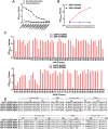
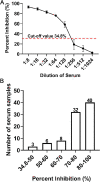
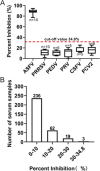
Results: The present study developed a new method for detection of ASFV specific antibodies using nanobody-horseradish peroxidase (Nb-HRP) fusion proteins as probe. By using camel immunization, phage library construction and phage display technology, five nanobodies against K205R protein were screened. Then, Nb-HRP fusion proteins were produced using genetic modification technology. Based on the Nb-HRP fusion protein as specific antibodies against K205R protein, a new type of cELISA was established to detect ASFV antibodies in pig serum. The cut-off value of the cELISA was 34.8%, and its sensitivity, specificity, and reproducibility were good. Furthermore, the developed cELISA exhibited 99.3% agreement rate with the commercial available ELISA kit (kappa value = 0.98).
Conclusions: The developed cELISA method has the advantages of simple operation, rapid and low-costing, and can be used for monitoring of ASFV infection in pigs, thus providing a new method for the prevention and control of ASF.
Keywords: ASFV; Antibody; K205R; Nanobody-HRP; cELISA.
© 2022. The Author(s).
Conflict of interest statement
All the authors approved the final manuscript and they have no competing interests to declare.
Figures


Similar articles
-
HRP-conjugated-nanobody-based cELISA for rapid and sensitive clinical detection of ASFV antibodies.Appl Microbiol Biotechnol. 2022 Jun;106(11):4269-4285. doi: 10.1007/s00253-022-11981-4. Epub 2022 May 25. Appl Microbiol Biotechnol. 2022. PMID: 35612629 Free PMC article.
-
A simple nanobody-based competitive ELISA to detect antibodies against African swine fever virus.Virol Sin. 2022 Dec;37(6):922-933. doi: 10.1016/j.virs.2022.09.004. Epub 2022 Sep 8. Virol Sin. 2022. PMID: 36089216 Free PMC article.
-
Nanobodies against African swine fever virus p72 and CD2v proteins as reagents for developing two cELISAs to detect viral antibodies.Virol Sin. 2024 Jun;39(3):478-489. doi: 10.1016/j.virs.2024.04.002. Epub 2024 Apr 6. Virol Sin. 2024. PMID: 38588947 Free PMC article.
-
Research progress on the proteins involved in African swine fever virus infection and replication.Front Immunol. 2022 Jul 22;13:947180. doi: 10.3389/fimmu.2022.947180. eCollection 2022. Front Immunol. 2022. PMID: 35935977 Free PMC article. Review.
-
[African swine fever in Russian Federation].Vopr Virusol. 2012 Sep-Oct;57(5):4-10. Vopr Virusol. 2012. PMID: 23248852 Review. Russian.
Cited by
-
Bridging the Gap: Can COVID-19 Research Help Combat African Swine Fever?Viruses. 2023 Sep 15;15(9):1925. doi: 10.3390/v15091925. Viruses. 2023. PMID: 37766331 Free PMC article. Review.
-
A review on camelid nanobodies with potential application in veterinary medicine.Vet Res Commun. 2024 Aug;48(4):2051-2068. doi: 10.1007/s11259-024-10432-x. Epub 2024 Jun 13. Vet Res Commun. 2024. PMID: 38869749 Review.
-
Identification and characterization of nanobodies specifically against African swine fever virus major capsid protein p72.Front Microbiol. 2022 Oct 13;13:1017792. doi: 10.3389/fmicb.2022.1017792. eCollection 2022. Front Microbiol. 2022. PMID: 36312984 Free PMC article.
-
Screening and affinity optimization of single domain antibody targeting the SARS-CoV-2 nucleocapsid protein.PeerJ. 2024 Aug 30;12:e17846. doi: 10.7717/peerj.17846. eCollection 2024. PeerJ. 2024. PMID: 39224822 Free PMC article.
References
-
- Zhao D, Liu R, Zhang X, Li F, Wang J, Zhang J, Liu X, Wang L, Zhang J, Wu X, Guan Y, Chen W, Wang X, He X, Bu Z. Replication and virulence in pigs of the first African swine fever virus isolated in China. Emerg Microbes Infect. 2019;8:438–447. doi: 10.1080/22221751.2019.1590128. - DOI - PMC - PubMed
MeSH terms
Substances
LinkOut - more resources
Full Text Sources

