

Small molecule photocatalysis enables drug target identification via energy transfer
- PMID: 35969791
- PMCID: PMC9407219
- DOI: 10.1073/pnas.2208077119
Small molecule photocatalysis enables drug target identification via energy transfer
Abstract
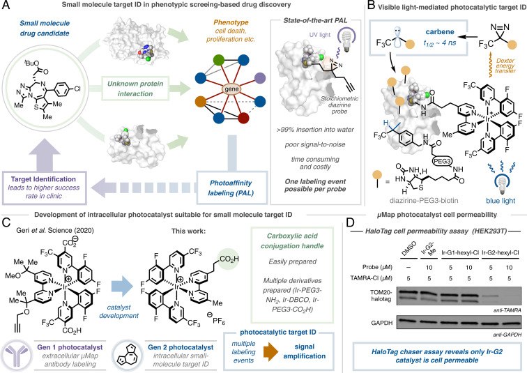
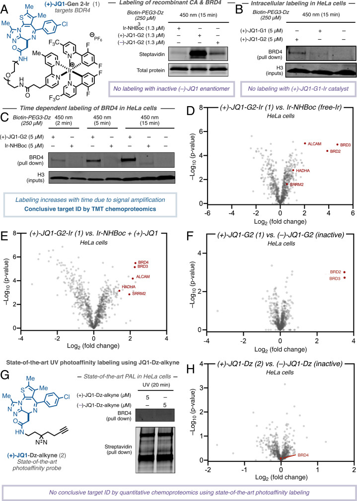
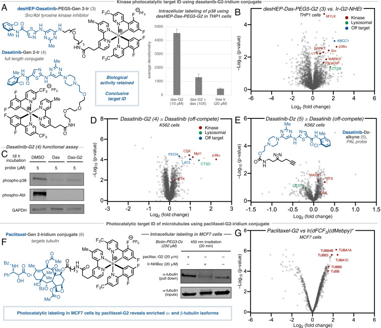
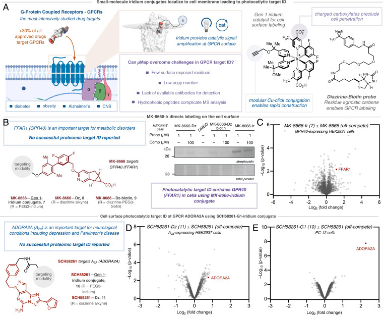
Over half of new therapeutic approaches fail in clinical trials due to a lack of target validation. As such, the development of new methods to improve and accelerate the identification of cellular targets, broadly known as target ID, remains a fundamental goal in drug discovery. While advances in sequencing and mass spectrometry technologies have revolutionized drug target ID in recent decades, the corresponding chemical-based approaches have not changed in over 50 y. Consigned to outdated stoichiometric activation modes, modern target ID campaigns are regularly confounded by poor signal-to-noise resulting from limited receptor occupancy and low crosslinking yields, especially when targeting low abundance membrane proteins or multiple protein target engagement. Here, we describe a broadly general platform for photocatalytic small molecule target ID, which is founded upon the catalytic amplification of target-tag crosslinking through the continuous generation of high-energy carbene intermediates via visible light-mediated Dexter energy transfer. By decoupling the reactive warhead tag from the small molecule ligand, catalytic signal amplification results in unprecedented levels of target enrichment, enabling the quantitative target and off target ID of several drugs including (+)-JQ1, paclitaxel (Taxol), dasatinib (Sprycel), as well as two G-protein-coupled receptors-ADORA2A and GPR40.
Keywords: photocatalysis; proteomics; target identification.
Conflict of interest statement
Competing interest statement: A provisional US patent has been filed by A.D.T., C.P.S., and D.W.C.M. based in part on this work (62/982,366; 63/076,658). International Application PCT/US2021/019959. D.W.C.M. declares an ownership interest, and A.D.T. and C.P.S. declare an affiliation interest, in the company Dexterity Pharma LLC, which has commercialized materials used in this work. D.W.C.M. declares an ownership interest in Penn PhD, which has commercialized materials used in this work.
Figures
Similar articles
-
[Recent advances in protein precipitation-based methods for drug-target screening].Se Pu. 2024 Jul;42(7):613-622. doi: 10.3724/SP.J.1123.2023.11019. Se Pu. 2024. PMID: 38966970 Free PMC article. Review. Chinese.
-
Target identification of natural and traditional medicines with quantitative chemical proteomics approaches.Pharmacol Ther. 2016 Jun;162:10-22. doi: 10.1016/j.pharmthera.2016.01.010. Epub 2016 Jan 22. Pharmacol Ther. 2016. PMID: 26808165 Review.
-
[Chemical proteomics and discovery of drug targets].Yao Xue Xue Bao. 2011 Aug;46(8):877-82. Yao Xue Xue Bao. 2011. PMID: 22007509 Review. Chinese.
-
[Development of antituberculous drugs: current status and future prospects].Kekkaku. 2006 Dec;81(12):753-74. Kekkaku. 2006. PMID: 17240921 Review. Japanese.
-
Photochemical Methods Applied to DNA Encoded Library (DEL) Synthesis.Acc Chem Res. 2023 Feb 7;56(3):385-401. doi: 10.1021/acs.accounts.2c00778. Epub 2023 Jan 19. Acc Chem Res. 2023. PMID: 36656960 Free PMC article.
Cited by
-
Etomoxir repurposed as a promiscuous fatty acid mimetic chemoproteomic probe.iScience. 2024 Aug 2;27(9):110642. doi: 10.1016/j.isci.2024.110642. eCollection 2024 Sep 20. iScience. 2024. PMID: 39252970 Free PMC article.
-
µMap proximity labeling in living cells reveals stress granule disassembly mechanisms.Nat Chem Biol. 2024 Aug 30:10.1038/s41589-024-01721-2. doi: 10.1038/s41589-024-01721-2. Online ahead of print. Nat Chem Biol. 2024. PMID: 39215100
-
Targeted proximity-labelling of protein tyrosines via flavin-dependent photoredox catalysis with mechanistic evidence for a radical-radical recombination pathway.Chem Sci. 2023 May 17;14(26):7327-7333. doi: 10.1039/d3sc00638g. eCollection 2023 Jul 5. Chem Sci. 2023. PMID: 37416718 Free PMC article.
-
Ligand-Directed Photocatalysts and Far-Red Light Enable Catalytic Bioorthogonal Uncaging inside Live Cells.J Am Chem Soc. 2023 Mar 22;145(11):6067-6078. doi: 10.1021/jacs.2c10655. Epub 2023 Mar 7. J Am Chem Soc. 2023. PMID: 36881718 Free PMC article.
-
Tracking chromatin state changes using nanoscale photo-proximity labelling.Nature. 2023 Apr;616(7957):574-580. doi: 10.1038/s41586-023-05914-y. Epub 2023 Apr 5. Nature. 2023. PMID: 37020029 Free PMC article.
References
-
- Arrowsmith J., Miller P., Trial watch: Phase II and phase III attrition rates 2011-2012. Nat. Rev. Drug Discov. 12, 569 (2013). - PubMed
-
- Chan J. N. Y., Nislow C., Emili A., Recent advances and method development for drug target identification. Trends Pharmacol. Sci. 31, 82–88 (2010). - PubMed
-
- Hart C. P., Finding the target after screening the phenotype. Drug Discov. Today 10, 513–519 (2005). - PubMed
-
- Williams M., Target validation. Curr. Opin. Pharmacol. 3, 571–577 (2003). - PubMed
Publication types
MeSH terms
Grants and funding
LinkOut - more resources
Full Text Sources
Other Literature Sources
Research Materials
