Review
. 2022 Jul 27:13:946428.
doi: 10.3389/fimmu.2022.946428. eCollection 2022.

Antimicrobial Proteins and Peptides in Avian Eggshell: Structural Diversity and Potential Roles in Biomineralization

Affiliations
Review

Antimicrobial Proteins and Peptides in Avian Eggshell: Structural Diversity and Potential Roles in Biomineralization

Thierry Moreau et al. Front Immunol. .

Abstract

The calcitic avian eggshell provides physical protection for the embryo during its development, but also regulates water and gaseous exchange, and is a calcium source for bone mineralization. The calcified eggshell has been extensively investigated in the chicken. It is characterized by an inventory of more than 900 matrix proteins. In addition to proteins involved in shell mineralization and regulation of its microstructure, the shell also contains numerous antimicrobial proteins and peptides (AMPPs) including lectin-like proteins, Bacterial Permeability Increasing/Lipopolysaccharide Binding Protein/PLUNC family proteins, defensins, antiproteases, and chelators, which contribute to the innate immune protection of the egg. In parallel, some of these proteins are thought to be crucial determinants of the eggshell texture and its resulting mechanical properties. During the progressive solubilization of the inner mineralized eggshell during embryonic development (to provide calcium to the embryo), some antimicrobials may be released simultaneously to reinforce egg defense and protect the egg from contamination by external pathogens, through a weakened eggshell. This review provides a comprehensive overview of the diversity of avian eggshell AMPPs, their three-dimensional structures and their mechanism of antimicrobial activity. The published chicken eggshell proteome databases are integrated for a comprehensive inventory of its AMPPs. Their biochemical features, potential dual function as antimicrobials and as regulators of eggshell biomineralization, and their phylogenetic evolution will be described and discussed with regard to their three-dimensional structural characteristics. Finally, the repertoire of chicken eggshell AMPPs are compared to orthologs identified in other avian and non-avian eggshells. This approach sheds light on the similarities and differences exhibited by AMPPs, depending on bird species, and leads to a better understanding of their sequential or dual role in biomineralization and innate immunity.

Keywords: 3D protein structure; antimicrobial peptides and proteins; avian egg; biomineralizing properties; calcite; eggshell.


Conflict of interest statement

The authors declare that the research was conducted in the absence of any commercial or financial relationships that could be construed as a potential conflict of interest.

Figures
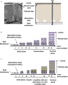
Figure 1
Figure 1
Avian eggshell structure and kinetics of its mineralization events. (A) Scanning electron microscope photography (left) and diagram (right) showing the six different layers composing the avian eggshell. From inside to outside are the two proteinaceous eggshell membrane layers supporting the calcified region of the shell (mammillary, palisade and vertical crystal layers). The most external zone is the thin organic cuticle layer. (B) Scheme representing shell mineralization pivotal stages that determine the eggshell ultrastructure and crystallographic texture in most bird species. They correspond to the initial stage dominated by amorphous calcium carbonate (ACC) deposition on eggshell membranes, its progressive transformation to form calcite aggregates on mammillary knobs, and the growth of large calcite units. The last stages correspond to the formation of the columnar calcite crystals and the deposition of cuticle. Black arrows describe the orientation of crystals that compete for the available space and only those favorably oriented nearly perpendicular to the shell surface (along the c-axis), are selected and contribute to the development of a preferential orientation of crystals. Timing (hours post-ovulation) are given for the kinetics of chicken eggshell fabrication. (C) Specific bilayer structure of the Guinea fowl eggshell. Guinea fowl eggshell mineralization initially follows the same pattern described in chicken resulting in the characteristic columnar structure (black arrows). However, a sharp change in the size and orientation of crystals occurs (yellow arrows) at 11 hours post ovulation, when one-third of the final eggshell thickness has been deposited. At this point, the large columnar calcite units break up into smaller crystal units with varying crystallographic orientations forming a microstructure with an intricate interlacing of calcite crystals, leading to the formation of a secondary layer with misoriented crystals.
Figure 2
Figure 2
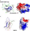
3D structures of eggshell lectin-like proteins, ovocleidin-17 and VMO1. 3D structure of chicken ovocleidin-17 (A) and chicken VMO1 (B). The left panels correspond to the cartoon representation of 3D structure while the right panels show the color-coded electrostatic potential molecular surface. Color scheme ranges from red (negatively charged regions) to deep blue (positively charged regions). The Ala60-Gly68 exposed loop of ovocleidin-17 containing the two phosphorylatable serine residues (Ser61 and Ser67) is indicated. The figure was prepared using Pymol software (48) and APBS (Adaptive Poisson-Boltzmann Solver) plugin (49) for electrostatic calculations using atomic coordinates of 1GZ2 and 1VMO PDB files (www.rcsb.org) for OC-17 and VMO1, respectively.
Figure 3
Figure 3
3D structures of human BPI and chicken eggshell BPI/LBP/PLUNC proteins, ovocalyxin-36 and TENP. 3D structure of human BPI (A), chicken ovocalyxin-36 (B), and chicken TENP (C). The left panels correspond to the cartoon representation of 3D structure while the right panels show the color-coded electrostatic potential molecular surface. Color scheme ranges from red (negatively charged regions) to deep blue (positively charged regions). The figure was prepared using Pymol software (48) and APBS (Adaptive Poisson-Boltzmann Solver) plugin (49) for electrostatic calculations using atomic coordinates of 1BP1 PDB file (www.rcsb.org) for human BPI. The structure of ovocalyxin-36 was predicted by homology modeling using SwissModel server (swissmodel.expasy.org) and human BPI as a template. The 3D structure of TENP was modeled using the AlphaFold algorithm and was retrieved in the AlphaFold protein structure database under accession number AF-O42273-F1 (72, 73).
Figure 4
Figure 4
3D structures of chicken defensins AvBD11 and OvoDA1. 3D structure of chicken AvBD11 (A) and chicken OvoDA1 (B). The left panels correspond to the cartoon representation of 3D structure while the right panels show the color-coded electrostatic potential molecular surface. Color scheme ranges from red (negatively charged regions) to deep blue (positively charged regions). The figure was prepared using Pymol software (48) and APBS (Adaptive Poisson-Boltzmann Solver) plugin (49) for electrostatic calculations using atomic coordinates of 6QEU and 2MJK PDB files (www.rcsb.org) for AvBD11 and OvoDA1 structure, respectively.
Figure 5
Figure 5
3D structures of avian lysozymes, lysozyme C and lysozyme G 3D structure of chicken lysozyme C (A) and goose lysozyme G (B). The left panels correspond to the cartoon representation of 3D structure while the right panels show the color-coded electrostatic potential molecular surface. Color scheme ranges from red (negatively charged regions) to deep blue (positively charged regions). The figure shows a disaccharide of N-acetyl-glucosamine bound in the active site of lysozyme C while the structure of lysozyme G is shown as a complex with a trisaccharide of N-acetyl glucosamine (green and red, balls-and-stick representation). The figure was prepared using Pymol software (48) and APBS (Adaptive Poisson-Boltzmann Solver) plugin (49) for electrostatic calculations using atomic coordinates of 1SF4 and 154L PDB file for hen egg white lysozyme C and goose lysozyme G, respectively.
Figure 6
Figure 6
3D structures of egg antiproteases with cystatin fold, chicken cystatin and ovocalyxin-32. 3D structure of chicken cystatin (A) and chicken ovocalyxin-32 (B). The left panels correspond to the cartoon representation of 3D structure while the right panels show the color-coded electrostatic potential molecular surface. Color scheme ranges from red (negatively charged regions) to deep blue (positively charged regions). The figure was prepared using Pymol software (48) and APBS (Adaptive Poisson-Boltzmann Solver) plugin (49) for electrostatic calculations using X-ray atomic coordinates of 1CEW PDB file for chicken cystatin. The 3D structure of OCX-32 was modeled using the AlphaFold method and was retrieved in the AlphaFold protein structure database under accession number AF-Q90YI1-F1 (72, 73).
Figure 7
Figure 7
3D structure of the serpin ovalbumin-related protein X (OVAX). The left panel corresponds to the cartoon representation of 3D structure of chicken OVAX while the right panel shows the color-coded electrostatic potential molecular surface. Color scheme ranges from red (negatively charged regions) to deep blue (positively charged regions). Fondaparinux is shown in stick representation (carbon: green, oxygen: red, sulfur: orange, nitrogen: blue). The figure was prepared using Pymol software (48) and APBS (Adaptive Poisson-Boltzmann Solver) plugin (49) for electrostatic calculations using X-ray atomic coordinates of OVAX kindly provided by F. Coste (CBM, CNRS, UPR4301, Orléans, France) prior to the release of these data in the PDB under 7QRN accession code.
Figure 8
Figure 8
3D structure of egg proteins with chelating activities: avidin and ovotransferrin. 3D structure of chicken avidin (A) and chicken ovotransferrin (B). The left panels correspond to the cartoon representation of 3D structure while the right panels illustrate the color-coded electrostatic potential molecular surface. Color scheme ranges from red (negatively charged regions) to deep blue (positively charged regions). Avidin (A) is complexed to the vitamin biotin through numerous interactions including H-bonds and hydrophobic interactions. Ovotransferrin (B) possesses two lobes, each containing two domains. Each lobe binds a Fe3+ ion and a carbonate ion. Location of the antibacterial peptide OTAP-92 is also shown. The figure was prepared using Pymol software (48) and APBS (Adaptive Poisson-Boltzmann Solver) plugin (49) for electrostatic calculations using atomic coordinates of 2AVI and 1N04 PDB file for avidin-biotin complex and ovotransferrin, respectively.
Figure 9
Figure 9
3D structure of the iron-chelator Ex-FABP. The left panel corresponds to the cartoon representation of 3D structure of chicken Ex-FABP while the right panel shows the color-coded electrostatic potential molecular surface. Color scheme ranges from red (negatively charged regions) to deep blue (positively charged regions). The figure shows an Fe3+ ion (colored orange)-dihydoxybenzoate complex bound in one of the three subcavities of Ex-FABP calyx. Dihydroxybenzoate (DHB) and lysophosphatidic acid (LPA) are shown in stick representation. The figure was prepared using Pymol software (48) and APBS (Adaptive Poisson-Boltzmann Solver) plugin (49) for electrostatic calculations using 3SAO PDB file for chicken Ex-FABP structure.
Figure 10
Figure 10
3D structures of β-microseminoprotein-like MSMB3. 3D structure of chicken MSMB3 monomer (A) and dimer (B). The left panels correspond to the cartoon representation of 3D structure while the right panels show the color-coded electrostatic potential molecular surface. Color scheme ranges from red (negatively charged regions) to deep blue (positively charged regions). The figure was prepared using Pymol software (48) and APBS (Adaptive Poisson-Boltzmann Solver) plugin (49) for electrostatic calculations using atomic coordinates of 6RWC PDB file.
Figure 11
Figure 11
3D structure of pleiotrophin. The left panel (A) corresponds to the cartoon representation of chicken pleiotrophin 3D structure while the right panel (B) illustrates the color-coded electrostatic potential molecular surface of N-ter and C-ter domains. Color scheme ranges from red (negatively charged regions) to deep blue (positively charged regions). Pleiotrophin has two thrombospondin type-1 repeat domains (TSR), one with a two-stranded β-sheet (N-ter domain) and the second with a three-stranded β-sheet (C-ter domain) linked by a flexible hinge region. Both TSR domains are flanked by unstructured termini. The structure of chicken pleiotrophin was predicted by comparative modeling using SwissModel server (swissmodel.expasy.org) and human pleiotrophin (PDB code 2N6F) as a template. The figure was prepared using Pymol software (48) and APBS (Adaptive Poisson-Boltzmann Solver) plugin (49) for electrostatic calculations.
Figure 12
Figure 12
Graphical representation of the innate immune functions of eggshell AMPPs in eggshell mineralization and antibacterial protection. (A) During the formation of the eggshell in the uterus, ions of the mineral phase (calcium, carbonate ions) and proteins (including AMPPs) are secreted by the uterine epithelial cells into the extracellular milieu (uterine fluid). Some AMPPs can interact with ions and participate in the stabilization of ACC and/or regulate the growth of calcite crystals. These AMPPs become progressively embedded in the eggshell mineral during the mineralization process and are assumed to be inactive once immobilized. If bacterial contamination of the uterine fluid occurs during eggshell formation, the presence of soluble AMPPs can provide an antimicrobial defense to keep the interior of the egg free from pathogens. (B) Once the egg is laid, the eggshell exterior is exposed to environmental bacterial species that colonize the egg cuticle surface. In contrast to AMPPs embedded in the calcite layer, surface-exposed cuticle AMPPs may directly interact with bacteria and modulate the eggshell microbiome. (C) During egg incubation, the embryo-derived chorioallantoic membrane progressively develops at the inner surface of the eggshell membranes and creates a local acidification that dissolves the mammillary layer mineral to release calcium and carbonate ions required for embryonic bone calcification, and also to liberate the numerous mineral-associated proteins. Once solubilized, eggshell AMPPs are thought to recover their biological activity and locally reinforce innate immune protection to resist bacterial contamination following the penetration of pathogens through eggshell pores or microcracks. (Original artwork by TM, NG and HDF).

Similar articles

References

    1. Gautron J, Stapane L, Le Roy N, Nys Y, Rodriguez-Navarro AB, Hincke MT. Avian Eggshell Biomineralization: An Update on its Structure, Mineralogy and Protein Tool Kit. BMC Mol Cell Biol (2021) 22(1):11. doi: 10.1186/s12860-021-00350-0 - DOI - PMC - PubMed
    1. Hincke MT, Nys Y, Gautron J, Mann K, Rodriguez-Navarro AB, McKee MD. The Eggshell: Structure, Composition and Mineralization. Front Biosci (Landm Ed) (2012) 17:1266–80. doi: 10.2741/3985 - DOI - PubMed
    1. Kulshreshtha G, D'Alba L, Dunn IC, Réhault-Godbert S, Rodriguez-Navarro AB, Hincke MT. Properties, Genetics and Innate Immune Function of the Cuticle in Egg-Laying Species. Front Immunol (2022) 13:838525. doi: 10.3389/fimmu.2022.838525 - DOI - PMC - PubMed
    1. Wisocki PA, Kennelly P, Rojas Rivera I, Cassey P, Burkey ML, Hanley D. The Global Distribution of Avian Eggshell Colours Suggest a Thermoregulatory Benefit of Darker Pigmentation. Nat Ecol Evol (2020) 4(1):148–55. doi: 10.1038/s41559-019-1003-2 - DOI - PubMed
    1. Marie P, Labas V, Brionne A, Harichaux G, Hennequet-Antier C, Nys Y, et al. . Quantitative Proteomics and Bioinformatic Analysis Provide New Insight Into Protein Function During Avian Eggshell Biomineralization. J Proteomics (2015) 113:178–93. doi: 10.1016/j.jprot.2014.09.024 - DOI - PubMed

Publication types

LinkOut - more resources