

Regulation of microglial activation in stroke in aged mice: a translational study
- PMID: 35963621
- PMCID: PMC9417226
- DOI: 10.18632/aging.204216
Regulation of microglial activation in stroke in aged mice: a translational study
Abstract
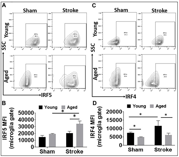
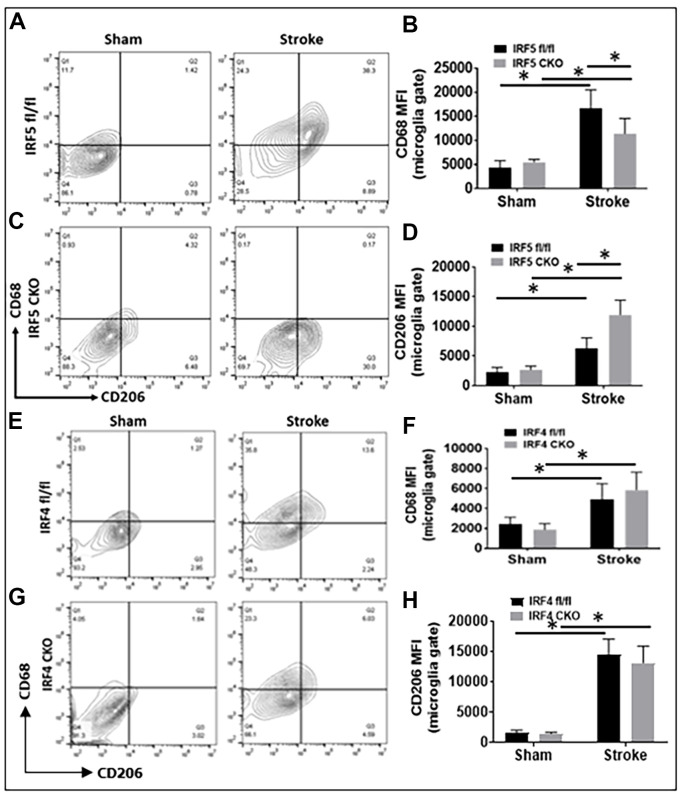
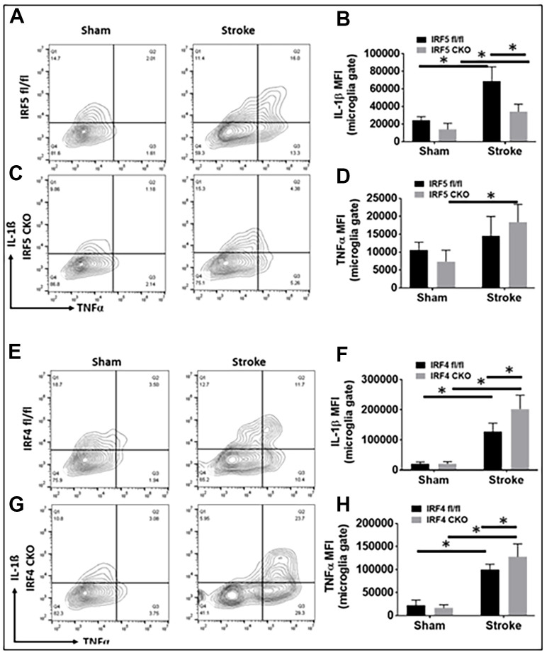
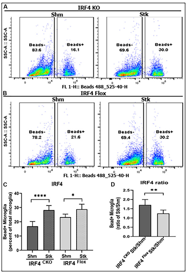
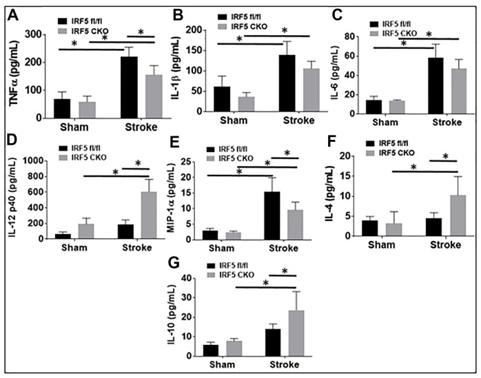
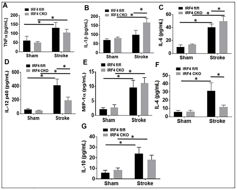
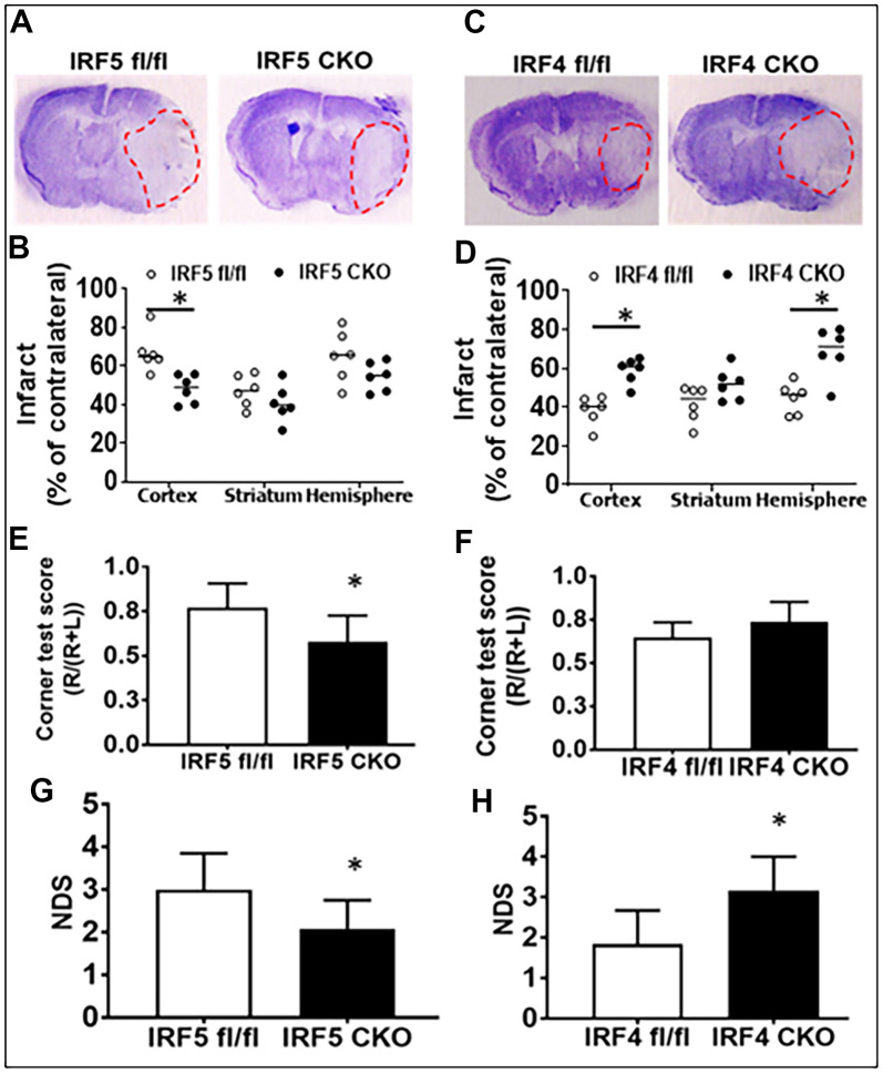
Numerous neurochemical changes occur with aging and stroke mainly affects the elderly. Our previous study has found interferon regulatory factor 5 (IRF5) and 4 (IRF4) regulate neuroinflammation in young stroke mice. However, whether the IRF5-IRF4 regulatory axis has the same effect in aged brains is not known. In this study, aged (18-20-month-old), microglial IRF5 or IRF4 conditional knockout (CKO) mice were subjected to a 60-min middle cerebral artery occlusion (MCAO). Stroke outcomes were quantified at 3d after MCAO. Flow cytometry and ELISA were performed to evaluate microglial activation and immune responses. We found aged microglia express higher levels of IRF5 and lower levels of IRF4 than young microglia after stroke. IRF5 CKO aged mice had improved stroke outcomes; whereas worse outcomes were seen in IRF4 CKO vs. their flox controls. IRF5 CKO aged microglia had significantly lower levels of IL-1β and CD68 than controls; whereas significantly higher levels of IL-1β and TNF-α were seen in IRF4 CKO vs. control microglia. Plasma levels of TNF-α and MIP-1α were decreased in IRF5 CKO vs. flox aged mice, and IL-1β/IL-6 levels were increased in IRF4 CKO vs. controls. The anti-inflammatory cytokines (IL-4/IL-10) levels were higher in IRF5 CKO, and lower in IRF4 CKO aged mice vs. their flox controls. IRF5 and IRF4 signaling drives microglial pro- and anti-inflammatory response respectively; microglial IRF5 is detrimental and IRF4 beneficial for aged mice in stroke. IRF5-IRF4 axis is a promising target for developing new, effective therapeutic strategies for the cerebral ischemia.
Keywords: IRF; aging; inflammation; microglia; stroke.
Conflict of interest statement
Figures
Similar articles
-
Central IRF4/5 Signaling Are Critical for Microglial Activation and Impact on Stroke Outcomes.Transl Stroke Res. 2024 Aug;15(4):831-843. doi: 10.1007/s12975-023-01172-2. Epub 2023 Jul 11. Transl Stroke Res. 2024. PMID: 37432594 Free PMC article.
-
Microglial IRF5-IRF4 regulatory axis regulates neuroinflammation after cerebral ischemia and impacts stroke outcomes.Proc Natl Acad Sci U S A. 2020 Jan 21;117(3):1742-1752. doi: 10.1073/pnas.1914742117. Epub 2019 Dec 31. Proc Natl Acad Sci U S A. 2020. PMID: 31892541 Free PMC article.
-
Interferon regulatory factor 4/5 signaling impacts on microglial activation after ischemic stroke in mice.Eur J Neurosci. 2018 Jan;47(2):140-149. doi: 10.1111/ejn.13778. Eur J Neurosci. 2018. PMID: 29131464 Free PMC article.
-
Regulation of microglial activation in stroke.Acta Pharmacol Sin. 2017 Apr;38(4):445-458. doi: 10.1038/aps.2016.162. Epub 2017 Mar 6. Acta Pharmacol Sin. 2017. PMID: 28260801 Free PMC article. Review.
-
Bystanders or not? Microglia and lymphocytes in aging and stroke.Neural Regen Res. 2023 Jul;18(7):1397-1403. doi: 10.4103/1673-5374.360345. Neural Regen Res. 2023. PMID: 36571333 Free PMC article. Review.
Cited by
-
Updates of the role of B-cells in ischemic stroke.Front Cell Neurosci. 2024 Mar 14;18:1340756. doi: 10.3389/fncel.2024.1340756. eCollection 2024. Front Cell Neurosci. 2024. PMID: 38550918 Free PMC article. Review.
-
Physical exercise regulates microglia in health and disease.Front Neurosci. 2024 Jun 7;18:1420322. doi: 10.3389/fnins.2024.1420322. eCollection 2024. Front Neurosci. 2024. PMID: 38911597 Free PMC article. Review.
-
Progesterone Receptor Agonist, Nestorone, Exerts Long-Term Neuroprotective Effects Against Permanent Focal Cerebral Ischemia in Adult and Aged Male Rats.Transl Stroke Res. 2024 Aug 22. doi: 10.1007/s12975-024-01288-z. Online ahead of print. Transl Stroke Res. 2024. PMID: 39172309
-
The Aging Immune System: A Critical Attack on Ischemic Stroke.Mol Neurobiol. 2024 Sep 14. doi: 10.1007/s12035-024-04464-2. Online ahead of print. Mol Neurobiol. 2024. PMID: 39271626 Review.
-
Escape of Kdm6a from X chromosome is detrimental to ischemic brains via IRF5 signaling.Res Sq [Preprint]. 2024 Sep 27:rs.3.rs-4986866. doi: 10.21203/rs.3.rs-4986866/v1. Res Sq. 2024. Update in: Transl Stroke Res. 2025 Jan 3. doi: 10.1007/s12975-024-01321-1. PMID: 39399684 Free PMC article. Updated. Preprint.
References
Publication types
MeSH terms
Substances
Grants and funding
LinkOut - more resources
Full Text Sources
Medical
Molecular Biology Databases
