. 2022 Aug 8;23(15):8804.
doi: 10.3390/ijms23158804.

The Effects of Nutrient Signaling Regulators in Combination with Phytocannabinoids on the Senescence-Associated Phenotype in Human Dermal Fibroblasts

Affiliations

The Effects of Nutrient Signaling Regulators in Combination with Phytocannabinoids on the Senescence-Associated Phenotype in Human Dermal Fibroblasts

Marta Gerasymchuk et al. Int J Mol Sci. .

Abstract

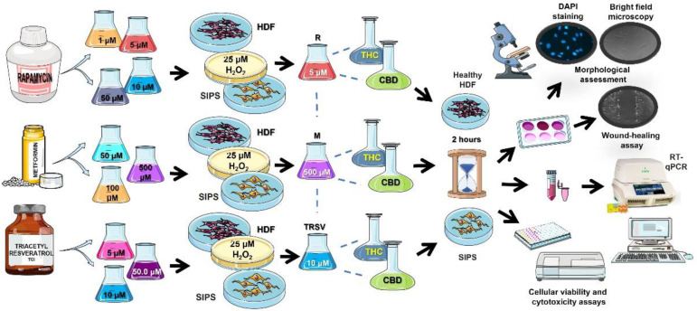
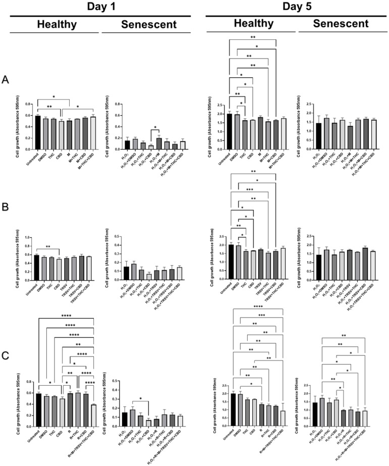
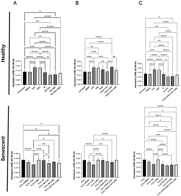
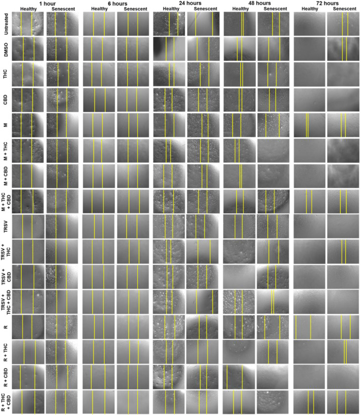
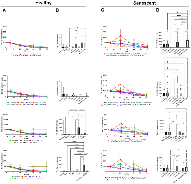
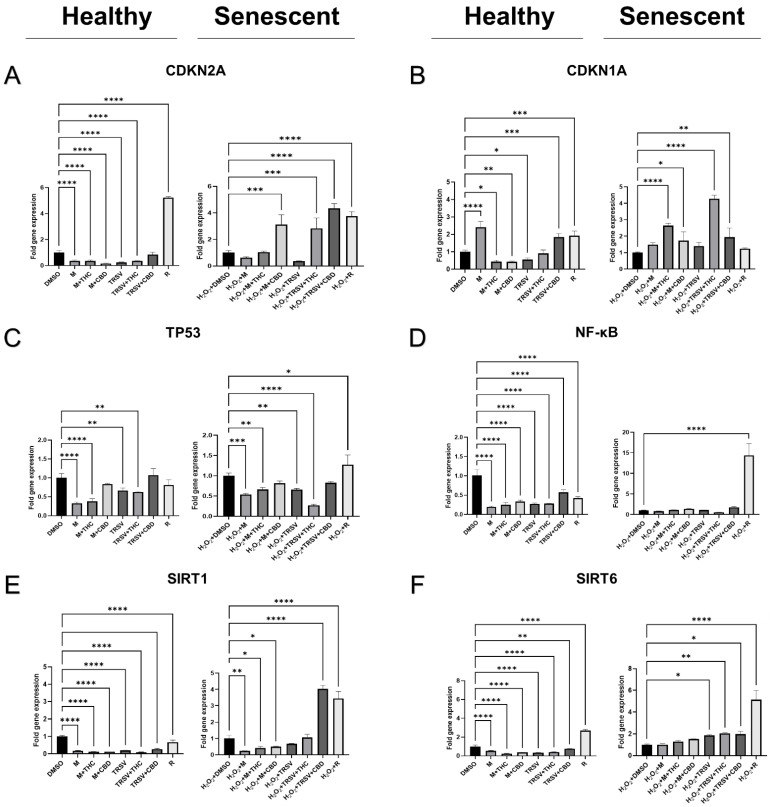
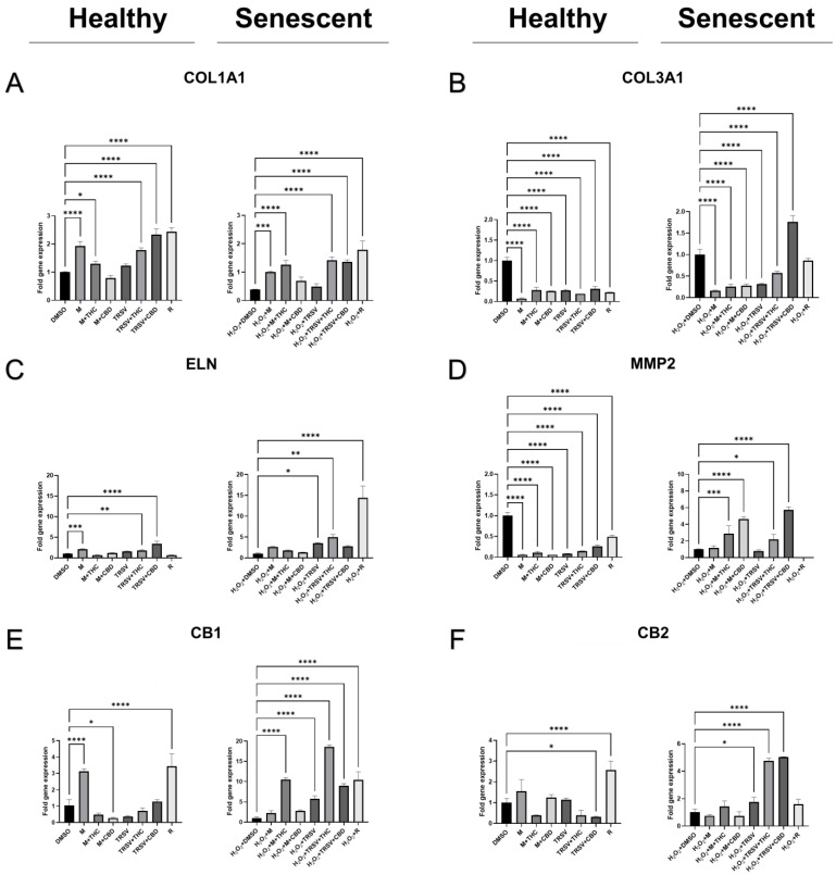
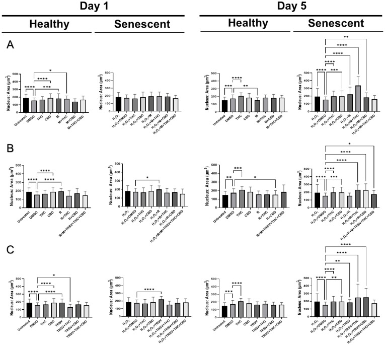
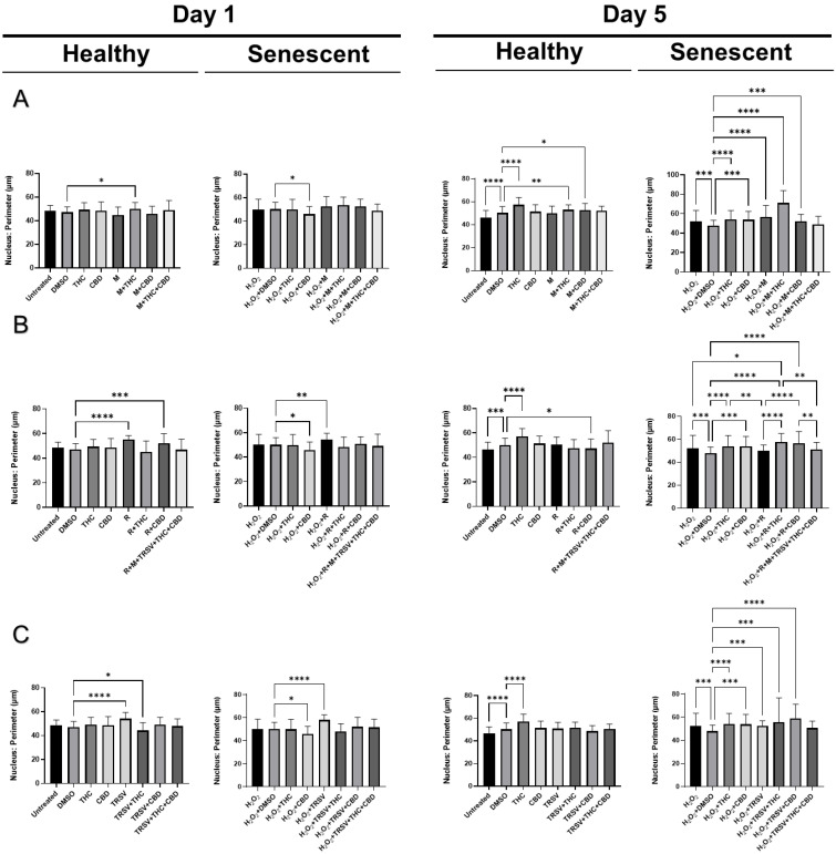
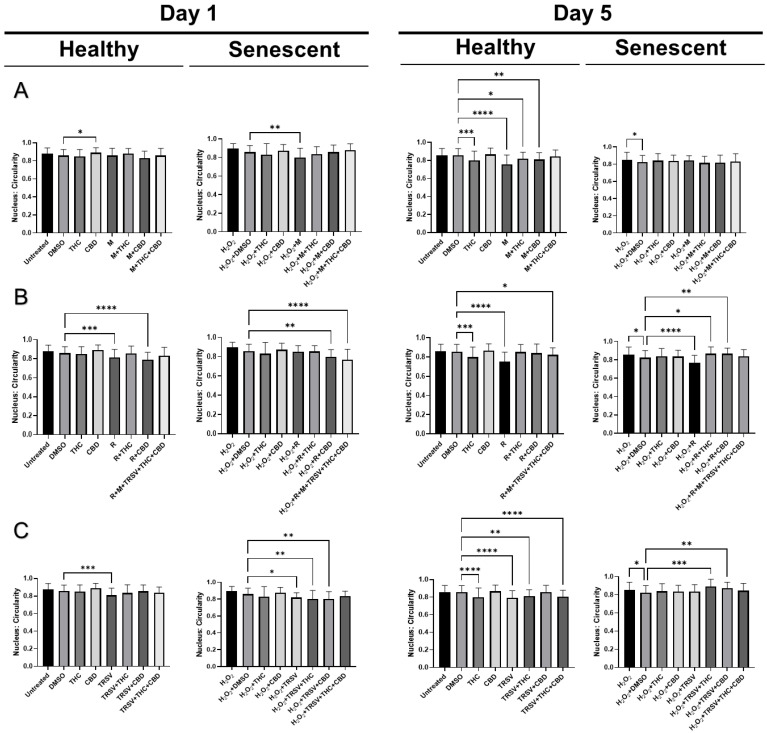
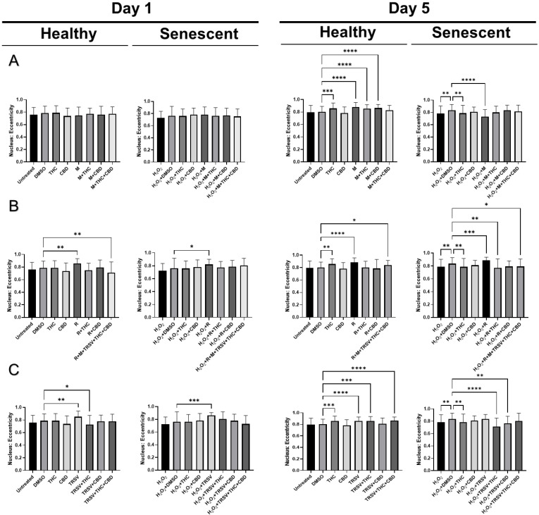
Identifying effective anti-aging compounds is a cornerstone of modern longevity, aging, and skin-health research. There is considerable evidence of the effectiveness of nutrient signaling regulators such as metformin, resveratrol, and rapamycin in longevity and anti-aging studies; however, their potential protective role in skin aging is controversial. In light of the increasing appearance of phytocannabinoids in beauty products without rigorous research on their rejuvenation efficacy, we decided to investigate the potential role of phytocannabinoids in combination with nutrient signaling regulators in skin rejuvenation. Utilizing CCD-1064Sk skin fibroblasts, the effect of metformin, triacetylresveratrol, and rapamycin combined with phytocannabinoids on cellular viability, functional activity, metabolic function, and nuclear architecture was tested. We found triacetylresveratrol combined with cannabidiol increased the viability of skin fibroblasts (p < 0.0001), restored wound-healing functional activity (p < 0.001), reduced metabolic dysfunction, and ameliorated nuclear eccentricity and circularity in senescent fibroblasts (p < 0.01). Conversely, metformin with or without phytocannabinoids did not show any beneficial effects on functional activity, while rapamycin inhibited cell viability (p < 0.01) and the speed of wound healing (p < 0.001). Therefore, triacetylresveratrol and cannabidiol can be a valuable source of biologically active substances used in aging and more studies using animals to confirm the efficacy of cannabidiol combined with triacetylresveratrol should be performed.

Keywords: CBD; THC; aging; fibroblast; metformin; rapamycin; skin; stress-induced premature senescence; triacetylresveratrol.


Conflict of interest statement

The authors declare no conflict of interest.

Figures

Figure 1
Figure 1
Testing of different concentrations of anti-aging drugs on prematurely senescent dermal fibroblasts. The figure shows steps to discover the efficient dose in treating senescent fibroblasts among three popular anti-aging drugs: rapamycin (R), metformin (M), and triacetylresveratrol (TRSV). CBD, cannabidiol; HDF, human dermal fibroblasts; H2O2, hydrogen peroxide; SIPS, stress-induced premature senescence; THC, Δ-9-tetrahydrocannabinol. Dashed lines show a combination of treatments. This figure was created using images from Servier Medical Art Commons Attribution 3.0 Unported License (http://smart.servier.com (accessed on 26 September 2020)).
Figure 2
Figure 2
Viability of dermal fibroblasts CCD-1064Sk (p.11) treated with nutrient-signaling regulators combined with phytocannabinoids. The graphs represent cell viability of skin fibroblasts analyzed by MTT assay after experimental treatments with nutrient-signaling regulators combined with THC and CBD. Graphs show cell viability after 1 and 5 days of exposure with (A) metformin, (B) triacetylresveratrol, and (C) rapamycin alone or in combination with phytocannabinoids compared to controls and phytocannabinoids in both the healthy and senescent state. Data were analyzed with a one-way ANOVA test followed by a Tukey post hoc multiple comparison test. Bars represent mean ± SD. Significance is indicated within the figures using the following scale: * p < 0.05, ** p < 0.01, *** p < 0.001, **** p < 0.0001. CBD, cannabidiol; DMSO, dimethyl sulfoxide (vehicle), M, metformin; R, rapamycin; THC, Δ-9-tetrahydrocannabinol; TRSV, triacetylresveratrol.
Figure 3
Figure 3
Viability of dermal fibroblasts CCD-1064Sk (p.11) treated with potential anti-aging compounds combined with phytocannabinoids. The graphs represent cell viability of skin fibroblasts estimated by NR assay after 72 h of exposure with (A) metformin, (B) triacetylresveratrol, and (C) rapamycin alone or in combination with phytocannabinoids compared to controls and phytocannabinoids in both the healthy and senescent state. Data were analyzed with a one-way ANOVA test followed by a Tukey post hoc multiple comparison test. Bars represent mean ± SD. Significance is indicated within the figures using the following scale: * p < 0.05, ** p < 0.01, *** p < 0.001, **** p < 0.0001. CBD, cannabidiol; DMSO, dimethyl sulfoxide (vehicle), M, metformin; R, rapamycin; THC, Δ-9-tetrahydrocannabinol; TRSV, triacetylresveratrol.
Figure 4
Figure 4
The effect of nutrient signaling regulators combined with phytocannabinoids on human skin fibroblasts CCD-1064Sk (p.11) in the wound-healing assay. Images show size of wounds after exposure to NSRs and/or pCBs for 1, 6, 24, 48, and 72 h in healthy and SIPS fibroblasts. Yellow lines delineate the edge of the wound. CBD, cannabidiol; DMSO, dimethyl sulfoxide (vehicle), M, metformin; R, rapamycin; THC, Δ-9-tetrahydrocannabinol; TRSV, triacetylresveratrol. Images were captured at 80X magnification using a light microscope.
Figure 5
Figure 5
The effect of nutrient signaling regulators combined with phytocannabinoids on wound healing. Graphs represented wound healing assay results in CCD-1064Sk (p.11) dermal fibroblasts. (A) the percentage of an unhealed wound in healthy fibroblasts treated with metformin, TRSV and rapamycin combined with pCBs. (B) the percentage of the unhealed wound after 72 h of experiment in healthy cells with metformin, TRSV and rapamycin combined with pCBs. (C) the percentage of an unhealed wound in senescent fibroblasts culture treated with metformin, TRSV and rapamycin combined with pCBs. (D) the percentage of the unhealed wound after 72 h in prematurely aged fibroblasts metformin, TRSV and rapamycin combined with pCBs. Data were analyzed with a one-way ANOVA test followed by a Tukey post hoc multiple comparison test. Bars represent mean ± SD. Significance is indicated within the figures using the following scale: * p < 0.05, ** p < 0.01, *** p < 0.001, **** p < 0.0001. CBD, cannabidiol; DMSO, dimethyl sulfoxide (vehicle), M, metformin; R, rapamycin; THC, Δ-9-tetrahydrocannabinol; TRSV, triacetylresveratrol.
Figure 6
Figure 6
The expression of cellular checkpoint regulators in CCD-1064Sk (p.11) fibroblasts treated with nutrient signaling regulators combined with cannabinoids. Changes of mRNA expression as measured by RT-qPCR for (A) CDKN2A (p16), (B) CDKN1A (p21), (C) TP53, (D) NF-κB, (E) SIRT1, and (F) SIRT6 in healthy and senescent fibroblasts. Data were analyzed with a one-way ANOVA test followed by a Dunnett post hoc test compared to the vehicle. Bars represent mean ± SD. Significance is indicated within the figures using the following scale: * p < 0.05, ** p < 0.01, *** p < 0.001, **** p < 0.0001. CBD, cannabidiol; DMSO, dimethyl sulfoxide (vehicle), M, metformin; R, rapamycin; THC, Δ-9-tetrahydrocannabinol; TRSV, triacetylresveratrol.
Figure 7
Figure 7
Effects of phytocannabinoids and nutrient signaling regulators on the expression of genes involved in the production of extracellular matrix in CCD-1064Sk (p.11). Changes of mRNA expression as measured by RT-qPCR for (A) COL1A1; (B) COL3A1, (C) ELN (elastin), (D) MMP2, (E) CB1, and (F) CB2 in healthy and senescent fibroblasts. Data were analyzed with a one-way ANOVA test followed by a Dunnett post hoc multiple comparison test. Bars represent mean ± SD. Significance is indicated within the figures using the following scale: * p < 0.05, ** p < 0.01, *** p < 0.001, **** p < 0.0001. CBD, cannabidiol; DMSO, dimethyl sulfoxide (vehicle), M, metformin; R, rapamycin; THC, Δ-9-tetrahydrocannabinol; TRSV, triacetylresveratrol.
Figure 8
Figure 8
DAPI-stained nuclei area parameters of skin fibroblasts CCD-1064Sk (p.11) exposed to metformin, triacetylresveratrol, and rapamycin combined with pCBs. Nuclear parameters were observed by immunofluorescence microscopy for healthy and senescent fibroblasts treated with (A) 500 μM metformin, (B) 5 μM rapamycin, or (C) 10 μM triacetylresveratrol and combinations with pCBs compared to 2 μM of THC, 2 μM of CBD, DMSO, and untreated fibroblasts after 1 day and 5 days of exposure. Data were analyzed with a one-way ANOVA test followed by a Dunnett’s post hoc test compared to the DMSO or H2O2 + DMSO control. Bars represent mean ± SD. Significance is indicated within the figures using the following scale: * p < 0.05, ** p < 0.01, *** p < 0.001, **** p < 0.0001. CBD, cannabidiol; DMSO, dimethyl sulfoxide (vehicle), M, metformin; R, rapamycin; THC, Δ-9-tetrahydrocannabinol; TRSV, triacetylresveratrol.
Figure 9
Figure 9
DAPI-stained nuclei perimeter parameters of skin fibroblasts CCD-1064Sk (p.11) exposed to metformin, triacetylresveratrol, and rapamycin combined with pCBs. Nuclear parameters were observed by immunofluorescence microscopy for: healthy and senescent fibroblasts treated with (A) 500 μM metformin, (B) 5 μM rapamycin, or (C) 10 μM triacetylresveratrol and combinations with pCBs compared to 2 μM of THC, 2 μM of CBD, DMSO, and untreated fibroblasts after 1 day and 5 days of exposure. Data were analyzed with a one-way ANOVA test followed by a Dunnett’s post hoc test compared to the DMSO or H2O2 + DMSO control. Bars represent mean ± SD. Significance is indicated within the figures using the following scale: * p < 0.05, ** p < 0.01, *** p < 0.001, **** p < 0.0001. CBD, cannabidiol; DMSO, dimethyl sulfoxide (vehicle), M, metformin; R, rapamycin; THC, Δ-9-tetrahydrocannabinol; TRSV, triacetylresveratrol.
Figure 10
Figure 10
DAPI-stained nuclei circularity parameters of skin fibroblasts CCD-1064Sk (p.11) exposed to metformin, triacetylresveratrol, and rapamycin combined with pCBs. Nuclear parameters were observed by immunofluorescence microscopy for: healthy and senescent fibroblasts treated with (A) 500 μM metformin, (B) 5 μM rapamycin, or (C) 10 μM triacetylresveratrol and combinations with pCBs compared to 2 μM of THC, 2 μM of CBD, DMSO, and untreated fibroblasts after 1 day and 5 days of exposure. Data were analyzed with a one-way ANOVA test followed by a Dunnett’s post hoc test compared to the DMSO or H2O2 + DMSO control. Bars represent mean ± SD. Significance is indicated within the figures using the following scale: * p < 0.05, ** p < 0.01, *** p < 0.001, **** p < 0.0001. CBD, cannabidiol; DMSO, dimethyl sulfoxide (vehicle), M, metformin; R, rapamycin; THC, Δ-9-tetrahydrocannabinol; TRSV, triacetylresveratrol.
Figure 11
Figure 11
DAPI stained nuclei eccentricity parameters of skin fibroblasts CCD-1064Sk (p.11) exposed to metformin, triacetylresveratrol, and rapamycin combined with pCBs. Nuclear parameters were observed by immunofluorescence microscopy for: healthy and senescent fibroblasts treated with (A) 500 μM metformin, (B) 5 μM rapamycin, or (C) 10 μM triacetylresveratrol and combinations with pCBs compared to 2 μM of THC, 2 μM of CBD, DMSO, and untreated fibroblasts after 1 day and 5 days of exposure. Data were analyzed with a one-way ANOVA test followed by a Dunnett’s post hoc test compared to the DMSO or H2O2 + DMSO control. Bars represent mean ± SD. Significance is indicated within the figures using the following scale: * p < 0.05, ** p < 0.01, *** p < 0.001, **** p < 0.0001. CBD, cannabidiol; DMSO, dimethyl sulfoxide (vehicle), M, metformin; R, rapamycin; THC, Δ-9-tetrahydrocannabinol; TRSV, triacetylresveratrol.

Similar articles

Cited by

References

    1. Shetty A.K., Kodali M., Upadhya R., Madhu L.N. Emerging Anti-Aging Strategies—Scientific Basis and Efficacy. Aging Dis. 2018;9:1165. doi: 10.14336/AD.2018.1026. - DOI - PMC - PubMed
    1. Gerasymchuk M., Cherkasova V., Kovalchuk O., Kovalchuk I. The Role of microRNAs in Organismal and Skin Aging. Int. J. Mol. Sci. 2020;21:5281. doi: 10.3390/ijms21155281. - DOI - PMC - PubMed
    1. Zhu M., Meng P., Ling X., Zhou L. Advancements in therapeutic drugs targeting of senescence. Ther. Adv. Chronic Dis. 2020;11:2040622320964125. doi: 10.1177/2040622320964125. - DOI - PMC - PubMed
    1. Thoppil H., Riabowol K. Senolytics: A Translational Bridge Between Cellular Senescence and Organismal Aging. Front. Cell Dev. Biol. 2019;7:367. doi: 10.3389/fcell.2019.00367. - DOI - PMC - PubMed
    1. Cummings N.E., Lamming D.W. Regulation of metabolic health and aging by nutrient-sensitive signaling pathways. Mol. Cell. Endocrinol. 2017;455:13–22. doi: 10.1016/j.mce.2016.11.014. - DOI - PMC - PubMed

LinkOut - more resources