. 2022 Aug 8;14(15):6202-6226.
doi: 10.18632/aging.204211. Epub 2022 Aug 8.

SIAH1-mediated RPS3 ubiquitination contributes to chemosensitivity in epithelial ovarian cancer

Affiliations

SIAH1-mediated RPS3 ubiquitination contributes to chemosensitivity in epithelial ovarian cancer

Lu Chen et al. Aging (Albany NY). .

Abstract

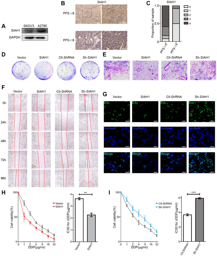
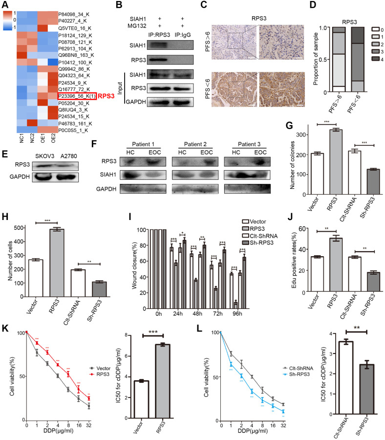
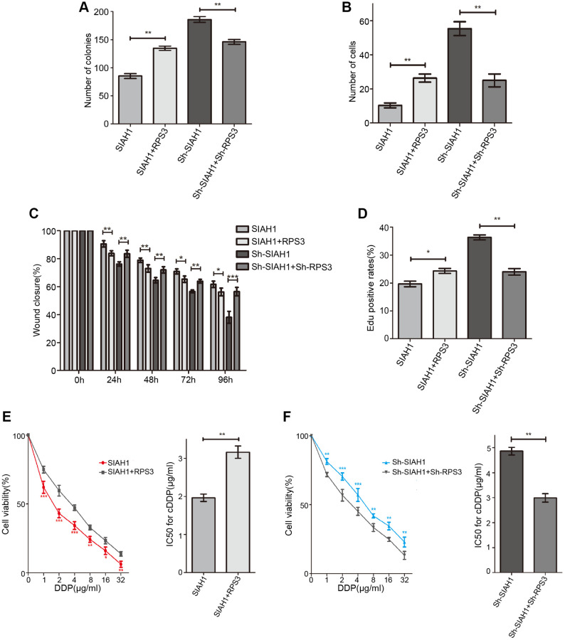
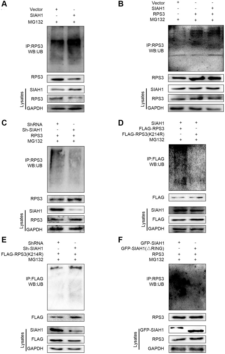
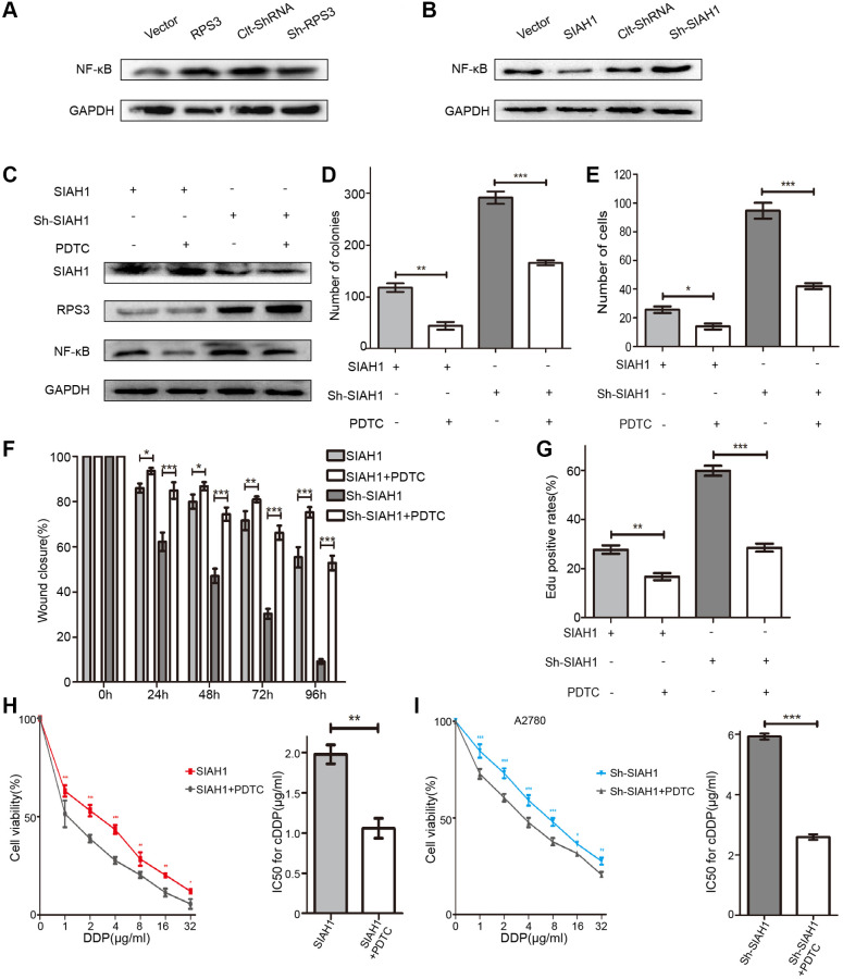
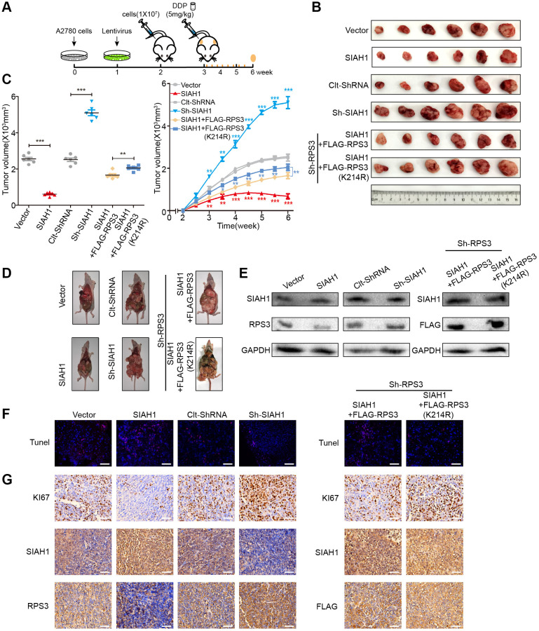
The E3 ligase SIAH1 is deregulated in human cancers and correlated with poor prognosis, but its contributions to chemoresistance in epithelial ovarian cancer (EOC) are not evident. Herein we found that SIAH1 was decreased in EOC tumour tissues and cell lines and negatively correlated with the RPS3 levels. SIAH1 overexpression suppressed tumour cell growth, colony formation, invasion, metastasis, and cisplatin resistance in vivo and in vitro. SIAH1 promoted RPS3 ubiquitination and degradation using the RING-finger domain, and these steps were required for RPS3 localization to the cytoplasm, which led to subsequent NF-κB inactivation and thereby conferred chemosensitivity. Moreover, ectopic expression of RPS3 or depletion of RPS3 ubiquitination mediated by SIAH1 via the K214R mutant significantly impaired cisplatin-induced tumour suppression in cells stably expressing SIAH1. Together, our findings reveal a tumour suppressor function of SIAH1 and provide evidence showing that the SIAH1-RPS3-NF-κB axis may act as an appealing strategy for tackling treatment resistance in EOC.

Keywords: EOC; NF-κB; RPS3; SIAH1; chemoresistance.


Conflict of interest statement

CONFLICTS OF INTEREST: The authors declare no conflicts of interest related to this study.

Figures

Figure 1
Figure 1
SIAH1 sensitizes ovarian cancer cells to cDDP. (A) Western blotting for SIAH1 in SKOV3 and A2780 cells. (B) Representative images of immunohistochemical staining for SIAH1 in tumour specimens from ovarian cancer patients with PFS > 6 months vs. PFS < 6 months. Scale bar: 200 μm (left) and 100 μm (right). (C) Staining was assessed and scored on a scale of 0 (<5% staining) to 4 (>75% staining). The quantification of IHC staining (n = 24; PFS > 6, n = 12; PFS < 6, n = 12) was shown. The results from a Cell Colony formation assay (D) and Transwell assay (E) of A2780 cells transfected with Vector, SIAH1, Clt-shRNA and sh-SIAH1 were shown. Scale bar: 400 μm. A wound-healing assay was used to assess the effects of SIAH1 on cellular motility over time as shown (0, 24, 48, 72 and 96 h; Scale bar: 400 μm) (F). The results from a Cell Edu assay (Scale bar: 200 μm) (G), Cell Viability (H, I left panels) and IC50 for cDDP (H, I right panels) in A2780 cells were shown.*p < 0.05, **p < 0.01, ***p < 0.001.
Figure 2
Figure 2
Identification of RPS3 as an interaction partner of SIAH1. (A) The heatmap showed that the protein level of RPS3 was down-regulated after SIAH1 overexpression in EOC cells. (B) Pull down of the RPS3 protein with SIAH1 antibody and IgG antibody in A2780 cells transfected with SIAH1. Cells were treated with 10 μM MG132 for 6 h before cell lysis. (C) Representative images of immunohistochemical staining for RPS3 in tumour specimens from ovarian cancer patients with PFS > 6 months vs. PFS < 6 months. Scale bar: 200 μm (left) and 100 μm (right). (D) The staining was assessed and scored on a scale of 0 (<5% staining) to 4 (>75% staining). The quantification of IHC staining (n = 24; PFS > 6, n = 12; PFS < 6, n = 12) was shown. (E) Protein level of RPS3 in SKOV3 and A2780 cells. (F) Protein levels of SIAH1 and RPS3 in serum samples from healthy controls (HC) and EOC patients (EOC). A2780 cells were separately transfected with Vector, RPS3, Clt-shRNA and sh-RPS3 for 48 h, the number of Cell Colonies was determined (G), the cell number from the Transwell assay was obtained (H), the Wound closure percentage was calculated (0, 24, 48, 72 and 96 h) (I), and the Edu positive rates (J), Cell Viability (K, L left panels), IC50 for cDDP (K, L right panels) were obtained. *p < 0.05, **p < 0.01, ***p < 0.001.
Figure 3
Figure 3
SIAH1 down-regulates the protein level of RPS3. A2780 cells were separately transfected with SIAH1, RPS3, sh-SIAH1 or sh-RPS3 for 48 h, the number of Cell Colonies was determined (A), the cell number from the Transwell assay was obtained (B), the Wound closure percentage was calculated (0, 24, 48, 72 and 96 h) (C), and the Edu positive rates (D), cell viability (E, F left panels), and IC50 for cDDP (E, F right panels) were measured. *p < 0.05, **p < 0.01, ***p < 0.001.
Figure 4
Figure 4
SIAH1 induces degradation of RPS3 protein. (A) A2780 cells were transfected with the Vector, SIAH1. Cells were treated with or without 10 μM MG132 for 6 h before cell lysis. and the resulting cell lysates were subjected to Western blotting. (B) RPS3 mRNA levels was detected in A2780 cells with SIAH1 overexpression. (C, D) The RPS3 protein half-life was assayed by using CHX (30 μg/ml) in HEK293T cells transfected with GFP or GFP-SIAH1 plasmid. The relative remaining RPS3 protein levels following CHX treatment at each time point were calculated accordingly. (E) Colocalization of RPS3 and SIAH1. The GFP-SIAH1 or Cherry-RPS3 plasmids were transfected into A2780 cells. Cells were treated with or without 10 μM MG132 for 6 h before cell lysis. GFP-SIAH1 was detected using a fluorescence microscope with an excitation wavelength of 488 nm. Cherry-RPS3 was detected with an excitation wavelength of 556 nm. The cell nuclei were stained with DAPI. Scale bar: 50 μm. (F) A2780 cells were transfected with plasmids as indicated, and the RPS3 protein was pull down with GFP antibody. Cell lysates were subjected to Western blotting. *p < 0.05, ***p < 0.001.
Figure 5
Figure 5
SIAH1 is an E3 ligase to induce RPS3 ubiquitination. (AF) HEK293T cells were transfected plasmids as indicated for 36 h. Cell lysates were subjected to denatured immunoprecipitation and Western blotting. Cells were treated with MG132 (10 μM) for 6 h before cell lysis. (A) SIAH1 overexpression induced ubiquitination and degradation of endogenous RPS3. (B) SIAH1 overexpression induced ubiquitination and degradation of exogenous RPS3. (C) When SIAH1 was knocked down, the ubiquitination level of RPS3 was significantly reduced. (D) Upon SIAH1 expression, FLAG-RPS3 (K214R) had a significantly lower ubiquitination than the wild type (FLAG-RPS3). (E) SIAH1 KD, induced no significant decrease in the ubiquitination level of FLAG-RPS3 (K214R). (F) In SIAH1-inhibited A2780 cells, mutation of the RING finger domain of SIAH1 significantly reduced the ubiquitination level of RPS3.
Figure 6
Figure 6
SIAH1 inhibits the NF-κB pathway via RPS3 downregulation. (A) Western blotting for NF-κB p65 in A2780 cells separately transfected with Vector, SIAH1, Clt-shRNA and sh-SIAH1 for 48 h. (B) Western blotting for NF-κB p65 in A2780 cells separately transfected with Vector, RPS3, Clt-shRNA and sh-RPS3 for 48 h. (C) Western blotting for SIAH1, RPS3 and NF-κB p65 in A2780 cells separately transfected with SIAH1and sh-SIAH1 for 48 h, and treated with PDTC (200 μM) for 24 h. A2780 cells were separately transfected with SIAH1 and sh-SIAH1 for 48 h and treated with PDTC for 24 h. The number of Cell Colonies was determined (D), the cell number of Transwell assay was obtained (E), the Wound closure percentage was calculated (0, 24, 48, 72 and 96 h) (F), and the Edu positive rates (G), cell viability (H, I left panels), and IC50 for cDDP (H, I right panels) were measured in A2780 cells. *p < 0.05, **p < 0.01, ***p < 0.001.
Figure 7
Figure 7
SIAH1 enhances the sensitivity of ovarian cancer cells to cDDP by down-regulating RPS3 in vivo. A2780 cells were transfected with Vector, SIAH1, Clt-shRNA, sh-SIAH1, SIAH1+FLAG-RPS3 and SIAH1+FLAG-RPS3 (K214R) lentivirus and were then subcutaneously and intraperitoneally injected into female BALB/c nude mice, and potential tumours were allowed to grow for one week (n = 6). All the groups were administered DDP (5 mg/kg) by intraperitoneal injection three times a week for a total of 8 times before sacrifice. (A) Flow chart of the animal experiment. (B) Representative images of the excised tumours on day 35 after tumour cell injection. (C) Tumour volumes of the excised tumours on day 35 (left) and the tumor growth curves (right) after tumour cell injection. (D) Typical pictures of tumours in the abdominal cavity after intraperitoneal injection in each group of mice. (E) The levels of SIAH1 and RPS3 proteins in mouse xenograft tumour tissues were assessed by Western blotting. (F) The apoptosis level of tumour tissue in each group was detected by TUNEL assay. Scale bar: 100 μm. (G) Immunohistochemistry analyses for KI67, SIAH1, RPS3 and FLAG staining were carried out with A2780 xenograft tumour sections. Representative staining is shown. Scale bar: 100 μm. **p < 0.01, ***p < 0.001. Scale bar: 100 μm.
Figure 8
Figure 8
Schematic diagram of this study.

Similar articles

Cited by

References

    1. Liu D, Zhang XX, Li MC, Cao CH, Wan DY, Xi BX, Tan JH, Wang J, Yang ZY, Feng XX, Ye F, Chen G, Wu P, et al.. C/EBPβ enhances platinum resistance of ovarian cancer cells by reprogramming H3K79 methylation. Nat Commun. 2018; 9:1739. 10.1038/s41467-018-03590-5 - DOI - PMC - PubMed
    1. Konecny GE, Wang C, Hamidi H, Winterhoff B, Kalli KR, Dering J, Ginther C, Chen HW, Dowdy S, Cliby W, Gostout B, Podratz KC, Keeney G, et al.. Prognostic and therapeutic relevance of molecular subtypes in high-grade serous ovarian cancer. J Natl Cancer Inst. 2014; 106:dju249. 10.1093/jnci/dju249 - DOI - PMC - PubMed
    1. Agarwal R, Kaye SB. Ovarian cancer: strategies for overcoming resistance to chemotherapy. Nat Rev Cancer. 2003; 3:502–16. 10.1038/nrc1123 - DOI - PubMed
    1. Wang A, Li J, Zhou T, Li T, Cai H, Shi H, Liu A. CUEDC2 Contributes to Cisplatin-Based Chemotherapy Resistance in Ovarian Serious Carcinoma by Regulating p38 MAPK Signaling. J Cancer. 2019; 10:1800–7. 10.7150/jca.29889 - DOI - PMC - PubMed
    1. Du L, Li CR, He QF, Li XH, Yang LF, Zou Y, Yang ZX, Zhang D, Xing XW. Downregulation of the ubiquitin ligase KBTBD8 prevented epithelial ovarian cancer progression. Mol Med. 2020; 26:96. 10.1186/s10020-020-00226-7 - DOI - PMC - PubMed

Publication types