. 2022 Aug 10;17(8):e0271695.
doi: 10.1371/journal.pone.0271695. eCollection 2022.

Newly synthesized mRNA escapes translational repression during the acute phase of the mammalian unfolded protein response

Affiliations

Newly synthesized mRNA escapes translational repression during the acute phase of the mammalian unfolded protein response

Mohammed R Alzahrani et al. PLoS One. .

Abstract

Endoplasmic Reticulum (ER) stress, caused by the accumulation of misfolded proteins in the ER, elicits a homeostatic mechanism known as the Unfolded Protein Response (UPR). The UPR reprograms gene expression to promote adaptation to chronic ER stress. The UPR comprises an acute phase involving inhibition of bulk protein synthesis and a chronic phase of transcriptional induction coupled with the partial recovery of protein synthesis. However, the role of transcriptional regulation in the acute phase of the UPR is not well understood. Here we analyzed the fate of newly synthesized mRNA encoding the protective and homeostatic transcription factor X-box binding protein 1 (XBP1) during this acute phase. We have previously shown that global translational repression induced by the acute UPR was characterized by decreased translation and increased stability of XBP1 mRNA. We demonstrate here that this stabilization is independent of new transcription. In contrast, we show XBP1 mRNA newly synthesized during the acute phase accumulates with long poly(A) tails and escapes translational repression. Inhibition of newly synthesized RNA polyadenylation during the acute phase decreased cell survival with no effect in unstressed cells. Furthermore, during the chronic phase of the UPR, levels of XBP1 mRNA with long poly(A) tails decreased in a manner consistent with co-translational deadenylation. Finally, additional pro-survival, transcriptionally-induced mRNAs show similar regulation, supporting the broad significance of the pre-steady state UPR in translational control during ER stress. We conclude that the biphasic regulation of poly(A) tail length during the UPR represents a previously unrecognized pro-survival mechanism of mammalian gene regulation.


Conflict of interest statement

The authors have declared that no competing interests exist.

Figures
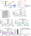
Fig 1
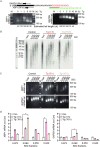
Fig 1. ER stress-induced translation inhibition stabilizes XBP1u mRNA during the acute UPR.
(A) (Left) Unconventional splicing of XBP1 mRNA in the cytoplasm. Upon accumulation of misfolded proteins, the cytoplasmic endoribonuclease IRE1α is activated and cleaves a 26-nucleotide (nt) intron from the coding region of the XBP1u mRNA. This process occurs in close proximity to the ER membrane. The resulting XBP1s mRNA is translated in the cytoplasm, and the XBP1s protein migrates to the nucleus to induce transcription of genes that protect cells from ER stress. The figure was created by Biorender.com (Right) Schematic of temporal responses to ER stress. This response includes an acute phase up to 3 h, and a chronic phase, beyond 6 h of treatment with stressors. (B) Western blot analysis of the indicated proteins in MEFs treated with Tg (400 nM) for the indicated durations. (C) (Left) The half-life of the spliced XBP1 mRNA (XBP1s) was determined by treating MEFs with Tg for the indicated durations. Actinomycin D (10 μg/ml) was added for 0, 0.5, 1, 2, 3, and 4 h at each indicated time of Tg-treatment to inhibit transcription and measure the mRNA decay rate at different time points of ER stress. (Right) The XBP1s mRNA levels were quantified by RT-qPCR. (D) The half-life and levels of XBP1u mRNA were assessed as in C, except MEF cells were treated with Tg in the presence of a selective inhibitor of the IRE1α endoribonuclease, 4μ8C (50 μM), for the indicated durations. In the absence of Tg treatment in the control condition, MEFs were treated with only 4μ8C for 1 h. (E) The half-life and levels of XBP1u mRNA were assessed in MEFs deficient in IRE1α protein (IRE1α-deficient MEFs). The evaluation of half-life and levels of XBP1u mRNA was performed as in C. (F) The half-life and levels of the XBP1u mRNA were determined in eIF2α-P-deficient MEFs in the presence of 4μ8C as described in C. (G) Protein synthesis was measured by [35S]-Met/Cys metabolic labeling in eIF2α-P-deficient MEFs in the presence of 4μ8C alone (Con) or together with Puromycin (Puro, 5 μg/ml) for 1 h to inhibit translation elongation. (H) The half-life of the XBP1u mRNA was measured in eIF2α-P-deficient MEFs treated with either 4μ8C for 1 h or Puro and 4μ8C for 1 h. Actinomycin D was added at the indicated times in the presence of 4μ8C or Puro and 4μ8C (t1/2 = 4μ8C: 1.2 +/- 0.2 h, 4μ8C+Puro: 3.5 +/-0.5 h).
Fig 2
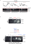
Fig 2. Acute UPR does not involve an increase of global poly(A) tail length of mRNAs.
(A) (top) Experimental diagram of the PCR-based poly(A) tailing assay [49] for XBP1 and GAPDH mRNAs. The 3’-end of total RNA was modified with GMP/IMP residues by poly(A) polymerase to form a GI-oligo tail connected to the poly(A) tail. Following RNA isolation and RT, universal primers were used as a reverse primer for cDNA amplification of targets. (Bottom) PCR-based poly(A) tailing assay in MEFs treated with Tg for the indicated durations. Estimated tail lengths are shown. As a negative control (-), a tail reaction was performed on cDNA derived from RNA not tagged with the GI-oligo tail. (B) mRNA fractionation based on poly(A) tail length in MEFs treated with Tg for the indicated durations. 3 h of Tg treatment represents acute ER stress, and 16 h of Tg treatment represents chronic ER stress. (C) The poly(A) tail length of XBP1 and GAPDH mRNA was estimated using the PCR-based poly(A) tailing assay in the fractionated mRNA pools from B. (D) The level of XBP1 and GAPDH mRNA was determined using RT-qPCR and compared to their own internal controls in all fractionated mRNA pools.
Fig 3
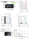
Fig 3. The long tail XBP1 mRNA escapes translational repression during the acute UPR.
(A) Polysome profiles of MEFs treated with Tg for 0, 1 and 16 h. Following RNA isolation and integrity check on agarose gels, RNA was combined into 3 pools as shown; ribosome-free {F}, light polyribosomes {L}, heavy polyribosomes {H}. (B) PCR-based poly(A) tailing assay for XBP1 mRNAs estimated in the pooled RNA from F, L, or H of Tg-treated MEFs for 0, 1, and 16 h. (C) Experimental schematic and PCR-based poly(A) tailing assay of XBP1 mRNA was estimated in 5-Ethynyl Uridine (5EU)-labeled RNA isolated from MEFs. Cells were treated with Tg for 1, or 16 h, and 5EU (400 μM) was added for the last 1 h of treatments. Vehicle (DMSO) was added in control cells for 1 h in the presence of 5EU. Following the click chemistry reaction, 5EU-labeled RNA was immobilized by streptavidin beads and was eluted {E}. The flow-through {FT} was also collected as unlabeled RNA. The PCR-based poly(A) tailing assay was performed on both E and FT pools as previously described and analyzed on agarose gels. As a negative control, MEFs were not treated with Tg or 5EU. (-) represents a sample of RNA without the GI-tail and serves as a negative control for the data as shown in Fig 2.
Fig 4
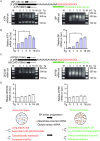
Fig 4. Polyadenylation of newly synthesized RNA during acute ER stress promotes cell survival.
(A) MEFs were treated with CPA for the indicated durations. Following RNA isolation and the PCR-based poly(A) tailing assay, the poly(A) tail length of XBP1 mRNA was measured in the indicated treatments. (B) MEFs were treated with DMSO (Con) or CPA (100 uM) for 3 h and cell viability was measured. Following CPA treatment, cells were allowed to recover in fresh media in the absence of CPA for 24 h past the 3 h treatment of CPA. (C) Cell survival was assayed in MEFs treated with DMSO (Con), CPA, 10 μg/ml cordycepin, or combined CPA and cordycepin for 3 h. (D) RT-qPCR analysis of XBP1 mRNA levels in MEFs treated with DMSO (Con), CPA, cordycepin, or combined treatment of CPA and cordycepin for 3 h. (E) MEFs were treated with DMSO (Con), CPA, cordycepin, or combined treatment of CPA with either cordycepin or Actinomycin D for 3 h. XBP1 mRNA poly(A) tail length was measured using the PCR-based poly(A) tailing assay. (F) Cells were treated with DMSO (Con), CPA, cordycepin, or combined treatment of CPA and cordycepin for 3 h followed by Actinomycin D treatment for the indicated durations. t1/2 = Con: 2 +/- 0.3 h, cordycepin: 2.3 +/- 0.2 h, CPA: 3.5 +/- 0.3 h, and CPA+cordycepin: 5.3 +/- 0.1 h.
Fig 5
Fig 5. Temporal regulation of poly(A) tail length of UPR-regulated genes.
(A) (top) Experimental diagram of the PCR-based poly(A) tailing assay of ATF4 and BiP mRNAs and (bottom) their dynamic poly(A) tail length in MEFs treated with Tg for the indicated durations. The poly(A) tail length of ATF4 and BiP mRNAs was estimated and a negative control (-) was used to monitor the tail reactions as previously explained. (**) indicates an artifact band. (B) The levels of ATF4 and BiP mRNA were assessed using RT-qPCR in Tg-treated MEFs for the indicated durations. (C) (top) Experimental diagram of the PCR-based poly(A) tailing assay of SEC24D and ATP5B mRNA and (bottom) their dynamic poly(A) tail length in MEFs treated with Tg for the indicated times As in Fig 5A. (D) The level of SEC24D and ATP5B mRNA was assessed using RT-qPCR in Tg-treated MEFs for the indicated durations. (E) Model of XBP1 mRNA regulation during acute and chronic ER stress. The steady state of the acute ER stress consists of different populations of XBP1 mRNA. In the acute phase, the pre-ER stress (old) mRNA is translationally repressed and stabilized. However, the newly synthesized (new) mRNA has long poly(A) tails and escapes translational repression. In chronic ER stress, the pre-ER stress (old) mRNA is partially translationally derepressed and becomes unstable. Meanwhile, the newly synthesized (new) mRNA is subject to poly(A) tail shortening due to co-translational deadenylation. The figure was created by Biorender.com.

Similar articles

Cited by

References

    1. Adams BM, Canniff NP, Guay KP, Larsen ISB, Hebert DN. Quantitative glycoproteomics reveals cellular substrate selectivity of the ER protein quality control sensors UGGT1 and UGGT2. Elife. 2020. Dec 15;9. doi: 10.7554/eLife.63997 - DOI - PMC - PubMed
    1. Guan BJ, van Hoef V, Jobava R, Elroy-Stein O, Valasek LS, Cargnello M, et al.. A Unique ISR Program Determines Cellular Responses to Chronic Stress. Mol Cell. 2017. Dec 7;68(5):885–900 e6. doi: 10.1016/j.molcel.2017.11.007 - DOI - PMC - PubMed
    1. Hetz C, Papa FR. The Unfolded Protein Response and Cell Fate Control. Mol Cell. 2018. Jan 18;69(2):169–81. doi: 10.1016/j.molcel.2017.06.017 - DOI - PubMed
    1. Wek RC. Role of eIF2alpha Kinases in Translational Control and Adaptation to Cellular Stress. Cold Spring Harb Perspect Biol. 2018. Jul 2;10(7). doi: 10.1101/cshperspect.a032870 - DOI - PMC - PubMed
    1. Costa-Mattioli M, Walter P. The integrated stress response: From mechanism to disease. Science. 2020. Apr 24;368(6489). doi: 10.1126/science.aat5314 - DOI - PMC - PubMed

Publication types

MeSH terms