. 2022 Aug 30;13(4):e0104222.
doi: 10.1128/mbio.01042-22. Epub 2022 Aug 10.

The Akt Forkhead Box O Transcription Factor Axis Regulates Human Cytomegalovirus Replication

Affiliations

The Akt Forkhead Box O Transcription Factor Axis Regulates Human Cytomegalovirus Replication

Hongbo Zhang et al. mBio. .

Abstract

The protein kinase Akt broadly impacts many cellular processes, including mRNA translation, metabolism, apoptosis, and stress responses. Inhibition of phosphatidylinositol 3-kinase (PI3K), a lipid kinase pivotal to Akt activation, triggers various herpesviruses to reactivate from latency. Hence, decreased Akt activity may promote lytic replication. Here, we show that Akt accumulates in an inactive form during human cytomegalovirus (HCMV) infection of permissive fibroblasts, as indicated by hypophosphorylation of sites that activate Akt, decreased phosphorylation of PRAS40, and pronounced nuclear localization of FoxO3a, a substrate that remains cytoplasmic when Akt is active. HCMV strongly activates mTORC1 during lytic infection, suggesting a potential mechanism for Akt inactivation, since mTORC1 negatively regulates PI3K. However, we were surprised to observe that constitutive Akt activity, provided by expression of Akt fused to a myristoylation signal (myr-Akt), caused a 1-log decrease in viral replication, accompanied by defects in viral DNA synthesis and late gene expression. These results indicated that Akt inactivation is required for efficient viral replication, prompting us to address which Akt substrates underpin this requirement. Interestingly, we found that short interfering RNA knockdown of FoxO3a, but not FoxO1, phenocopied the defects caused by myr-Akt, corroborating a role for FoxO3a. Accordingly, a chimeric FoxO3a-estrogen receptor fusion protein, in which nuclear localization is regulated by 4-hydroxytamoxifen instead of Akt, reversed the replication defects caused by myr-Akt. Collectively, our results reveal a role for FoxO transcription factors in HCMV lytic replication and argue that this single class of Akt substrates underpins the requirement for Akt inactivation during productive infection. IMPORTANCE Evidence from diverse herpesvirus infection models suggests that the PI3K/Akt signaling pathway suppresses reactivation from latency and that inactivation of the pathway stimulates viral lytic replication. Here, we show that Akt accumulates in an inactive state during HCMV infection of lytically permissive cells while the presence of constitutive Akt activity causes substantial viral replication defects. Although Akt phosphorylates a diverse array of cellular substrates, we identify an important role for the Forkhead box class O transcription factors. Our findings show that when FoxO3a nuclear localization is decoupled from its negative regulation by Akt, the viral replication defects observed in the presence of constitutively active Akt are reversed. Collectively, our results reveal that HCMV inactivates Akt to promote the nuclear localization of FoxO transcription factors, which strongly implies that FoxOs play critical roles in transactivating cellular and/or viral genes during infection.

Keywords: AKT signaling; cytomegalovirus; herpesviruses; human herpesviruses; metabolism; protein kinases; stress response; transcription factors.


Conflict of interest statement

The authors declare no conflict of interest.

Figures
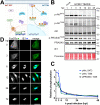
FIG 1
FIG 1
AKT is inactivated during HCMV infection. (A) Schematic denoting the influence of Akt activity on two key substrates, PRAS40 and FoxO transcription factors (FoxO TF). Left panel, when Akt is inactive, FoxO TF are able to enter the nucleus. Right panel, when Akt is recruited to membranes, such as during PI3K signaling, Akt is phosphorylated at activating residues Thr308 (T308) and Ser473 (S473). Once activated, Akt phosphorylates many downstream substrates, including PRAS40 and FoxO TF. Akt phosphorylation of FoxO TF recruits binding of 14-3-3 proteins, which sequester FoxO TF in the cytoplasm. (B) Fibroblasts were infected with HCMV strain TB40/E at an MOI of 2 or were mock infected (M) in the presence of 5% newborn calf serum; lysates were harvested at the indicated times postinfection (hours postinfection [hpi]) and analyzed by Western blotting for proteins immunoreactive to antibodies specific for the following antigens: p-AktS473 and p-AktT308, Akt phosphorylated at T308 or S473, respectively; Akt, total Akt; p-PRAS40T246, PRAS phosphorylated at T246; PRAS40, total PRAS40; IE1, the 72-kDa HCMV immediate early nuclear antigen. A section of nitrocellulose membrane was also subjected to Ponceau S staining (Ponceau) to assess total protein loading across lanes. (C) Fluorescent signals from far-red dye-labeled secondary antibodies used for Western blot detection of p-AktT308, p-AktS473, and p-PRAS40T246 were normalized to the detection signal for total Akt or total PRAS40, respectively. The mean signal is plotted relative to the baseline signal from a 1-h mock infection treatment, and data shown are the average of two independent biological replicates with error bars indicating standard deviations. (D) Fibroblasts were infected with HCMV strain TB40/E at an MOI of 1. The cells were fixed in 4% PFA at the indicated times postinfection and stained with antibodies specific for FoxO3a (green) or HCMV IE1 antigen (blue). Scale bar, 25 μm.
FIG 2
FIG 2
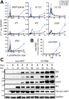
Constitutive AKT activity causes a viral replication defect. (A to C) Fibroblasts carrying doxycycline-inducible (tet-on) expression cassettes for either myristoylated Akt1 (myr-Akt) or a kinase-dead control (K179M) were induced for 24 h using 100 ng/mL doxycycline hyclate (Dox) and subsequently infected at an MOI of 1 with either HCMV strain AD169rv (A) or TB40/E (B and C). Supernatants were harvested at the indicated times postinfection (days postinfection [dpi]) and measured for the infectious titer by the TCID50 assay. (C) Results of an independent biological replicate of the infection experiment described in the legend for panel B but with additional controls: untransduced fibroblasts and tet-on myr-Akt and K179M fibroblasts in the absence of the transgene inducing agent (Dox). (D) Representative confocal microscope images of formalin-fixed fibroblasts at 24 h postinfection (MOI, 1; TB40/E) (see Fig. S1 in the supplemental material for a full time course from 0 to 72 h postinfection). (E and F) At least 30 cells per condition were scored at 72 hpi for the effect of HCMV infection (E) or myr-Akt or myr-Akt K179M expression (F) on nuclear localization of endogenous FoxO3a, as indicated on the y axis. HCMV infection was scored by detection of viral IE1 nuclear antigen in indirect immunofluorescent staining. Similarly, detection of HA epitope staining was used to score cells positive for expression of myr-Akt or K179M (the myr-Akt transgene carries an HA tag).
FIG 3
FIG 3
Constitutive Akt activity causes defects in viral gene expression and viral DNA synthesis. (A) Fibroblasts were induced for expression of either myr-Akt or myr-Akt-K179M (kinase dead) for 24 h and then were infected with HCMV strain TB40/E at an MOI of 1, and total RNA samples were isolated from either myr-Akt or K179M settings at 2, 4, 6, 8, 10, 12, 16, 24, 48, and 72 hpi, including an on-column DNase I digestion step. RNA samples were reverse transcribed into cDNA and assayed by qPCR for the abundance of the indicated viral transcripts relative to levels of cellular GAPDH mRNA. (B) Total DNA was isolated at the indicated times postinfection (hpi) from cells infected exactly as described for panel A, and copies of viral DNA were enumerated using quantitative PCR for the HCMV UL69 gene normalized to copies of cellular 18S rDNA loci. (C) Infections were set up as described for panel A. Cell lysates collected at the indicated times postinfection were assayed by Western blotting for the expression of the indicated viral proteins, as well as for the expression of myr-Akt (HA tag) or GAPDH, as a loading control.
FIG 4
FIG 4
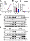
siRNA knockdown of FoxO3a causes viral replication defects. siRNA complexes targeting FoxO1, FoxO3a, or both FoxO1 and FoxO3a were reverse transfected into fibroblasts 24 h prior to infection with HCMV strain TB40/E at an MOI of 1. (A) Supernatants were harvested at the indicated times postinfection (days postinfection [dpi]). (B) Viral yield at day 5 postinfection for 3 independent biological replicates of the experiment described for panel A. An asterisk indicates a P value of 0.0276 using one-way analysis of variance (ANOVA) with Dunnett’s posttest to compare the mean of the results of each siRNA treatment condition to that of the nontargeting control (NTC) setting. (C) Viral DNA copies were enumerated by qPCR using a primer pair specific for HCMV UL69 and are shown normalized to copies of cellular 18S rDNA loci. Results are shown for samples collected at 2 h postinfection (hpi) and 96 hpi to accurately indicate input levels of viral genomes against replicated viral DNA. (D and E) Protein lysates were obtained at the indicated times postinfection and analyzed by Western blotting to validate knockdown of FoxO3a or FoxO1, respectively, and to assess for effects on viral protein expression. Cellular GAPDH protein was detected as a loading control. NTC, nontargeting control siRNA.
FIG 5
FIG 5
Hormone-regulated chimeric FoxO3a activity reverses Akt-dependent viral replication defects. (A) Cartoon depiction (created with BioRender) of chimeric FoxO3a “triple Akt phosphoacceptor site mutant” (TM), FoxO3a-TM-ER, in which FoxO3a-TM is fused to a mutant mouse estrogen receptor (ER) in which the antiestrogen 4-hydroxytamoxifen (4-OHT) activates its nuclear localization. Therefore, the FoxO3a-TM-ER protein allows FoxO3a activity to be decoupled from Akt regulation. (B) Western blot analysis of ARPE-19 epithelial cells carrying a doxycycline-inducible myr-Akt expression cassette (HA tagged) and constitutively expressing FoxO3a-TM-ER, also HA tagged. (C) Confocal immunofluorescence microscopy was used to validate 4-OHT-induced FoxO3a-TM-ER nuclear localization upon addition of 1 μM 4-OHT for 1 h. HA staining was used to detect the FoxO3a-TM-ER fusion protein; Hoechst 33342 signal is shown to counterstain nuclei in the merged image. (D) One-step viral replication kinetics at an MOI of 2 for HCMV strains TB40/E and AD169 repaired for UL131 (AD169 rUL131) in ARPE-19 cells induced for myr-Akt expression (100 ng/mL doxycycline [Dox]) and with or without 4-OHT treatment (1 μM) to induce nuclear localization of FoxO3a-TM-ER, as indicated. (E) Results for viral replication yield defect at day 8 postinfection from 3 independent biological replicates of strain AD169 rUL131 in ARPE-19 cells; the double asterisk indicates a P value of 0.0062 by an unpaired, two-tailed t test. (F) Viral DNA synthesis results for the same time points shown in panel D, comparing qPCR-detected copies of the HCMV UL69 gene normalized to detection of cellular 18S rDNA copies. Note, the legend shown below panel F applies to panels D and F. dpi, days postinfection. (G) Western blot analysis of viral protein expression following AD169 rUL131 infection (MOI, 2 TCID50/mL) in ARPE-19 cells in the presence or absence of Dox and/or 4-OHT treatments, as described above.

Similar articles

Cited by

References

    1. Manning BD, Toker A. 2017. AKT/PKB signaling: navigating the network. Cell 169:381–405. doi:10.1016/j.cell.2017.04.001. - DOI - PMC - PubMed
    1. Sancak Y, Thoreen CC, Peterson TR, Lindquist RA, Kang SA, Spooner E, Carr SA, Sabatini DM. 2007. PRAS40 is an insulin-regulated inhibitor of the mTORC1 protein kinase. Mol Cell 25:903–915. doi:10.1016/j.molcel.2007.03.003. - DOI - PubMed
    1. Inoki K, Li Y, Zhu T, Wu J, Guan K-L. 2002. TSC2 is phosphorylated and inhibited by Akt and suppresses mTOR signalling. Nat Cell Biol 4:648–657. doi:10.1038/ncb839. - DOI - PubMed
    1. Datta SR, Dudek H, Tao X, Masters S, Fu H, Gotoh Y, Greenberg ME. 1997. Akt phosphorylation of BAD couples survival signals to the cell-intrinsic death machinery. Cell 91:231–241. doi:10.1016/S0092-8674(00)80405-5. - DOI - PubMed
    1. Hoxhaj G, Manning BD. 2020. The PI3K-AKT network at the interface of oncogenic signalling and cancer metabolism. Nat Rev Cancer 20:74–88. doi:10.1038/s41568-019-0216-7. - DOI - PMC - PubMed

Publication types

MeSH terms

Substances