. 2022 Nov;100(11):2055-2076.
doi: 10.1002/jnr.25111. Epub 2022 Aug 2.

Chronic muscle recordings reveal recovery of forelimb function in spinal injured female rats after cortical epidural stimulation combined with rehabilitation and chondroitinase ABC

Affiliations

Chronic muscle recordings reveal recovery of forelimb function in spinal injured female rats after cortical epidural stimulation combined with rehabilitation and chondroitinase ABC

Eleni Sinopoulou et al. J Neurosci Res. 2022 Nov.

Abstract

Cervical level spinal cord injury (SCI) can severely impact upper limb muscle function, which is typically assessed in the clinic using electromyography (EMG). Here, we established novel preclinical methodology for EMG assessments of muscle function after SCI in awake freely moving animals. Adult female rats were implanted with EMG recording electrodes in bicep muscles and received bilateral cervical (C7) contusion injuries. Forelimb muscle activity was assessed by recording maximum voluntary contractions during a grip strength task and cortical motor evoked potentials in the biceps. We demonstrate that longitudinal recordings of muscle activity in the same animal are feasible over a chronic post-injury time course and provide a sensitive method for revealing post-injury changes in muscle activity. This methodology was utilized to investigate recovery of muscle function after a novel combination therapy. Cervical contused animals received intraspinal injections of a neuroplasticity-promoting agent (lentiviral-chondroitinase ABC) plus 11 weeks of cortical epidural electrical stimulation (3 h daily, 5 days/week) and behavioral rehabilitation (15 min daily, 5 days/week). Longitudinal monitoring of voluntary and evoked muscle activity revealed significantly increased muscle activity and upper limb dexterity with the combination treatment, compared to a single treatment or no treatment. Retrograde mapping of motor neurons innervating the biceps showed a predominant distribution across spinal segments C5-C8, indicating that treatment effects were likely due to neuroplastic changes in a mixture of intact and injured motor neurons. Thus, longitudinal assessments of muscle function after SCI correlate with skilled reach and grasp performance and reveal functional benefits of a novel combination therapy.

Keywords: EMG; chondroitinase ABC; electrical stimulation; motor evoked potential; physical rehabilitation.


Conflict of interest statement

The authors declare no competing financial interests.

Figures

FIGURE 1
FIGURE 1
Experimental design. (a) Experimental timeline demonstrating experimental and surgical procedures (gray boxes) and experimental time points (in red). (b) Schematic shows a bilateral contusion injury at level C7 of the spinal cord (red circle), unilateral cortical epidural stimulation (M1 stim), EMG muscle recordings in the contralateral bicep muscle, and injections of LV‐ChABC rostral and caudal to injury. (c) Stimulating cortical epidural electrode over the motor cortex. (d) Whishaw window reaching success rate pre‐injury baseline (BSL) scores prior to implantation surgery (dark gray bar), and postimplantation surgery (light gray bar) and post‐contusion injury surgery (light blue bar) showing that implants do not affect the animals' performance in behavioral tasks (F (3,12) = 1.700, p = .089 one‐way ANOVA), individual animals are plotted as symbols in the bar graph. (e) color‐coded experimental groups used for this study.
FIGURE 2
FIGURE 2
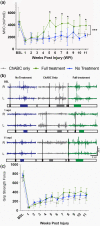
A triple combination treatment promotes increased muscle activity during voluntary behavior. (a) Weekly maximum voluntary contraction (MVC) recordings measured in awake animals during a behavioral (grip strength) task reveals a significant increase in MVC in the triple treatment group, which begins to emerge at 4 weeks post‐injury and is significantly increased compared to the untreated or LV‐ChABC only treatment groups at all subsequent time points (Time: F (4.793,71.90) = 15.96, p < .0001; Treatment: F (2,15) = 16.08, p < .001; Interaction: F (22,165) = 3.075, p < .0001, two‐way RM ANOVA, Dunnet's post‐hoc). (b) Raw traces during MVC recordings for right (R) and left (L) forelimbs at three time points (BSL, 1 week post‐injury and 11 weeks post‐injury) recorded from the same animal over time, averages of three trials. Colored traces indicate the different groups, outlined boxes indicate the response duration, and black boxes indicate the timestamped start of each trial. (c) In contrast to the electrophysiological evaluations, weekly grip strength behavioral testing revealed no differences between groups in the force measurement values evaluated over the 11 week time course (Time: F (11,55) = 90.77, p < .0001, Treatment: F (2,10) = 5.276, p < .05; Interaction: F (22,110) = 1.789, p < .05 two‐way RM ANOVA, Bonferroni's post‐hoc).
FIGURE 3
FIGURE 3
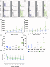
A triple combination treatment promotes increased muscle activity during evoked cortical stimulation. Weekly motor evoked potential (MEP) recordings acquired in awake freely moving animals. Rectified average traces (15 triggers average) of MEPs from the different groups at three different time points (BSL, 1 week post‐injury and 11 weeks post‐injury) at (a) threshold intensity and at (b) 1.5 times the threshold intensity recorded from the same animal over the time course of 11 weeks. Note different y‐axis scale bar for 11 weeks post‐injury at threshold values to accommodate for better trace representation. Quantified MEP weekly recordings, at (c) threshold intensity and at (d) 1.5 times the threshold intensity. The triple treatment group showed a significant increase in MEPs when compared to the no treatment or LV‐ChABC only treatment groups (Time: F (11,55) = 3.422, p = .001; Treatment: F (2,10) = 7.809, p < .01; Interaction: F (22,110) = 1.679, p < .05 two‐way RM ANOVA, Bonferroni's post‐hoc) when stimulated at higher current intensity (d). Duration of MEP AUC response compared between the treatment groups at BSL and at 11 weeks post‐injury at (e) threshold intensity and at (f) 1.5 times the threshold intensity. The triple treatment group at 11 weeks post‐injury showed responses with significantly longer duration than the no treatment or LV‐ChABC only treatment groups (e, F (5,30) = 1.978, p < .0001, one‐way ANOVA, Bonferroni's post‐hoc; f, F (5,30) = 1.822, p < .0001, one‐way ANOVA, Bonferroni's post‐hoc;). (g) Current (mA) threshold intensities used for therapeutic stimulation remained stable throughout the experiment (Time: F (2,45) = 73.79, p < .0001; Treatment: F (2,45) = 12.98, p < .001; Interaction: F (4,45) = 2.266, p = .0768, two‐way RM ANOVA, Tukey's post‐hoc).
FIGURE 4
FIGURE 4
Increased muscle activity corresponds to recovery of skilled forelimb function. (a) no significant variation in the contusion force was observed, confirming consistent injuries between the groups (F (2,15) = 1.342, p = .291, one‐way ANOVA). (b) Weekly behavioral assessments of reaching success rates in the Whishaw window reaching task indicate that the triple treatment group showed a significant increase in the number of sugar pellets retrieved, compared to the no treatment or LV‐ChABC only treatment groups (Time: F (12,180) = 32.11, p < .0001; Treatment: F (2,15) = 3.315, p = .064; Interaction: F (24,180) = 1.712, p < .05 two‐way RM ANOVA, Tukey's post‐hoc). (c) Weekly behavioral assessments in the Montoya staircase task measuring the number of total pellets eaten indicated that the triple treatment group reached for the most pellets using the side contralateral to stimulation (Stim side) compared to the side ipsilateral to stimulation (Non‐stim side) (Time: F (4.876,97.53) = 26.09, p < .0001; Treatment: F (3,20) = 5.125, p < .01 Interaction: F (33,220) = 1.705, p < .01, two‐way RM ANOVA, Tukey's post‐hoc) (d) Grayscale heat map indicating the degree of displacement and/or dropped pellets during the Montoya staircase task shows more accurate reach and grasp in the triple treatment group, compared to the no treatment and LV‐ChABC only groups (white = 0 pellets dropped/displaced; black = more than 6 pellets dropped/displaced). Error bars = Standard Deviation.
FIGURE 5
FIGURE 5
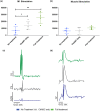
A triple combination treatment promotes increased evoked muscle activity during terminal cortical stimulation. Terminal electrophysiology performed 12 weeks after contusion injury under anesthesia. (a) Quantified area under the curve (AUC) (mV*mS) measurements from motor evoked potentials (MEPs) recorded from percutaneous EMG electrodes by stimulating the implanted M1 cortical epidural electrode. The triple treatment group and the LV‐ChABC only treatment group showed a significant increase in muscle activity compared to the no treatment group (F (2,12) = 9.288, p = .004 one‐way ANOVA, Tukey's post‐hoc). (b) Quantified AUC (mV*mS) measurements from MEPs recorded from percutaneous EMG electrodes by stimulating the implanted EMG electrode through the implanted brain interface board. No differences in muscle activity were observed between the treatment groups when the muscle was being stimulated (F (2,12) = 1.551, p = .252 one‐way ANOVA). (c) Colored representative rectified average traces (10 triggers/average) of MEPs from the different groups, obtained by stimulating the M1. The no treatment group has a different y‐axis scale bar to achieve better trace representation. (d) Colored representative rectified average traces (10 triggers average) of MEPs from the different groups, obtained by peripheral muscle stimulation.
FIGURE 6
FIGURE 6
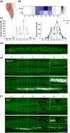
Biceps brachii spinal motor neuronal pool mapping. (a) Schematic showing deep (white) and superficial (blue) injection sites in the two heads of the right biceps. The short head of the biceps received two deep and two superficial injections aiming for the motor endplates (dashed line) and two deep injections aiming at the region halfway between the proximal end of the muscle and the motor endplates. The long head of the biceps received one deep and one superficial injection aiming for the motor endplates and one deep injection aiming at the region halfway between the proximal end of the muscle and the motor endplates. (b) Heat map graph showing distribution of biceps motor neurons in the naive and SCI groups. Biceps motor neurons were found between spinal levels C4 to T1, with highest concentration in C7. After spinal cord injury the numbers of biceps motor neurons were dramatically reduced in C7 (lesion epicenter) and slightly decreased in adjacent levels C6 and C8. (c) Bar and line graphs showing the number of biceps motor neurons counted at each spinal levels for the naïve and SCI groups. Individual animals were plotted as symbols in the bar graph and individual lines in the line graph. After SCI, the number of motor neurons in the lesion epicenter (C7) is reduced compared to naive. (d) CTB‐positive biceps motor neurons. Scale bar = 250 μm. (e,f) Horizontal sections of the spinal ventral horn of a naïve (e) and a SCI (f) rat that had the right biceps injected with CTB. C7 (lesion epicenter) is showed in detail at the top right of each image. The bottom image on each panel shows a 2D reconstruction of all counted CTB positive neurons (white dots) present on all analyzed slides of a representative animal. Biceps motor neurons were present from C4 to T1, more densely between C5 and C8. SCI reduced the amount of motor neurons present in the lesion epicenter (C7) and neighboring levels. Scale bar = 2 mm. Error bars = Standard Deviation.

Similar articles

Cited by

References

    1. Adkins, D. L. , Hsu, J. E. , & Jones, T. A. (2008). Motor cortical stimulation promotes synaptic plasticity and behavioral improvements following sensorimotor cortex lesions. Experimental Neurology, 212, 14–28. - PMC - PubMed
    1. Adkins‐Muir, D. L. , & Jones, T. A. (2013). Cortical electrical stimulation combined with rehabilitative training: Enhanced functional recovery and dendritic plasticity following focal cortical ischemia in rats. Neurological Research, 25, 780–788. - PubMed
    1. Aguilar, R. M. , Steward, O. , & Reeve, P. D. (2010). A bilateral cervical contusion injury model in mice: Assessment of gripping strength as a measure of forelimb motor function. Experimental Neurology, 221, 38–53. - PMC - PubMed
    1. Ahlborn, P. , Schachner, M. , & Irintchev, A. (2007). One hour electrical stimulation accelerates functional recovery after femoral nerve repair. Experimental Neurology, 208, 137–144. - PubMed
    1. Ahuja, C. S. , Wilson, J. R. , Nori, S. , Kotter, M. R. N. , Druschel, C. , Curt, A. , & Fehlings, M. G. (2017). Traumatic spinal cord injury. Nature Reviews Disease Primers, 3, 17018. - PubMed

Publication types

Substances