. 2022 Aug;14(8):884-890.
doi: 10.1038/s41557-022-01005-z. Epub 2022 Jul 29.

Pathogenic bacteria remodel central metabolic enzyme to build a cyclopropanol warhead

Affiliations

Pathogenic bacteria remodel central metabolic enzyme to build a cyclopropanol warhead

Felix Trottmann et al. Nat Chem. 2022 Aug.

Abstract

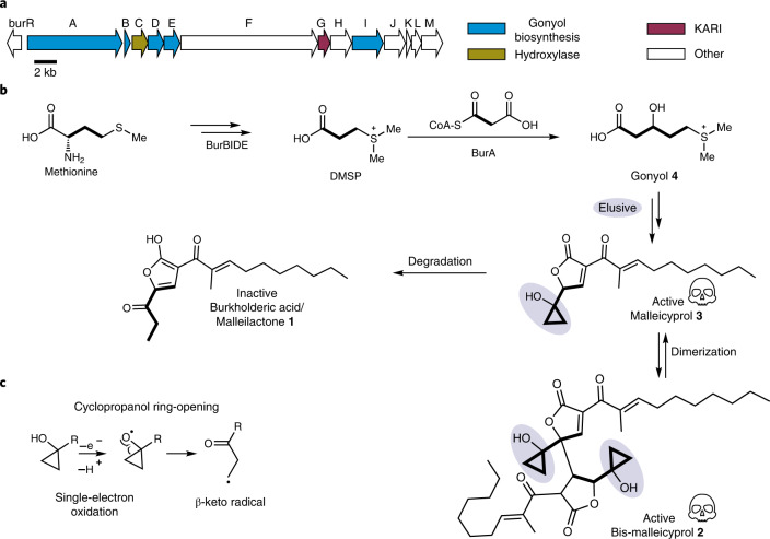
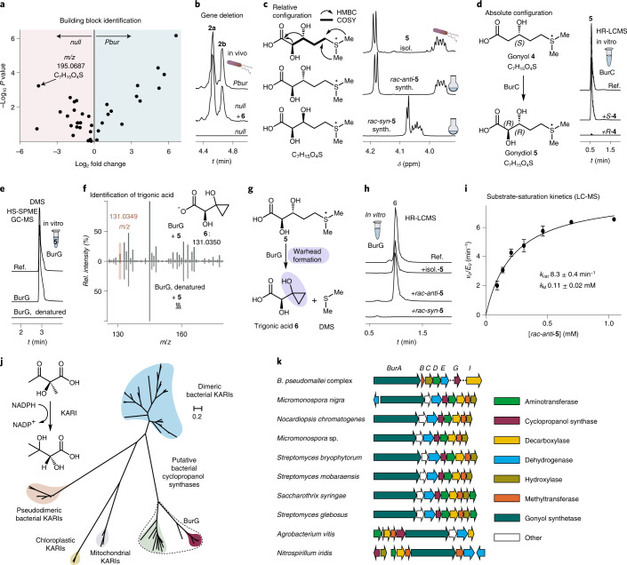
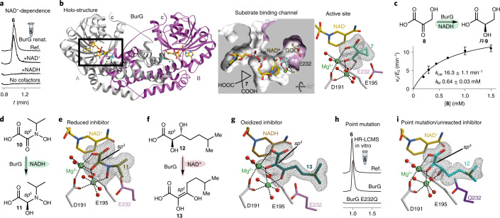
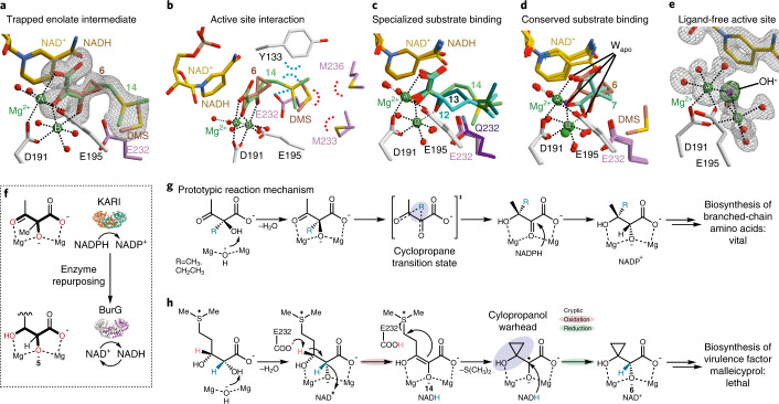
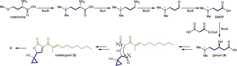
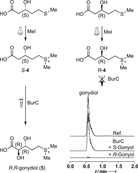
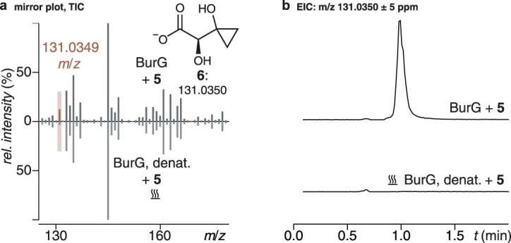
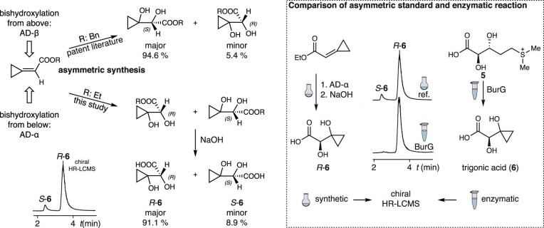
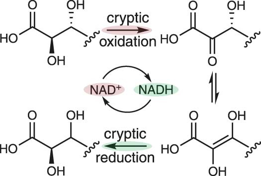
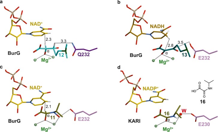
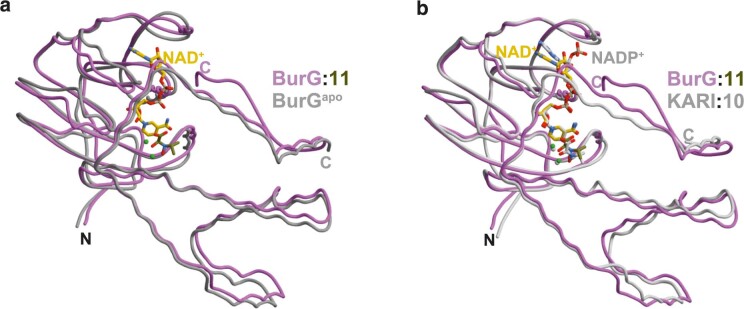
Bacteria of the Burkholderia pseudomallei (BP) group pose a global health threat, causing the infectious diseases melioidosis, a common cause of pneumonia and sepsis, and glanders, a contagious zoonosis. A trait of BP bacteria is a conserved gene cluster coding for the biosynthesis of polyketides (malleicyprols) with a reactive cyclopropanol unit that is critical for virulence. Enzymes building this warhead represent ideal targets for antivirulence strategies but the biochemical basis of cyclopropanol formation is unknown. Here we describe the formation of the malleicyprol warhead. We show that BurG, an unusual NAD+-dependent member of the ketol-acid reductoisomerase family, constructs the strained cyclopropanol ring. Biochemical assays and a suite of eight crystal structures of native and mutated BurG with bound analogues and inhibitors provide snapshots of each step of the complex reaction mechanism, involving a concealed oxidoreduction and a C-S bond cleavage. Our findings illustrate a remarkable case of neofunctionalisation, where a biocatalyst from central metabolism has been evolutionarily repurposed for warhead production in pathogens.


Conflict of interest statement

The authors declare no competing interests.

Figures

Fig. 1
Fig. 1. Malleicyprols, virulence factors encoded by conserved locus in BP group pathogens.
a, Gene cluster coding for malleicyprol biosynthesis. KARI, ketol-acid reductoisomerase b, Structures of malleicyprols 2 and 3 and their degradation product 1, and selected steps in the biosynthesis of the C5 building block. See Extended Data Fig. 1. for detailed biosynthetic pathway. c, Ring-opening of cyclopropanols mediated by single-electron oxidation.
Fig. 2
Fig. 2. Substrate identification and reconstitution of enzymatic cyclopropanol formation.
a, Metabolomics analysis of B. thailandensis Pbur versus B. thailandensis Pbur BurG::Kan (malleicyprol null mutant) cell extracts (biological triplicates, n = 3). Data filtered for sulfur-containing compounds; see Supplementary Fig. 1 for non-filtered analysis. b, Chemical complementation of the null mutant with 6 restores malleicyprol production (isoforms 2a and 2b) to wild-type level. c, Structure elucidation of isolated (isol.) gonydiol 5, and comparison of key 1H NMR shifts with synthetic (synth.) racemic syn- and anti-isoforms. COSY, correlated spectroscopy; HMBC, heteronuclear multiple bond correlation. d, Formation of 5 catalysed by hydroxylase BurC in assays with addition of R-4 or S-4; top trace: reference (5). e, Detection of DMS as product of BurG reactions by GC-MS. f, Mirror plot of enzymatic assay (top) of BurG incubated with 5 versus denatured BurG (bottom) indicates a compound with the m/z value of 6 as enzymatic product; see Extended Data Fig. 3 for chromatogram. g, BurG-catalysed transformation of 5 to 6. h, Monitoring of enzymatic turnover of isolated 5 and synthetic rac-syn-5 or rac-anti-5 into 6 via HR-LCMS. i, Steady-state kinetics of formation of 6 from rac-anti-5; mean and standard deviation obtained from two independent preparations (n = 2). j, Phylogenetic analysis of BurG, orthologues and canonical KARIs. The scale bar indicates amino acid substitutions per site. k, Alignment of biosynthetic gene cluster encoding for BurG orthologues clustered with the genes associated with biosynthesis of 5. Source data
Fig. 3
Fig. 3. Concealed redox cycle catalysed by BurG.
a, Gonydiol 5 transformation assay with NAD+-free BurG with addition of NAD+, NADH or in the absence of cofactor; top: 6 as reference. b, Full view of the 3D structure of BurG co-crystallized with the KARI inhibitor 7 (left). A and B, subunits; N and C, termini. The black rectangle displays the area for the zoom in the substrate binding channel (middle) and active site zoom (right) (PDB 7PCG). GOL, glycerol. c, Steady-state kinetics of NADH-mediated reduction of hydroxypyruvate 8 to glyceric acid 9 through BurG; mean and standard deviation as obtained from two independent protein preparations (n = 2). d, Scheme of the transformation of hydroxamate 10 to hemiaminal 11. e, 3D structure of BurG co-crystallized with 11 after incubation with 10 and NADH (PDB 7PCL). Two magnesium atoms (A and B) are shown as green spheres. f, BurG-mediated oxidation of carba-gonydiol 12 to the enolate 13. g, 3D structure of BurG with 13 obtained after co-crystallization with 12 and NAD+ (PDB 7PCM). h, Single amino acid substitution (E232Q) renders BurG inactive. i, 3D structure of BurG E232Q co-crystallized with 12 (PDB 7PCO). Source data
Fig. 4
Fig. 4. Parallels and differences between biocatalysis of BurG and canonical KARIs.
a, Structures of BurG with the reaction products 6 and DMS and the trapped enolate intermediate 14 after co-crystallization with gonydiol 5 (PDB 7PCN). Two magnesium atoms (A and B) are shown as green spheres. b, Interaction of active site residues of BurG with 6, 14 and DMS. c, Structural overlay of BurG bound to 12, 13 and 14 indicates a strong interaction of the positively charged sulfonium moiety of 14 with E232 as compared to neutral 12 and 13. d, Tight lock of the oxygen-functionalized compounds 6 and 7 through interaction with Mg2+ ions in the active centre of BurG. Wapo indicates the binding sites of water molecules as found in the apo structure that are replaced by ligand atoms in the depicted structures. e, Structure of unliganded holo-BurG illustrates a hydroxide ion bridged between two Mg2+ centres (PDB 7PCC). f, Comparison of BurG and KARIs displays a similar binding mode of substrates in the Mg2+ cluster. g, Proposed reaction mechanism for KARIs with cyclopropane transition state. h, Reaction mechanism proposed for BurG involving a concealed (transient) redox reaction.
Extended Data Fig. 1
Extended Data Fig. 1. Model of biosynthetic pathway to malleicyprol.
More detailed model of the biosynthetic pathway to malleicyprol.
Extended Data Fig. 2
Extended Data Fig. 2. Absolute configuration of gonydiol.
Synthesis of R- and S-gonyol (4) and subsequent enzymatic transformation to gonydiol (5) establishes the absolute configuration of naturally occurring gonyol and gonydiol. HR-LCMS extracted ion monitoring of gonydiol (m/z of 195.0686 in positive-ion mode) after addition of R-4 or S-4 to reaction mixtures containing the hydroxylase BurC. Source data
Extended Data Fig. 3
Extended Data Fig. 3. Identification of trigonic acid (6).
a, Mirror plot of enzymatic assay (top) of BurG incubated with 5 vs. denatured BurG (bottom) indicates a compound with the m/z value of 6 as enzymatic product. b, Extracted ion chromatogram (EIC) in negative ion mode of the enzymatic assay (top) vs. denatured BurG (bottom). Source data
Extended Data Fig. 4
Extended Data Fig. 4. Determination of stereochemistry of trigonic acid (6).
Left panel: stereoselectivity of the asymmetric dihydroxylation (AD)-mix β leads to mainly S-configurated products; Bn: benzyl protecting group. Consequently AD-mix α forms mainly R-configurated products. Chiral HR-LCMS (bottom left) validates the successful asymmetric synthesis with AD-mix α after hydrolysis of the formed ethyl ester (Et). Right panel: chiral HR-LCMS of asymmetric synthesis products (ref.) vs. enzymatically formed 6 shows the stereochemistry of enzymatically formed 6 to be of R-configuration. Source data
Extended Data Fig. 5
Extended Data Fig. 5. Binding mode of NAD+ in the active site of BurG.
The amino acid residues Asn48 and Asp51 exclude the negatively charged phosphate group of NADP+ (as commonly found in canonical KARIs) from binding in the active centre of BurG.
Extended Data Fig. 6
Extended Data Fig. 6. Cryptic redox cycle.
Scheme representing the general concept of a cryptic redox cycle.
Extended Data Fig. 7
Extended Data Fig. 7. Complex structure of BurG co-crystallized with hydroxypyruvate (8) (PDB ID: 7PCI).
The FO-FC electron density map (contoured to 3 σ) for 8 bound to the Mg-atoms is shown as grey mesh. The ligand has been omitted for phasing. E232 is located on subunit B and marked in pink.
Extended Data Fig. 8
Extended Data Fig. 8. Complex structure of BurG co-crystallized with gonydiol (5) (PDB ID: 7PCN).
The 2FO-FC electron density maps (grey meshes, contoured to 1.0 σ) and the FO-FC maps (green meshes, contoured to 3.0 σ) are depicted for the respective ligand. a. 6 and DMS (shadowed in grey) have been omitted for phasing. b. 14 (shadowed in grey) has been omitted for phasing. The FO-FC electron density maps clearly depict the excluded ligands, proving that 14, 6 and DMS are all bound to the active site. The occupancy of each ligand was set to 0.5. E232 is located on subunit B and marked in pink.
Extended Data Fig. 9
Extended Data Fig. 9. Cofactor and ligand orientation in the active site of BurG and KARIs.
The structural comparison between BurG and KARIs illustrates the preferred reaction trajectories of hydride transfer in the distinct enzymes. The interactions of the ligand with the two Mg2+ ions (black dots) indicate that Cα is the preferred carbon atom for catalysis. a, Structure of the catalytically inactive BurG E232Q mutant in complex with surrogate 12. The ligand adopts ideal orientation for hydride transfer to NAD+ (2.3 Å distance, angle between C(NAD+)HCα: 174°, PDB code: 7PCO). b, Structure of BurG co-crystallized with 12 and NAD+. The electron density shows the ligand oxidized to 13 resulting in NADH. Glu232 acts as base and is key to hydride transfer. Distance and angle between the cosubstrate and Cα (2.8 Å, C(NADH)HCα: 123°) are unfavourable for the back reaction. The replacement of sulfur by carbon in surrogate 13 prevents the elimination reaction and keeps the metabolite in its position (PDB code: 7PCM). c, Structure of BurG co-crystallized with 10 and NADH. The electron density shows the Cα-atom of the ligand in sp3 configuration. Therefore, the molecule represents the carbinolamine 11 and the cosubstrate is oxidized to NAD+. Distance and angle between the cosubstrate and Cα allow for a back reaction (2.1 Å, C(NAD+)HCα: 168°). However, formation of the alkoxide is favoured by the interactions with both Mg2+ ions (PDB code: 7PCL). d, Active site architecture of the KARI from Staphylococcus aureus (PDB code: 6AQJ). Crystallization of the enzyme was carried out in presence of 10 and NADPH. It is reported that hydride transfer from NADPH to the nitrogen of 10 takes place, hereby forming NADP+ and converting the ligand to 16 via water (W) release. The complex structure lacks defined H-bonds between cosubstrate and ligand.
Extended Data Fig. 10
Extended Data Fig. 10. Structural overlays.
a, Structural overlay of BurG (holo) bound to 11 (PDB code: 7PCL, coloured in pink) with apo BurG (PDB code: 7PCE, coloured in grey). The root mean square deviation (rmsd) between both structures is 1.2 Å over 283 Cα-atoms (353 residues in total). BurG (apo) displays increased flexibility (high temperature factors) and individual secondary structure elements are shifted compared to BurG (holo). As a result, the catalytic centre in apo BurG is accessible to cofactors and ligands (open conformation). In contrast, the active site of BurG (holo) is sequestered in a closed chamber. b, Structural overlay of BurG (holo) (PDB code: 7PCL, coloured in pink) with the ketol-acid reductoisomerase (KARI, holo) from Staphylococcus aureus (PDB code: 6AQJ, coloured in grey). The KARI from S. aureus is bound to 10 while BurG is bound to 11 (the reduced form of 10). Both structures adopt the closed conformation and display a rmsd of 1.4 Å over 288 Cα-atoms (sequence identity 35 %).

Similar articles

Cited by

References

    1. Galyov EE, Brett PJ, DeShazer D. Molecular insights into Burkholderia pseudomallei and Burkholderia mallei pathogenesis. Annu. Rev. Microbiol. 2010;64:495–517. - PubMed
    1. Cruz-Migoni A, et al. A Burkholderia pseudomallei toxin inhibits helicase activity of translation factor eIF4A. Science. 2011;334:821–824. - PMC - PubMed
    1. Franke J, Ishida K, Hertweck C. Evolution of siderophore pathways in human pathogenic bacteria. J. Am. Chem. Soc. 2014;136:5599–5602. - PubMed
    1. Franke J, Ishida K, Hertweck C. Plasticity of the malleobactin pathway and its impact on siderophore action in human pathogenic bacteria. Chem. Eur. J. 2015;21:8010–8014. - PubMed
    1. Franke J, Ishida K, Ishida-Ito M, Hertweck C. Nitro versus hydroxamate in siderophores of pathogenic bacteria: effect of missing hydroxylamine protection in malleobactin biosynthesis. Angew. Chem. Int. Ed. 2013;52:8271–8275. - PubMed

Publication types

LinkOut - more resources