. 2022 Oct 1;71(10):2166-2180.
doi: 10.2337/db21-1108.

Inhibition of CCL28/CCR10-Mediated eNOS Downregulation Improves Skin Wound Healing in the Obesity-Induced Mouse Model of Type 2 Diabetes

Affiliations

Inhibition of CCL28/CCR10-Mediated eNOS Downregulation Improves Skin Wound Healing in the Obesity-Induced Mouse Model of Type 2 Diabetes

Zhenlong Chen et al. Diabetes. .

Abstract

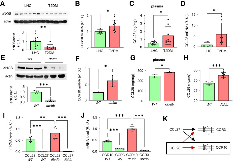
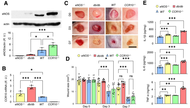
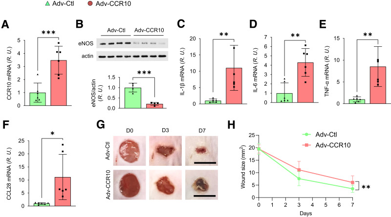
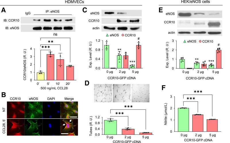
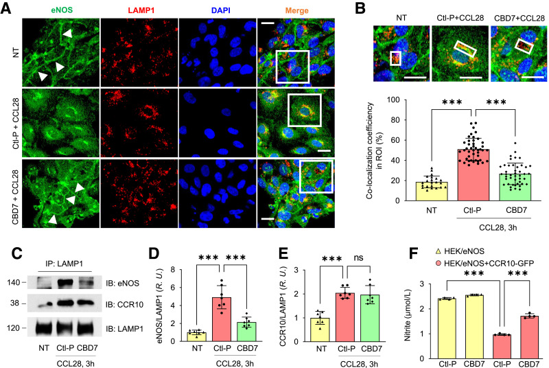
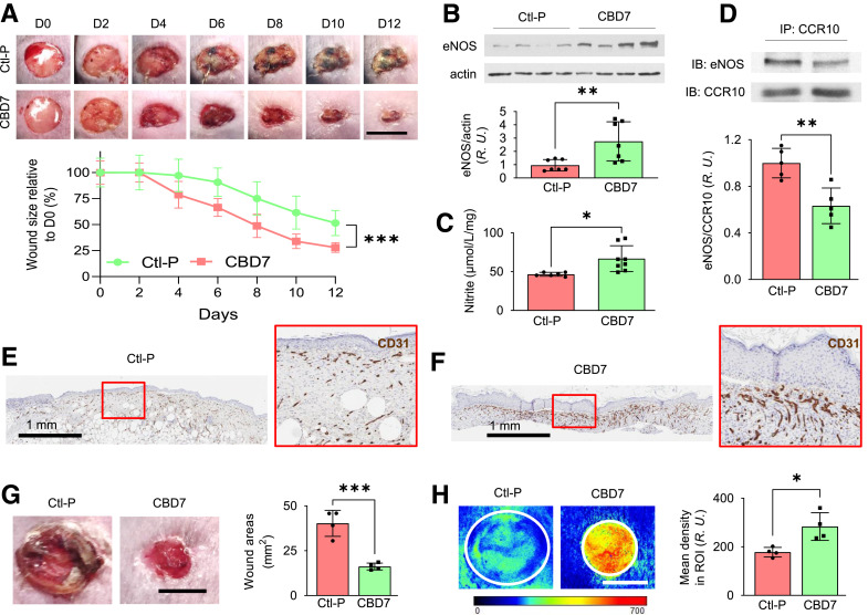
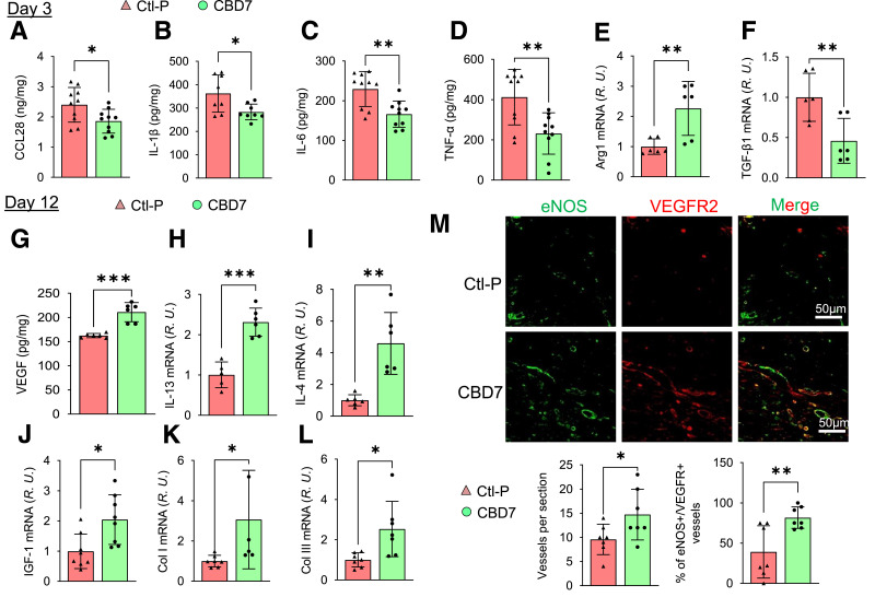
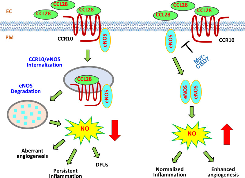
Chronic, nonhealing skin wounds, such as diabetic foot ulcers (DFUs), are common in patients with type 2 diabetes. Here, we investigated the role of chemokine (C-C motif) ligand 28 (CCL28) and its receptor C-C chemokine receptor type 10 (CCR10) in downregulation of endothelial nitric (NO) oxide synthase (eNOS) in association with delayed skin wound healing in the db/db mouse model of type 2 diabetes. We observed reduced eNOS expression and elevated CCL28/CCR10 levels in dorsal skin of db/db mice and subdermal leg biopsy specimens from human subjects with type 2 diabetes. Further interrogation revealed that overexpression of CCR10 reduced eNOS expression, NO bioavailability, and tube formation of human dermal microvascular endothelial cells (HDMVECs) in vitro, which was recapitulated in mouse dorsal skin. In addition, incubation of HDMVECs with CCL28 led to internalization of the CCR10/eNOS complex and colocalization with lysosome-associated membrane protein 1. Finally, topical application of myristoylated CCR10 binding domain 7 amino acid (Myr-CBD7) peptide prevented CCR10-eNOS interaction and subsequent eNOS downregulation, enhanced eNOS/NO levels, eNOS/VEGF-R2+ microvessel density, and blood perfusion, reduced inflammatory cytokine levels, and importantly, decreased wound healing time in db/db mice. Thus, endothelial cell CCR10 activation in genetically obese mice with type 2 diabetes promotes eNOS depletion and endothelial dysfunction, and targeted disruption of CCR10/eNOS interaction improves wound healing.


Figures

Figure 1
Figure 1
Reduced eNOS expression and elevated CCR10 level in punch biopsy specimens from human subjects and db/db mice with type 2 diabetes. A: Upper panel, subdermal biopsy specimens obtained from LHC donors and patients with type 2 diabetes were homogenized in RIPA buffer and prepared for Western blot. Bottom panel, normalized values of eNOS expression. B: Quantitative real-time RT-PCR revealed upregulated CCR10 mRNA in type 2 diabetes compared with LHC donors. Elevated plasma level of CCL28 determined by ELISA and mRNA by real-time RT-PCR in type 2 diabetes samples (C) compared with LHC (D). E: Less eNOS protein level in the dorsal skin of db/db mice. Mouse dorsal skin was collected and prepared for Western blotting, and normalized values are shown in the bar graph. F: Quantitative PCR revealed elevated CCR10 mRNA in db/db mouse dorsal skin compared with WT mice. Increased levels of CCL28 in plasma (G) and in dorsal skin (H) by ELISA measurement in db/db mice compared with WT mice. IK: Comparison of mRNA levels of CCL27 and CCL28 and their receptors CCR3 and CCR10 in dorsal skin tissue of WT and db/db mice. I: Dorsal skin was collected and prepared for real-time RT-PCR. CCL28 expression level was greater than CCL27 in both WT and db/db mouse skin, and CCL28 expression was greater in db/db dorsal skin compared with WT. J: CCR10 expression was greater than CCR3 in both WT and db/db mice, while higher CCR10 mRNA level was obtained in db/db dorsal skin compared with WT. R.U., relative units. K: The signaling pair of CCL28/CCR10 (black box), rather than CCL27/CCR3, was thus chosen for further study in db/db mouse wound healing. Data are mean ± SD. *P < 0.05, **P < 0.01, ***P < 0.001.
Figure 2
Figure 2
Reciprocal relationship between eNOS and CCR10 expression. A: Dorsal skin from eNOS−/−, db/db, WT, and CCR10−/− mice was collected and prepared for Western blotting to detect eNOS expression; normalized values are shown in the bottom panel. B: mRNA level of CCR10 in dorsal skin by real-time RT-PCR. Note the expression levels of eNOS and CCR10 are opposite in mouse skin. R.U., relative units. C: Representative photomicrographs of wounds in WT, CCR10−/−, eNOS−/−, and db/db mice. Four 5-mm full-thickness excisional wounds were made on the mouse dorsal skin, and wound images were acquired at indicated times. D, day. Scale bar, 5 mm. D: Normalized wound size over time in WT, CCR10−/−, eNOS−/−, and db/db mice. E: Levels of proinflammatory cytokines IL-1β, IL-6, and TNF-α in dorsal skin of CCR10−/−, eNOS−/−, db/db, and WT mice were determined by ELISA. Data are presented as mean ± SD. *P < 0.05, **P < 0.01, ***P < 0.001.
Figure 3
Figure 3
CCR10 overexpression delays dorsal skin wound healing. A: WT mice were injected subcutaneously in the four typical dorsal wound regions with 100 µL empty vector Adv control (Adv-Ctl) or CCR10-expressing Adv (Adv-CCR10; 1 × 10⁹ particles/mL), and the skin was subsequently harvested and homogenized after 72 h. CCR10 mRNA, as determined by RT-PCR, was overexpressed approximately threefold above basal control level. B: Adv-CCR10 reduced eNOS expression in dermal microvessels. Elevated mRNA levels of IL-1β (C), IL-6 (D), TNF-α (E), and CCL28 (F) were observed in Adv-CCR10 infected mouse skin. R.U., relative units. G and H: Delayed skin wound healing was also noted in Adv-CCR10 transfected mice. Four 5 mm full-thickness excisional wounds were produced on the mouse dorsal skin, and wound images were taken at indicated times. D, day. Scale bar, 5 mm. Data are presented as mean ± SD. Average wound area of four wounds/mouse is presented as mean ± SD (n = 6). *P < 0.05, **P < 0. 01, ***P < 0.001.
Figure 4
Figure 4
CCR10 overexpression downregulates eNOS and attenuates angiogenesis. A: Increased interaction between CCR10 and eNOS in HDMVECs following treatment with 500 ng/mL CCL28. After treatment, the cells were collected and immunoprecipitated (IP) with anti-eNOS Ab. Normalized ratios between CCR10 and eNOS are shown in the bottom panel. IB, immunoblot; R.U., relative units. Data are presented as mean ± SD. *P < 0.05, **P < 0.01, ***P < 0.001 vs. time 0. B: Confocal microscopy reveals colocalization (white arrows) between CCR10 (red) and eNOS (green) in HDMVECs following stimulation with 500 ng/mL CCL28 for 5 min. DAPI (blue) is a nuclear marker. NT, no treatment. Scale bar, 10 µm. Similar results were observed in five independent experiments. C: eNOS expression in HDMVECs transduced with CCR10-GFP cDNA. Cells were collected 48 h after transfection with different amounts of CCR10-GFP cDNA and lysed for Western blotting. Normalized expression of eNOS and CCR10 are shown in the bottom panel. D: Angiogenesis (tube formation) of HDMVECs transfected with CCR10-GFP cDNA. Top panel, photomicrographs of EC tubes on Matrigel-coated plates 2 days after transfection in culture medium supplemented with 500 ng/mL CCL28. Bottom panel, normalized EC tube number. Data are presented as mean ± SD. **P < 0.01, ***P < 0.001 vs. 0 μg CCR10-GFP; P < 0.001 vs. 2 μg CCR10-GFP. Reduced eNOS expression level (E) and NO production (F) in HEK/eNOS cells transduced with CCR10-GFP cDNA. Data are presented as mean ± SD. **P < 0.01, ***P < 0.001 vs. 0 μg CCR10-GFP; P < 0.001 vs. 2 μg CCR10-GFP.
Figure 5
Figure 5
Myr-CBD7 treatment prevents eNOS degradation by blocking eNOS-LAMP1 interaction in HDMVECs stimulated with CCL28. A: In cells pretreated with control peptide, eNOS (green) internalization and colocalization with LAMP1 was observed following stimulation with 500 ng/mL CCL28 for 3 h (middle panel), whereas eNOS remained associated with the plasma membrane of ECs pretreated with Myr-CBD7 peptide (bottom panel), similar to no treatment (NT) (top panel). Scale bar, 10 μm. B: Enlarged images of white boxes in panel A. Bar graph: colocalization coefficient between eNOS and LAMP1 in LAMP1+ ROI (white boxes). Scale bar, 10 μm. (C) After the same treatment as in A, cells were collected, and anti-LAMP1 Ab was used to immunoprecipitate (IP) cell lysates. IB, immunoblot. Normalized interaction between LAMP1 with eNOS (D) and CCR10 (E). R.U., relative units. (F) Effect of Myr-CBD7 peptide on NO production in HEK/eNOS cells transfected with CCR10-GFP cDNAs. After pretreatment with 50 μmol/L Myr-control or CBD7 peptide, cells were further stimulated with 5 μmol/L A23187 for 1 h at 37°C. Supernatants were assessed for total nitrite concentration. Data are presented as mean ± SD. *** P < 0.001.
Figure 6
Figure 6
Topical application of Myr-CBD7 elevated eNOS/NO level and microvessel density in association with reduced db/db mouse skin wound healing time. A: Representative photomicrographs of 8-mm full-thickness excisional wounds after treatment with 50 µL of 50 μmol/L Ctl-P or Myr-CBD7 peptide per wound. Scale bar, 8 mm. D, day. Myr-CBD7 peptide in Pluronic solution was topically applied twice during the first 24 h after wounding, which reduced wound size significantly from day 4 onward. Myr-CBD7 increased eNOS expression (B) and elevated NO production (Griess reaction) (C) on day 12 after treating db/db mouse wounds with Myr-CBD7. R.U., relative units. D: Myr-CBD7 reduced CCR10-eNOS interaction observed by co-IP from day 12 mouse wounds compared with control peptide. IB, immunoblot. Immunohistochemical staining of CD31 (brown color) in dorsal wounds of db/db mice after treatment with control peptide (E) compared with Myr-CBD7 (F) on day 12 (representative of at least eight independent experiments). Blood perfusion measured by laser speckle contrast analysis (G) in db/db mouse wounds (H) (same wounds that are shown in panel G) was significantly enhanced on day 10 in mice treated with Myr-CBD7 peptide. Scale bar, 5 mm. Data are presented as mean ± SD. *P < 0.05, **P < 0.01, ***P < 0.001.
Figure 7
Figure 7
Alteration of db/db mouse wound microenvironment by treatment with Myr-CBD7 peptide. Skin wounds of db/db mice treated with 50 μmol/L Myr-CBD7 or Myr- Ctl-P were collected on day 3 or day 12 and analyzed by ELISA or RT-PCR. By day 3, Myr-CBD7 reduced the levels of CCL28 (A) and proinflammatory cytokines TNF-α (B), IL-1β (C), and IL-6 (D), increased Arg1 mRNA (E), and decreased TGF-β1 mRNA (F). R.U., relative units. By day 12, Myr-CBD7 treatment resulted in the upregulation VEGF (G) and anti-inflammatory cytokines IL-13 (H) and IL-4 (I) as well as IGF-1 (J), Col I (K), and Col III (L) genes in db/db mouse wounds. M: Fluorescent immunohistochemistry of eNOS and VEGFR2 in formaldehyde-fixed paraffin-embedded wound tissue sections following initial treatment with Myr-Ctl-P or Myr-CBD7 at day 12. Note increase in vessel density and percentage of double-positive vessels in Myr-CBD7–treated wounds. Data are mean ± SD (n = 6–8). *P < 0.05, **P < 0.01, ***P < 0.001.
Figure 8
Figure 8
Schematic hypothesis: Excessive CCL28-dependent CCR10-mediated eNOS downregulation in dermal microvessels of subjects with type 2 diabetes. Left panel, overexpression of CCL28 activates its receptor CCR10, promoting direct binding to eNOS in ECs. eNOS is internalized together with CCR10, a GPCR, into EEA1+ and LAMP1+ structures where it is presumed to be degraded. Thus, the data indicate eNOS expression/NO production is reduced, leading to aberrant angiogenesis, inflammation, and delayed wound healing in type 2 diabetes. Right panel, treatment with Myr-CBD7 blocks CCR10-eNOS interaction, upregulates eNOS expression and NO production, and results in enhanced angiogenesis, normalized inflammation, and improved skin wound healing in subjects with type 2 diabetes. PM, plasma membrane.

Similar articles

Cited by

References

    1. Hoffstad O, Mitra N, Walsh J, Margolis DJ. Diabetes, lower-extremity amputation, and death. Diabetes Care 2015;38:1852–1857 - PubMed
    1. Boulton AJ. The diabetic foot: from art to science. The 18th Camillo Golgi lecture. Diabetologia 2004;47:1343–1353 - PubMed
    1. Walsh JW, Hoffstad OJ, Sullivan MO, Margolis DJ. Association of diabetic foot ulcer and death in a population-based cohort from the United Kingdom. Diabet Med 2016;33:1493–1498 - PubMed
    1. Jhamb S, Vangaveti VN, Malabu UH. Genetic and molecular basis of diabetic foot ulcers: clinical review. J Tissue Viability 2016;25:229–236 - PubMed
    1. Gallagher KA, Liu ZJ, Xiao M, et al. . Diabetic impairments in NO-mediated endothelial progenitor cell mobilization and homing are reversed by hyperoxia and SDF-1 alpha. J Clin Invest 2007;117:1249–1259 - PMC - PubMed

MeSH terms