Noninvasive Diagnosis of the Mitochondrial Function of Doxorubicin-Induced Cardiomyopathy Using In Vivo Dynamic Nuclear Polarization-Magnetic Resonance Imaging
- PMID: 35892655
- PMCID: PMC9331045
- DOI: 10.3390/antiox11081454
Noninvasive Diagnosis of the Mitochondrial Function of Doxorubicin-Induced Cardiomyopathy Using In Vivo Dynamic Nuclear Polarization-Magnetic Resonance Imaging
Abstract
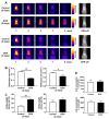
Doxorubicin (DOX) induces dose-dependent cardiotoxicity via oxidative stress and abnormal mitochondrial function in the myocardium. Therefore, a noninvasive in vivo imaging procedure for monitoring the redox status of the heart may aid in monitoring diseases and developing treatments. However, an appropriate technique has yet to be developed. In this study, we demonstrate a technique for detecting and visualizing the redox status of the heart using in vivo dynamic nuclear polarization-magnetic resonance imaging (DNP-MRI) with 3-carbamoyl-PROXYL (CmP) as a molecular imaging probe. Male C57BL/6N mice were administered DOX (20 mg/kg) or saline. DNP-MRI clearly showed a slower DNP signal reduction in the DOX group than in the control group. Importantly, the difference in the DNP signal reduction rate between the two groups occurred earlier than that detected by physiological examination or clinical symptoms. In an in vitro experiment, KCN (an inhibitor of complex IV in the mitochondrial electron transport chain) and DOX inhibited the electron paramagnetic resonance change in H9c2 cardiomyocytes, suggesting that the redox metabolism of CmP in the myocardium is mitochondrion-dependent. Therefore, this molecular imaging technique has the potential to monitor the dynamics of redox metabolic changes in DOX-induced cardiomyopathy and facilitate an early diagnosis of this condition.
Keywords: dynamic nuclear polarization–magnetic resonance imaging; heart; mitochondria; nitroxyl radical; oxidation reduction.
Conflict of interest statement
The authors declare no conflict of interest.
Figures


Similar articles
-
Development of 20 cm sample bore size dynamic nuclear polarization (DNP)-MRI at 16 mT and redox metabolic imaging of acute hepatitis rat model.Free Radic Biol Med. 2021 Jun;169:149-157. doi: 10.1016/j.freeradbiomed.2021.04.017. Epub 2021 Apr 15. Free Radic Biol Med. 2021. PMID: 33865961
-
Spatiotemporal imaging of redox status using in vivo dynamic nuclear polarization magnetic resonance imaging system for early monitoring of response to radiation treatment of tumor.Free Radic Biol Med. 2022 Feb 1;179:170-180. doi: 10.1016/j.freeradbiomed.2021.12.311. Epub 2021 Dec 27. Free Radic Biol Med. 2022. PMID: 34968704
-
In Vivo Dynamic Nuclear Polarization Magnetic Resonance Imaging for the Evaluation of Redox-Related Diseases and Theranostics.Antioxid Redox Signal. 2022 Jan;36(1-3):172-184. doi: 10.1089/ars.2021.0087. Epub 2021 Jul 7. Antioxid Redox Signal. 2022. PMID: 34015957 Review.
-
Noninvasive mapping of the redox status of dimethylnitrosamine-induced hepatic fibrosis using in vivo dynamic nuclear polarization-magnetic resonance imaging.Sci Rep. 2016 Sep 2;6:32604. doi: 10.1038/srep32604. Sci Rep. 2016. PMID: 27587186 Free PMC article.
-
[Novel redox molecular imaging "ReMI" with dual magnetic resonance].Yakugaku Zasshi. 2013;133(7):803-14. doi: 10.1248/yakushi.13-00139. Yakugaku Zasshi. 2013. PMID: 23811768 Review. Japanese.
Cited by
-
H2S Protects from Rotenone-Induced Ferroptosis by Stabilizing Fe-S Clusters in Rat Cardiac Cells.Cells. 2024 Feb 21;13(5):371. doi: 10.3390/cells13050371. Cells. 2024. PMID: 38474335 Free PMC article.
-
Pregnenolone Inhibits Doxorubicin-Induced Cardiac Oxidative Stress, Inflammation, and Apoptosis-Role of Matrix Metalloproteinase 2 and NADPH Oxidase 1.Pharmaceuticals (Basel). 2023 Apr 28;16(5):665. doi: 10.3390/ph16050665. Pharmaceuticals (Basel). 2023. PMID: 37242448 Free PMC article.
-
Primary Protection of Diosmin Against Doxorubicin Cardiotoxicity via Inhibiting Oxido-Inflammatory Stress and Apoptosis in Rats.Cell Biochem Biophys. 2024 Jun;82(2):1353-1366. doi: 10.1007/s12013-024-01289-7. Epub 2024 May 14. Cell Biochem Biophys. 2024. PMID: 38743136
References
-
- Ito H., Miller S.C., Billingham M.E., Akimoto H., Torti S.V., Wade R., Gahlmann R., Lyons G., Kedes L., Torti F.M. Doxorubicin selectively inhibits muscle gene expression in cardiac muscle cells in vivo and in vitro. Proc. Natl Acad. Sci. USA. 1990;87:4275–4279. doi: 10.1073/pnas.87.11.4275. - DOI - PMC - PubMed
Grants and funding
LinkOut - more resources
Full Text Sources

