. 2022 Jun 22;14(7):1322.
doi: 10.3390/pharmaceutics14071322.

Therapeutic Potential of Mesenchymal Stem Cells versus Omega n - 3 Polyunsaturated Fatty Acids on Gentamicin-Induced Cardiac Degeneration

Affiliations

Therapeutic Potential of Mesenchymal Stem Cells versus Omega n - 3 Polyunsaturated Fatty Acids on Gentamicin-Induced Cardiac Degeneration

Fatma Y Meligy et al. Pharmaceutics. .

Abstract

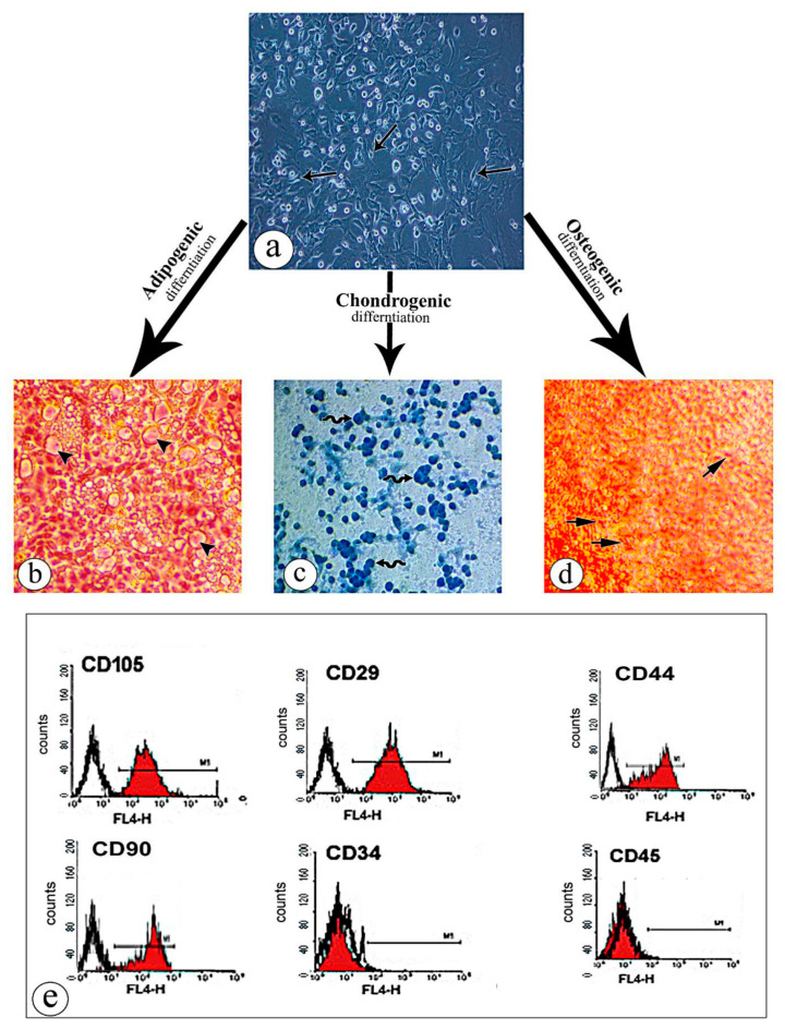
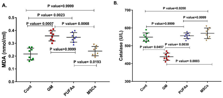
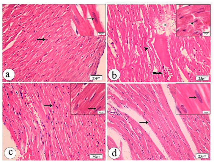
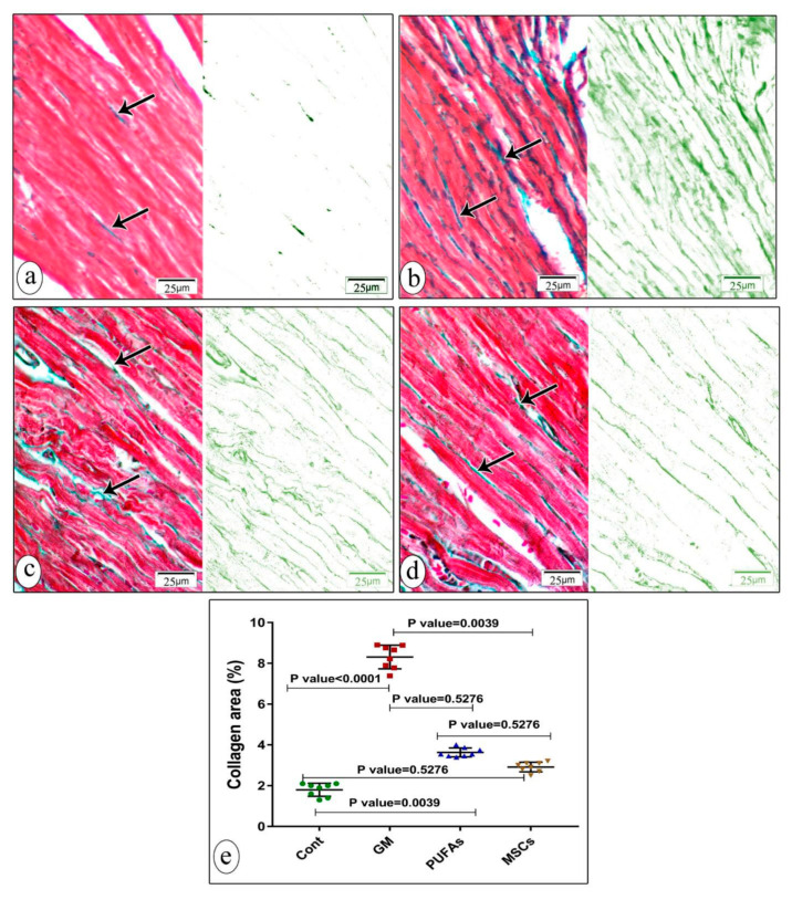
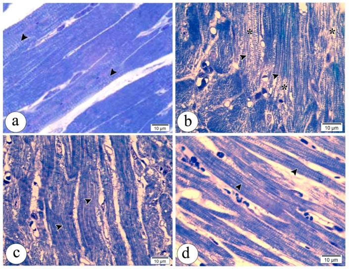
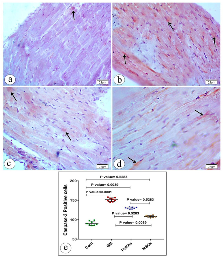
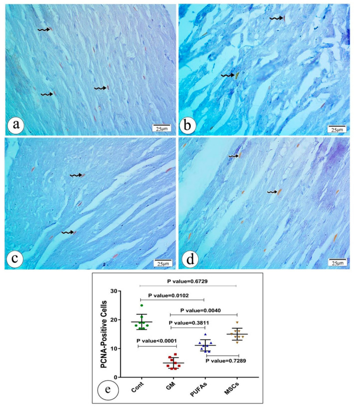
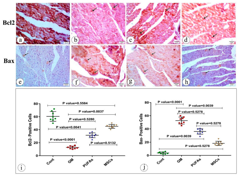
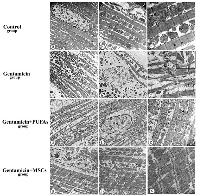
This study compared the cardioprotective action of mesenchymal stem cells (MSCs) and PUFAs in a rat model of gentamicin (GM)-induced cardiac degeneration. Male Wistar albino rats were randomized into four groups of eight rats each: group I (control group), group II (gentamicin-treated rats receiving gentamicin intraperitoneally (IP) at dose of 100 mg/kg/day for 10 consecutive days), group III (gentamicin and PUFA group receiving gentamicin IP at dose of 100 mg/kg/day for 10 consecutive days followed by PUFAs at a dose of 100 mg/kg/day for 4 weeks), and group IV (gentamicin and MSC group receiving gentamicin IP at dose of 100 mg/kg/day followed by a single dose of MSCs (1 × 10⁶)/rat IP). Cardiac histopathology was evaluated via light and electron microscopy. Immunohistochemical detection of proliferating cell nuclear antigen (PCNA), caspase-3 (apoptosis), Bcl2, and Bax expression was performed. Moreover, cardiac malonaldehyde (MDA) content, catalase activity, and oxidative stress parameters were biochemically evaluated. Light and electron microscopy showed that both MSCs and PUFAs had ameliorative effects. Their actions were mediated by upregulating PCNA expression, downregulating caspase-3 expression, mitigating cardiac MDA content, catalase activity, and oxidative stress parameters. MSCs and PUFAs had ameliorative effects against gentamicin-induced cardiac degeneration, with MSCs showing higher efficacy compared to PUFAs.

Keywords: MDA; PCNA; cardiac degeneration; caspase-3; catalase; mesenchymal stem cells; n − 3 polyunsaturated fatty acids.


Conflict of interest statement

The authors declare no conflict of interest.

Figures

Figure 1
Figure 1
Characterization of bone marrow-derived mesenchymal stem cells: (a) the adherent cells appeared fusiform in shape with an elliptical nucleus (arrow); (b) BM-MSCs differentiated into adipocytes and stained with Oil Red O exhibited intense cytoplasmic staining, signifying the accumulation of lipid vacuoles (head arrow); (c) BM-MSCs differentiated into chondrocytes, exhibiting positive Alcian Blue staining (wavy arrow); (d) BM-MSCs differentiated into osteocytes and stained with Alizarin red staining, indicating clusters of calcium depositions (thick arrow); (e) FACS analysis of isolated stem cells showing that the bone marrow-derived mesenchymal stem cells were strongly positive for CD105, CD29, CD44, and CD90 markers but negative for CD34 and CD45 markers.
Figure 2
Figure 2
Cardiac MDA content and catalase activity in all experimental groups. (A) MDA is a lipid peroxidation product and an important marker of oxidative stress. The MDA content was significantly enhanced in the gentamicin-induced cardiac degeneration model (p = 0.0007). Treatment with MSCs reduced MDA expression significantly (p = 0.0068). An insignificant change in MDA expression was observed in the PUFA-treated group (p > 0.9999). (B) Catalase is an antioxidant enzyme. Its activity was significantly reduced in the gentamicin-induced cardiac degeneration model (p = 0.0457). Treatment with either MSCs or PUFAs enhanced catalase activity significantly (p = 0.0003; p = 0.003, respectively). Nonparametric statistics were used. Data were analyzed using Kruskal–Wallis test followed by Dunn’s test. Results were expressed as medians and interquartile ranges. Differences were considered significant at p < 0.05.
Figure 3
Figure 3
Photomicrographs of heart sections stained by hematoxylin and eosin: (a) control group showing the normal histological structure of cardiac myocytes that appeared arranged in a linear array that branched and anastomosed with acidophilic sarcoplasm and oval, centrally located nuclei (arrow); (b) gentamycin-treated group showing that most of the cardiac muscle fibers were disorganized and lost the normal striations, while nuclei of the cardiomyocytes showed deformation in sizes and shapes, and others appeared pyknotic (Head arrow), in addition to cytoplasmic loss and fragmentation of cardiac muscle (star) and interstitial hemorrhage (tailed arrow); (c) gentamycin- and PUFA-treated group showing regular arrangement of the cardiac muscle fibers with oval, centrally located vesicular nucleus (arrow); (d) gentamycin- and MSC-treated group showing regularly arranged cardiac myofibers with oval, rounded nuclei (arrow). Note that inset images of higher magnification were added in all panels photos to show the nucleus.
Figure 4
Figure 4
Photomicrographs of sections of heart stained by Masson trichrome: (a) control group showing scanty collagen fibers in green color in between the cardiomyocytes which appear dark red in color (arrow); (b) gentamycin-treated group showing increased collagen fibers between the cardiomyocytes (arrow); (c) gentamycin- and PUFA-treated group showing less serious accumulation than gentamycin group(arrow); (d) gentamycin- and MSC-treated group showing reduced collagen accumulation (arrow). (e) Statistical analysis of collagen score. Nonparametric statistics were used. Data were analyzed using the Kruskal–Wallis test followed by Dunn’s test. Results are expressed as medians and interquartile ranges. Differences were considered significant at p < 0.05.
Figure 5
Figure 5
Semithin sections of cardiac muscles showing transverse striations: (a) Control group showing cylindrical cardiac muscle fibers and central, oval, and vesicular nuclei, along with visible transverse striations (head arrow); (b) gentamycin-treated group showing loss of striation in some parts of the cardiomyocytes( head arrow), along with areas of fibrinolysis (star); (c) gentamycin- and PUFA-treated group showing restored normal striation of cardiac muscle fibers but less prominent striations compared with the control group (head arrow); (d) gentamycin- and MSC-treated group showing more or less normal cardiac muscle fibers with visible transverse striations (head arrow).
Figure 6
Figure 6
Immunostained sections of heart tissue stained with caspase-3 antibody: (a) control group showing mild positive caspase-3 immunostaining in the sarcoplasm of the muscle fibers (arrow); (b) gentamycin-treated group showing highly positive caspase-3 immunostaining in the sarcoplasm of the muscle fibers(arrow); (c) gentamycin- and PUFA-treated group showing moderate positive caspase-3 immunostaining in the sarcoplasm section in the cardiac muscle of control group(arrow); (d) gentamycin- and MSC-treated group showing moderate to mild positive caspase-3 expression with lower caspase-3 expression than the gentamycin group(arrow). (e) Statistical analysis of caspase intensity in cardiac myocytes in all groups studied. Nonparametric statistics were used. Data were analyzed using the Kruskal–Wallis test followed by Dunn’s test. Results are expressed as medians and interquartile ranges. Differences were considered significant at p < 0.05.
Figure 7
Figure 7
Immunostained sections of heart tissue PCNA expression in cardiac myocytes in all studied groups. Positive staining is shown by a dark-brown color: (a) control group showing moderately positive PCNA immunoreactivity in some myocytes (wavy arrow); (b) gentamycin-treated group showing almost absent PCNA immunoreactivity expression (wavy arrow); (c) gentamycin- and PUFA-treated group showing moderately positive immunoreactivity (wavy arrow); (d) gentamycin- and MSC-treated group showing moderately positive PCNA immunoreactivity almost similar to the control (wavy arrow). (e) Statistical analysis of PCNA expression in cardiac myocytes in all groups studied. Nonparametric statistics were used. Data were analyzed using the Kruskal–Wallis test followed by Dunn’s test. Results are expressed medians and interquartile ranges. Differences were considered significant at p < 0.05.
Figure 8
Figure 8
Immunostained sections of heart tissue of Bcl2 and Bax expression in cardiac myocytes in all studied groups. Positive staining is shown by a brown or red color: (a) control group showing strong Bcl2 immunohistochemical staining of control heart tissue in most of cardiomyocytes (arrow); (b) gentamycin-treated group showing minor positive Bcl2 immunoreactivity in cardiomyocytes (arrow); (c) gentamycin- and PUFA-treated group showing moderately positive Bcl2 immunoreactivity in some of the cardiomyocytes (arrow); (d) gentamycin- and MSC-treated group showing strong positive Bcl2 immunoreactivity in most cardiomyocytes (arrow); (e) control group showing positive Bax immunohistochemical staining of control heart tissue in a few myocytes (arrow); (f) gentamycin-treated group showing strong positive Bax immunoreactivity (overexpression of Bax) in the cardiomyocytes (arrow); (g) gentamycin- and PUFA-treated group showing moderately positive Bax immunoreactivity in some of the cardiomyocytes (arrow); (h) gentamycin- and MSC-treated group showing slight dark-red positive Bax immunoreactivity in most myocytes almost similar to the control (arrow). (i,j) respectively showing Statistical analysis of Bcl2 and Bax expression in cardiac myocytes in all groups studied. Nonparametric statistics were used. Data were analyzed using the Kruskal–Wallis test followed by Dunn’s test. Results are expressed as medians and interquartile ranges. Differences were considered significant at p < 0.05.
Figure 9
Figure 9
Electron micrographs of heart sections from rats of the control group: (a) centrally located nucleus (N), regular arrangement of the myofibrils (F), and mitochondria (m); (b) normal cell membrane of cardiac myocytes (arrow) with invaginations at T-tubules (arrow heads); (c) Z lines (Z) and H lines (H). Electron micrographs of heart sections from rats of the gentamycin-treated group: (a,b) separation of myofibrils with increased interfibrillar spaces (star) and myofibrillar fragmentation and degeneration (F) and irregular nucleus (N); (c) discontinuation and disarrangement of Z lines (Z), with disorganization of mitochondria (m in the incet); Electron micrographs of heart sections from rats of the gentamycin- and PUFA-treated group: (a) normal arrangement of myofibrils (F) with slight loss of myofibrils (star); (b) normal appearance of nucleus (N); (c) well-arranged mitochondria with some disorganization (arrow head) and continuation of Z lines (Z) and H lines (H) in some areas but not others (arrow). Electron micrographs of heart sections from rats of the gentamycin- and MSC-treated group: (a) normal arrangement of myofibrils (F) with a nearly normal nucleus (N); (b) mitochondria with a normal appearance regained (M); (c) continuation of Z lines (Z).

Similar articles

Cited by

References

    1. Kowalewski M., Pawliszak W., Zaborowska K., Navarese E.P., Szwed K.A., Kowalkowska M.E., Kowalewski J., Borkowska A., Anisimowicz L. Gentamicin-collagen sponge reduces the risk of sternal wound infections after heart surgery: Meta-analysis. J. Thorac. Cardiovasc. Surg. 2015;149:1631–1640.e6. doi: 10.1016/j.jtcvs.2015.01.034. - DOI - PubMed
    1. Urbanowicz T., Straburzynska-Migaj E., Buczkowski P., Grajek S., Jemielity M. Novel Method of Infection Prophylaxis in Heart Transplantation by Retrosternal Gentamycin Sponge Application. Transpl. Proc. 2015;47:1954–1957. doi: 10.1016/j.transproceed.2015.02.026. - DOI - PubMed
    1. Khan Z.A., Hollenberg S.M. Valvular Heart Disease in Adults: Infective Endocarditis. FP Essent. 2017;457:30–38. - PubMed
    1. Germain M., Thibault L., Jacques A., Tremblay J., Bourgeois R. Heart valve allograft decontamination with antibiotics: Impact of the temperature of incubation on efficacy. Cell Tissue Bank. 2010;11:197–204. doi: 10.1007/s10561-009-9155-y. - DOI - PubMed
    1. Moffett B.S., Rossano J.W. Use of 4-mg/kg/24-hour empiric aminoglycoside dosing in preoperative neonates with congenital heart disease. Ann. Pharmacother. 2012;46:1193–1197. doi: 10.1345/aph.1Q792. - DOI - PubMed

Grants and funding

This research received no external funding.