

Transcriptional Profiling of the Candida albicans Response to the DNA Damage Agent Methyl Methanesulfonate
- PMID: 35886903
- PMCID: PMC9317300
- DOI: 10.3390/ijms23147555
Transcriptional Profiling of the Candida albicans Response to the DNA Damage Agent Methyl Methanesulfonate
Abstract
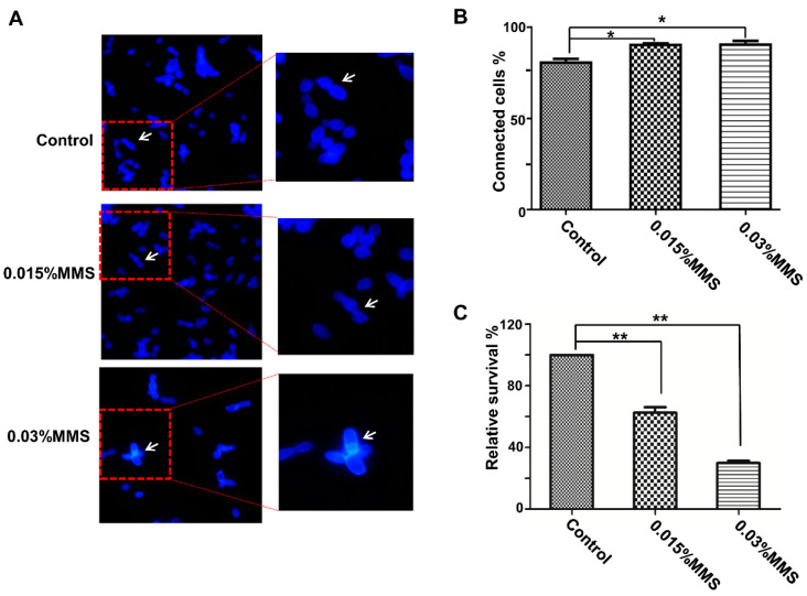
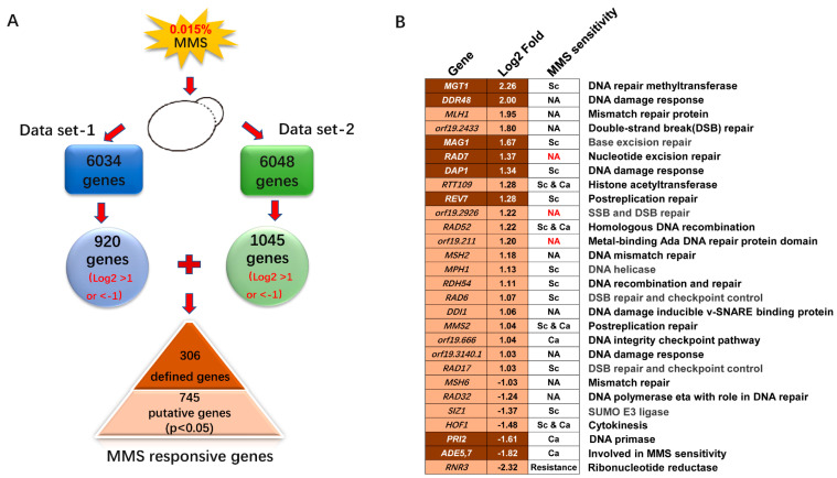
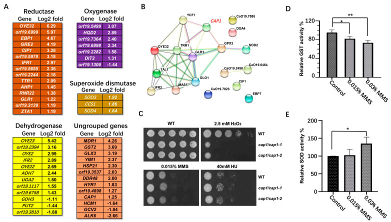
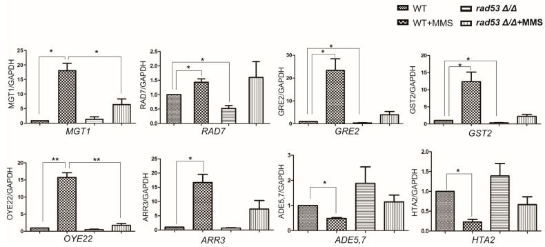
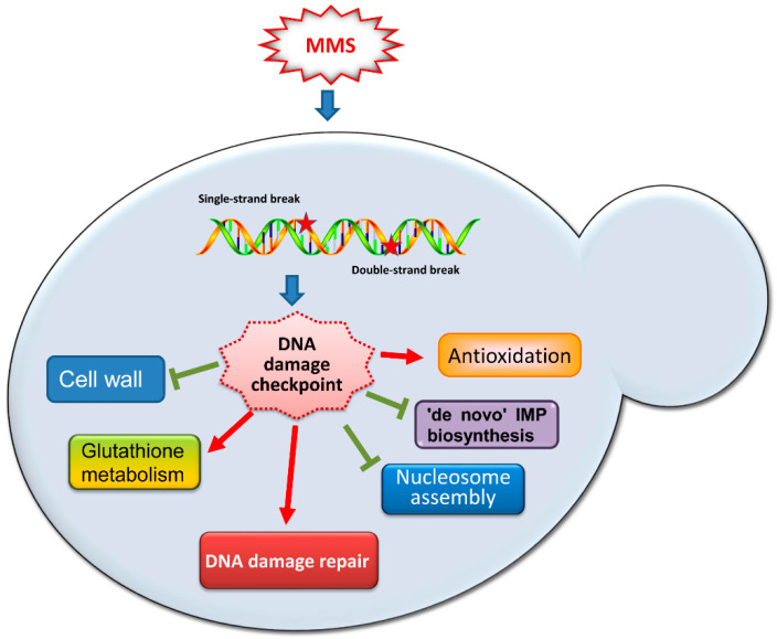
The infection of a mammalian host by the pathogenic fungus Candida albicans involves fungal resistance to reactive oxygen species (ROS)-induced DNA damage stress generated by the defending macrophages or neutrophils. Thus, the DNA damage response in C. albicans may contribute to its pathogenicity. Uncovering the transcriptional changes triggered by the DNA damage-inducing agent MMS in many model organisms has enhanced the understanding of their DNA damage response processes. However, the transcriptional regulation triggered by MMS remains unclear in C. albicans. Here, we explored the global transcription profile in response to MMS in C. albicans and identified 306 defined genes whose transcription was significantly affected by MMS. Only a few MMS-responsive genes, such as MGT1, DDR48, MAG1, and RAD7, showed potential roles in DNA repair. GO term analysis revealed that a large number of induced genes were involved in antioxidation responses, and some downregulated genes were involved in nucleosome packing and IMP biosynthesis. Nevertheless, phenotypic assays revealed that MMS-induced antioxidation gene CAP1 and glutathione metabolism genes GST2 and GST3 showed no direct roles in MMS resistance. Furthermore, the altered transcription of several MMS-responsive genes exhibited RAD53-related regulation. Intriguingly, the transcription profile in response to MMS in C. albicans shared a limited similarity with the pattern in S. cerevisiae, including COX17, PRI2, and MGT1. Overall, C. albicans cells exhibit global transcriptional changes to the DNA damage agent MMS; these findings improve our understanding of this pathogen's DNA damage response pathways.
Keywords: Candida albicans; DNA damage response; RNA-seq; Rad53; methyl methanesulfonate.
Conflict of interest statement
The authors declare no conflict of interest.
Figures


Similar articles
-
Homolog of Saccharomyces cerevisiae SLX4 is required for cell recovery from MMS-induced DNA damage in Candida albicans.FEMS Yeast Res. 2021 Mar 18;21(2):foab010. doi: 10.1093/femsyr/foab010. FEMS Yeast Res. 2021. PMID: 33606011
-
Pph3 dephosphorylation of Rad53 is required for cell recovery from MMS-induced DNA damage in Candida albicans.PLoS One. 2012;7(5):e37246. doi: 10.1371/journal.pone.0037246. Epub 2012 May 14. PLoS One. 2012. PMID: 22606354 Free PMC article.
-
Characterization of Pph3-mediated dephosphorylation of Rad53 during methyl methanesulfonate-induced DNA damage repair in Candida albicans.Biochem J. 2017 Mar 23;474(7):1293-1306. doi: 10.1042/BCJ20160889. Biochem J. 2017. PMID: 28183985
-
Co-regulation of pathogenesis with dimorphism and phenotypic switching in Candida albicans, a commensal and a pathogen.Int J Med Microbiol. 2002 Oct;292(5-6):299-311. doi: 10.1078/1438-4221-00215. Int J Med Microbiol. 2002. PMID: 12452278 Review.
-
A role for Rad23 proteins in 26S proteasome-dependent protein degradation?Mutat Res. 2002 Jan 29;499(1):53-61. doi: 10.1016/s0027-5107(01)00291-3. Mutat Res. 2002. PMID: 11804604 Review.
Cited by
-
Loss of Gst1 enhances resistance to MMS by reprogramming the transcription of DNA damage response genes in a Rad53-dependent manner in Candida albicans.Cell Commun Signal. 2024 Oct 14;22(1):495. doi: 10.1186/s12964-024-01865-7. Cell Commun Signal. 2024. PMID: 39402632 Free PMC article.
-
DNA Damage Checkpoints Govern Global Gene Transcription and Exhibit Species-Specific Regulation on HOF1 in Candida albicans.J Fungi (Basel). 2024 May 29;10(6):387. doi: 10.3390/jof10060387. J Fungi (Basel). 2024. PMID: 38921373 Free PMC article.
-
Analysis of transcriptional response in haploid and diploid Schizosaccharomyces pombe under genotoxic stress.G3 (Bethesda). 2024 Sep 4;14(9):jkae177. doi: 10.1093/g3journal/jkae177. G3 (Bethesda). 2024. PMID: 39120426 Free PMC article.
References
-
- van Berlo D., Wessels A., Boots A.W., Wilhelmi V., Scherbart A.M., Gerloff K., van Schooten F.J., Albrecht C., Schins R.P. Neutrophil-derived ROS contribute to oxidative DNA damage induction by quartz particles. Free Radic. Biol. Med. 2010;49:1685–1693. doi: 10.1016/j.freeradbiomed.2010.08.031. - DOI - PubMed
MeSH terms
Substances
Grants and funding
LinkOut - more resources
Full Text Sources
Molecular Biology Databases
Research Materials
Miscellaneous
