. 2022 Jul 8:13:941991.
doi: 10.3389/fmicb.2022.941991. eCollection 2022.

The Secreted Ribonuclease SRE1 Contributes to Setosphaeria turcica Virulence and Activates Plant Immunity

Affiliations

The Secreted Ribonuclease SRE1 Contributes to Setosphaeria turcica Virulence and Activates Plant Immunity

Shidao He et al. Front Microbiol. .

Abstract

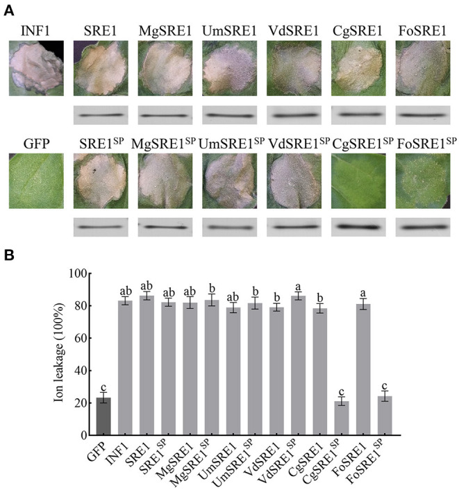
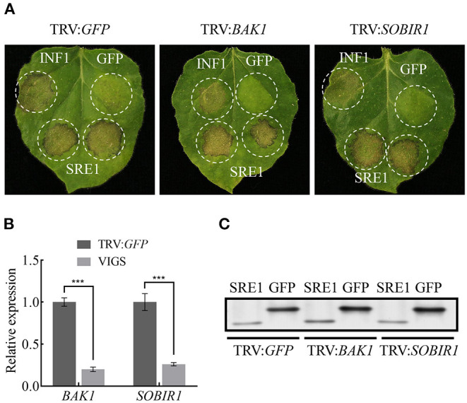
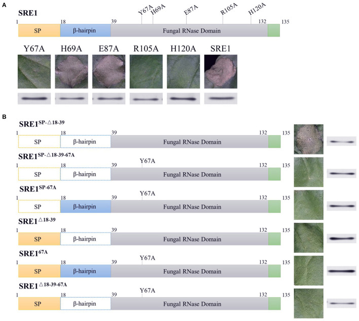
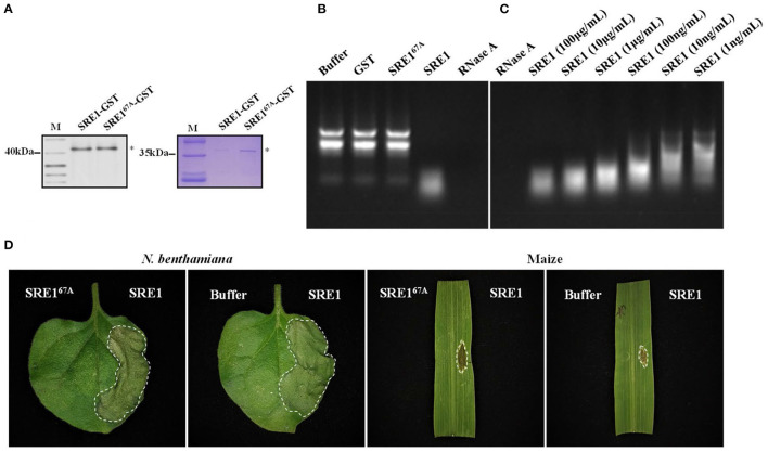
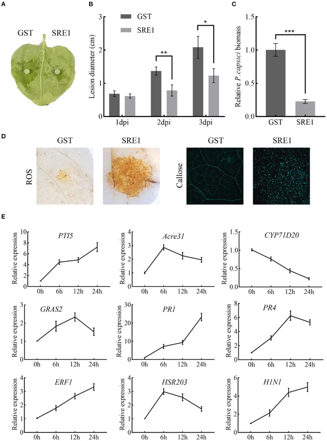
During the plant infection process, pathogens can secrete several effectors. Some of the effectors are well-known for their roles in regulating plant immunity and promoting successful pathogen colonization. However, there are few studies on the ribonuclease (RNase) effectors secreted by fungi. In the present study, we discovered a secretable RNase (SRE1) in the secretome of Setosphaeria turcica that was significantly upregulated during the early stages of S. turcica infection in maize. Knockdown of SRE1 significantly reduced the virulence of S. turcica. SRE1 can induce cell death in maize and Nicotiana benthamiana. However, unlike the conventional hypersensitive response (HR) caused by other effectors, SRE1 is not dependent on its signal peptide (SP) or plant receptor kinases (such as BAK1 and SOBIR1). SRE1-induced cell death depends upon its enzymatic activity and the N-terminal β-hairpin structure. SRE1 relies on its N-terminal β-hairpin structure to enter cells, and then degrades plant's RNA through its catalytic activity causing cytotoxic effects. Additionally, SRE1 enhances N. benthamiana's resistance to pathogenic fungi and oomycetes. In summary, SRE1 promotes the virulence of S. turcica, inducing plant cell death and activating plant immune responses.

Keywords: Setosphaeria turcica; cell death; maize; plant immunity; ribonuclease.


Conflict of interest statement

The authors declare that the research was conducted in the absence of any commercial or financial relationships that could be construed as a potential conflict of interest.

Figures

Figure 1
Figure 1
Identifying the S. turcica secreted proteins by liquid chromatography-mass spectrometry (LC-MS/MS). S. turcica was cultivated for 5 days in a potato liquid medium, the supernatant was collected, and the mycelium and impurities were removed through ultrafiltration tubes. The composition of S. turcica secretory proteome was analyzed by LC-MS/MS. The different colors in the column chart represent different protein families. The numbers and the percentages of different proteins are also shown.
Figure 2
Figure 2
Secretory function and phylogenetic analysis of S. turcica effector SRE1. (A) The yeast-signal peptide trap system was used to verify the signal peptide function of SRE1. A CMD-W medium screened the yeast strain YTK12 carrying the pSUC2 vector. The signal peptide with secretion function can make up the secretion defect of YTK12 invertase so that YTK12 can grow in the YPRAA medium. The invertase activity was detected by reducing TTC to insoluble red TPF. The Avr1b-SP and the empty vector pSUC2 were used as positive and negative controls, respectively. (B) Phylogenetic tree of SRE1 homologs in fungi of different trophic types. The sequences were derived from the Uniprot database and aligned using CLUSTALW. The phylogenetic tree was constructed in MEGA. The colored circles represent different trophic types of fungi, and the numbers represent the number of cysteines.
Figure 3
Figure 3
The homologous genes of SRE1 from different pathogenic fungi induces cell death in N. benthamiana. (A) Transient expression of SRE1, SRE1 homologous proteins, and signal peptide deletion mutants in N. benthamiana. INF1 and GFP were used as positive control and negative control, respectively. Western blot analysis of 3 × HA-tagged proteins expressed in N. benthamiana leaves. (B) Quantitative analysis of cell death by electrolyte leakage. The bars are the average of three independent experiments, and the error bars represent the standard deviations. Different letters at the top of the bars indicate significant differences (one-way ANOVA; P < 0.05).
Figure 4
Figure 4
SRE1-induced cell death is BAK1/SOBIR1-independent. (A) The TRV-mediated gene silencing system was used to verify that SRE1-induced cell death is independent of BAK1/SOBIR1. INF1 and GFP were positive and negative controls, respectively. (B) Expression levels of BAK1 and SOBIR1 after silencing were detected by qPCR. The Student's t-test was carried out to determine the significance of the difference. Error bars indicate the standard deviations of three biological replicates. ***Indicates a significant difference at a P-value of < 0.001. (C) Western blot analysis of proteins expressed in silenced N. benthamiana leaves.
Figure 5
Figure 5
The cytotoxicity of SRE1 depends on its enzymatic active site and N-terminal β-hairpin structure. (A) N. benthamiana leaves were infiltrated with A. tumefaciens carrying the SER1 enzyme active site mutants. Western blot analysis of 3 × HA-tagged proteins expressed in N. benthamiana leaves. (B) N. benthamiana leaves were infiltrated with A. tumefaciens carrying different mutants of the SER1. The superscripts SP, Δ18-39, and 67A, represent SRE1 missing the signal peptide, N-terminal β-hairpin, and catalytic site Y67, respectively. Western blot analysis of 3 × HA-tagged proteins expressed in N. benthamiana leaves.
Figure 6
Figure 6
SRE1 induces cell necrosis through the degradation of total plant RNA. (A) Coomassie brilliant blue detection and immunoblot analysis of SRE1-GST and SRE167A-GST. Asterisks represent the location of the target proteins. (B) Maize total RNA was used to determine the RNase activity of SRE1 and SRE167A recombinant proteins. Buffer and GST were negative controls, and RNase A was used as a positive control. The final concentrations of GST, RNase A, SRE1, and SRE167A were 100 μg/mL. (C) Effects of different concentrations of SRE1 on the degradation of maize total RNA. (D) N. benthamiana and maize leaves were treated with recombinant proteins SRE1 and SRE167A at a concentration of 0.4 mg/mL. The buffer solution was used as a negative control.
Figure 7
Figure 7
SRE1 triggers immune responses in N. benthamiana. (A) Pretreatment with 1 μg/mL SRE1 significantly increased resistance against the oomycetes pathogen P. capsica. The photograph was taken 2 days post-inoculation (dpi). (B) The lesion diameter was evaluated at 1, 2, and 3 dpi. The bars are the average of three independent experiments, and the error bars indicate standard deviations (* P < 0.05, ** P < 0.01, Student's t-test). (C) Infected leaves were collected 48 h post-inoculation, and total DNA was extracted for qPCR analysis. NbEF-1α was used as a reference gene, and the relative expression of PcActin was calculated as the relative pathogen biomass. The bars are the average of three independent experiments, and the error bars represent the standard deviations (*** P < 0.001, Student's t-test). (D) ROS and callose production in N. benthamiana leaves treated with 1 μg/ml SRE1. (E) Relative expression of the PTI-associated marker genes (PTI5, Acre31, CYP71D20, and GRAS2), pathogenesis-related genes (PR1, PR4, and ERF1), and HR genes (HSR203 and H1N1). Relative expression was quantified by qRT-PCR using NbEF-1α as a reference gene. Bars indicate mean fold changes (±SD).
Figure 8
Figure 8
SRE1 contributes to the virulence of S. turcica. (A) Comparison of colony, conidia, and appressorium morphology between wild-type TL-5 and silent transformants RNAi#18 and RNAi#20. The black arrow in the picture points to the newly-formed appressorium. (B) Pathogenicity assays to investigate the role of SRE1 gene silencing transformants RNAi#18 and RNAi#20 in the virulence of maize B73. The photograph was taken at 5 dpi. (C) The qPCR results showed the relative transcript levels of SRE1 in SRE1-silenced transformants. Average lesion diameters (D) and relative S. turcica biomass (E) were calculated from three independent experiments and the error bars indicate standard deviations (*P < 0.05, **P < 0.01, ***P < 0.001, Student's t-test).

Similar articles

Cited by

References

    1. Anderson J. P., Sperschneider J., Win J., Kidd B., Yoshida K., Hane J. (2017). Comparative secretome analysis of Rhizoctonia solani isolates with different host ranges reveals unique secretomes and cell death inducing effectors. Sci. Rep. 7, 10410. 10.1038/s41598-017-10405-y - DOI - PMC - PubMed
    1. Asai S., Yoshioka H. (2010). Nitric Oxide as a Partner of Reactive Oxygen Species Participates in Disease Resistance to Necrotrophic Pathogen Botrytis cinerea in Nicotiana benthamiana. Mol. Plant Microbe Interact. 22, 619–629. 10.1094/MPMI-22-6-0619 - DOI - PubMed
    1. Babilonia K., Wang P., Liu Z., Jamieson P., Mormile B., Rodrigues O. (2021). A nonproteinaceous Fusarium cell wall extract triggers receptor-like protein-dependent immune responses in Arabidopsis and cotton. New Phytol. 230, 275–289. 10.1111/nph.17146 - DOI - PubMed
    1. Bentolila S., Guitton C., Bouvet N., Sailland A., Nykaza S., Freyssinet G. (1991). Identification of an RFLP marker tightly linked to the Ht1 gene in maize. Tag. Theor. Appl. Genet. 82, 393–398. 10.1007/BF00588588 - DOI - PubMed
    1. Cai Q., Qiao L., Wang M., He B., Lin F. M., Palmquist J. (2018). Plants send small RNAs in extracellular vesicles to fungal pathogen to silence virulence genes. Science. 360, 1126–1129. 10.1126/science.aar4142 - DOI - PMC - PubMed

LinkOut - more resources