. 2022 Aug 12;50(14):8331-8348.
doi: 10.1093/nar/gkac620.

Zinc controls PML nuclear body formation through regulation of a paralog specific auto-inhibition in SUMO1

Affiliations

Zinc controls PML nuclear body formation through regulation of a paralog specific auto-inhibition in SUMO1

Mathieu Lussier-Price et al. Nucleic Acids Res. .

Abstract

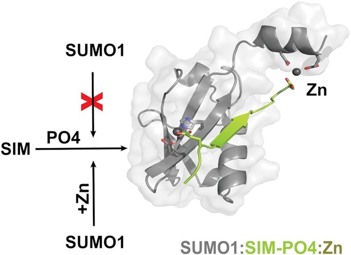
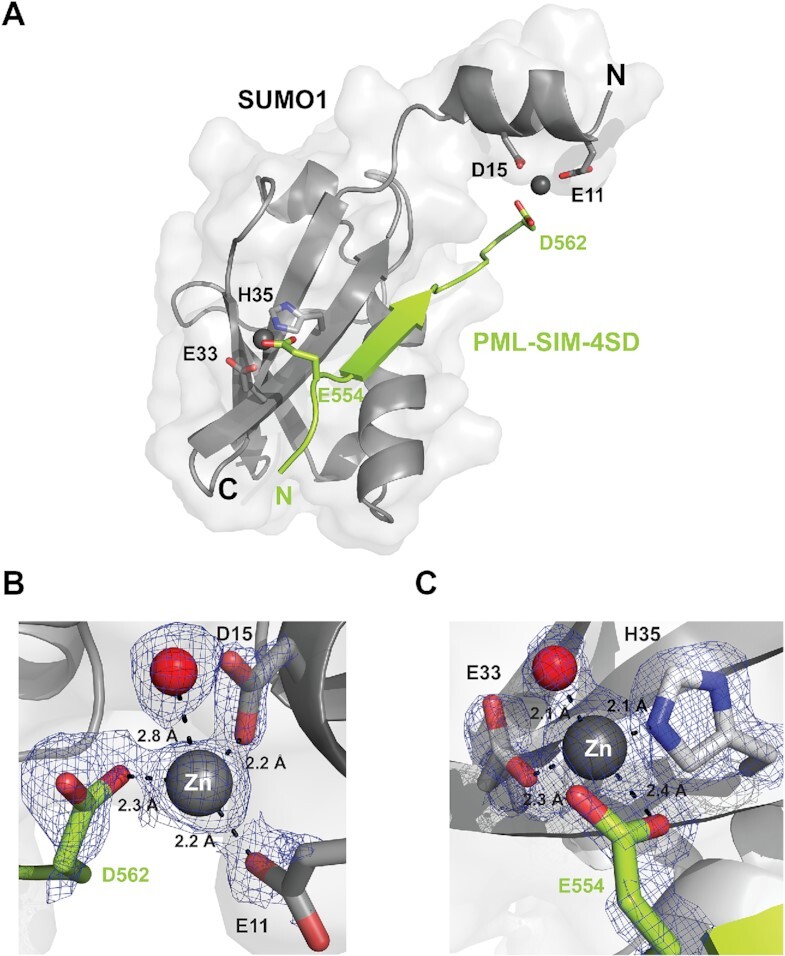
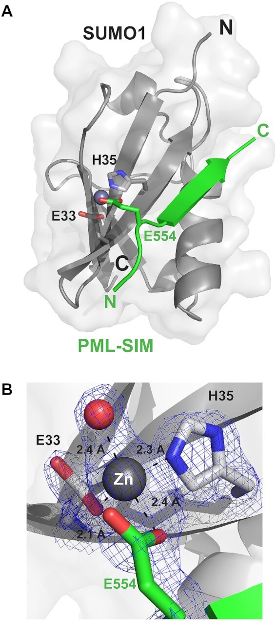
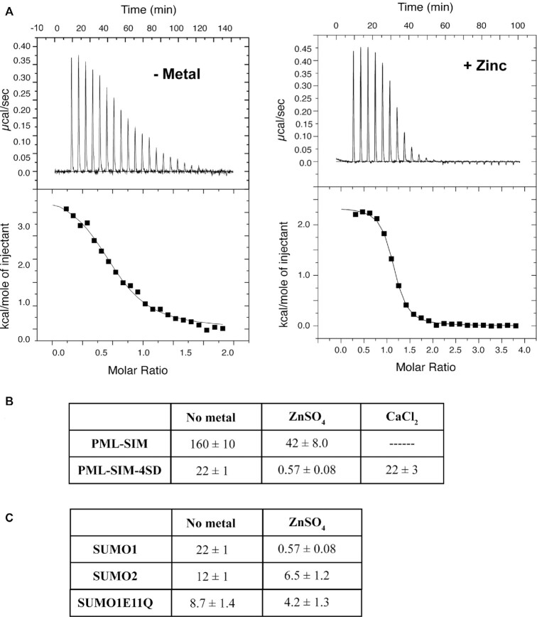
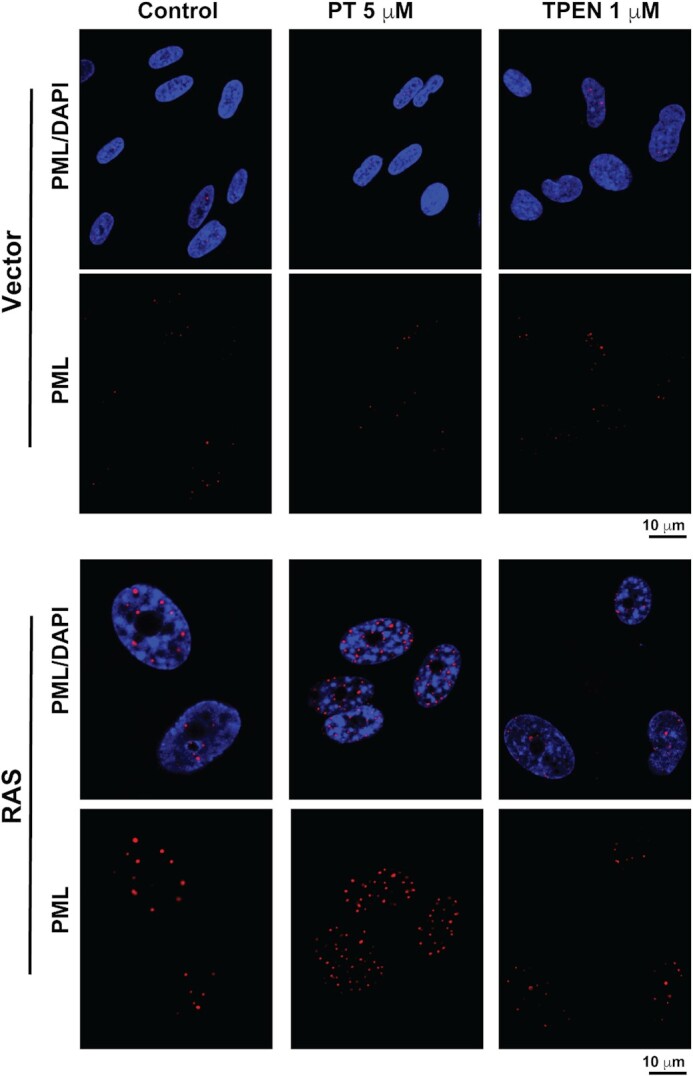
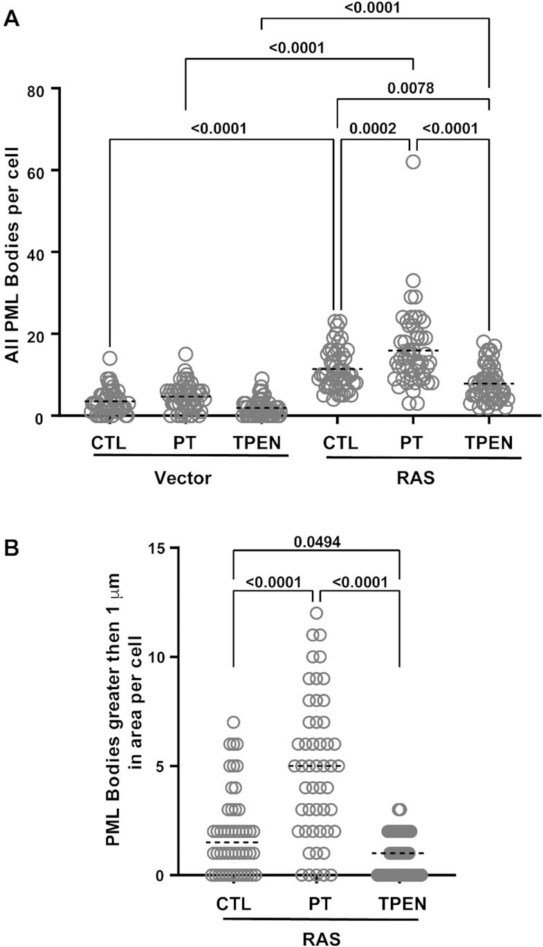
SUMO proteins are important regulators of many key cellular functions in part through their ability to form interactions with other proteins containing SUMO interacting motifs (SIMs). One characteristic feature of all SUMO proteins is the presence of a highly divergent intrinsically disordered region at their N-terminus. In this study, we examine the role of this N-terminal region of SUMO proteins in SUMO-SIM interactions required for the formation of nuclear bodies by the promyelocytic leukemia (PML) protein (PML-NBs). We demonstrate that the N-terminal region of SUMO1 functions in a paralog specific manner as an auto-inhibition domain by blocking its binding to the phosphorylated SIMs of PML and Daxx. Interestingly, we find that this auto-inhibition in SUMO1 is relieved by zinc, and structurally show that zinc stabilizes the complex between SUMO1 and a phospho-mimetic form of the SIM of PML. In addition, we demonstrate that increasing cellular zinc levels enhances PML-NB formation in senescent cells. Taken together, these results provide important insights into a paralog specific function of SUMO1, and suggest that zinc levels could play a crucial role in regulating SUMO1-SIM interactions required for PML-NB formation and function.


Figures

Graphical Abstract
Graphical Abstract
In the presence of zinc ions, the paralog specific auto-inhibition of SUMO1 is relieved to enhance binding to SUMO-interaction motifs (SIMs) in their phosphorylated forms.
Figure 1.
Figure 1.
The N-terminal region of SUMO1 inhibits binding of phosphorylated SIMs. (A) Amino-acid sequence of SIMs from PML and Daxx used for ITC studies. Underlined residues represent the hydrophobic core region of the SIM and those in bold represent either the phospho-mimetic or phosphorylated serine residues. (B) Representative ITC thermograms for the interaction between PML-SIM-4SD and either SUMO1 (left panel) or ΔN-SUMO1 (right panel). (C) Comparison of the KD values (in μM) obtained in ITC studies for the binding of either SUMO1 or ΔN-SUMO1 to the different SIM peptides and UBC9. All experiments were conducted in 20 mM Tris–HCl, pH 7.4.
Figure 2.
Figure 2.
The N-terminal of SUMO1 inhibits interactions with the SIM-binding interface. (A) Fluorescence spectra of bis-ANS binding to either SUMO1 (full line) or ΔN-SUMO1 (dashed line). (B) Graphical plot of the bis-ANS fluorescence signal recorded at 487 nm in the presence of ΔN-SUMO1 following addition of various amounts (0–4 molar equivalence) of the PML-SIM-4SD peptide.
Figure 3.
Figure 3.
The N-terminal of SUMO2 does not inhibit binding of phosphorylated SIMs. (A) ITC thermograms of the titration of PML-SIM-4SD with either SUMO2 (left panel) and ΔN-SUMO2 (right panel). (B) Comparison of KD values (in μM) obtained in ITC studies for the binding of either SUMO2 or ΔN-SUMO2 to the different SIM peptides. All experiments were conducted in 20 mM Tris–HCl, pH 7.4.
Figure 4.
Figure 4.
Structural characterization of a SUMO1:PML-SIM-4SD:Zn complex. (A) Cartoon representation of the structure of the SUMO1: PML-SIM-4SD complex in the presence of zinc highlighting the presence of an alpha helix in the N-terminal region of SUMO1. In the representation, SUMO1 is colored in gray and the PML-SIM-4SD peptide in lime green. The bound zinc atoms (Zn1 and Zn2) are represented as grey spheres and the side chains of the residues from SUMO1 and PML-SIM-4SD involved in chelating the zinc atoms are highlighted in stick representation. (BC) Close-up and metrics (in Å) of Zn1 bound to the N-terminal region (B) and Zn2 bound to the core region (C) of SUMO1 and the PML-SIM-4SD peptide. The residues involved in coordinating the zinc atoms are in stick representation with their respective electron density contoured at 1.5σ cut-off from the 2FoFc map generated from the crystal diffraction data. The red spheres represent water molecules that are coordinated by the zinc atoms.
Figure 5.
Figure 5.
Structural characterization of a SUMO1:PML-SIM:Zn complex. (A) Overall structure of the SUMO1: PML-SIM complex in the presence of zinc highlighting the zinc bound between the core region of SUMO1 and the PML-SIM peptide. The proteins are in cartoon representation with SUMO1 in gray and PML-SIM peptide in green. The bound zinc atom is represented as a grey sphere and the side chains of the residues from SUMO1 and PML-SIM peptide involved in chelating the zinc atom is highlighted in stick representation. (B) Close-up and metrics (in Å) of the zinc atom bound to the core region of SUMO1 and the PML-SIM peptide. The residues involved in coordinating the zinc atom are in stick representation with their respective electron density contoured at 1.5σ cut-off from the 2FoFc map generated from the crystal diffraction data. The red sphere represents a water molecule that is coordinated by the zinc atom.
Figure 6.
Figure 6.
Zinc ions specifically enhance binding of the phospho-mimetic SIM of PML to SUMO1. (A) ITC thermograms from the titration of the PML-SIM-4SD peptide with SUMO1 either in the absence (left panel) or the presence of zinc sulfate (right panel). (B) Comparison of the KD values (in μM) obtained in ITC studies for SUMO1 binding to PML-SIM and PML-SIM-4SD and SUMO1E11Q binding to PML-SIM-4SD in the presence and absence of zinc as well as the binding to PML-SIM-4SD in the presence of calcium. (C) Comparison of the KD values (in μM) obtained in ITC studies for PML-SIM-4SD binding to SUMO1 and SUMO2 in the presence and absence of zinc. All experiments were conducted in 20 mM Tris–HCl pH7.4, 50 mM NaCl in the absence or presence of 1 mM ZnSO4.
Figure 7.
Figure 7.
Increasing cellular zinc levels in senescent cells enhances PML-NB formation. Normal human fibroblasts IMR90 cells expressing a control vector or the activated RAS oncogene, to induce senescence were treated for 24h with either control vehicle, 5 μM PT (pyrithione) or 1 μM TPEN prior to fixation and indirect immunofluorescence with a specific anti-PML antibody (Bethyl # A301-167A) and with DAPI to stain DNA in the nucleus.
Figure 8.
Figure 8.
Increasing cellular zinc levels in senescent cells enhances PML-NB formation. Normal human fibroblasts IMR90 cells expressing a control vector or the activated RAS oncogene, to induce senescence, were treated for 24 h with either control vehicle, 5 μM pyrithione (PT) or 1 μM TPEN prior to fixation and quantitative analysis for the overall number of PML-NBs per cell (A) and the number of PML-NBs greater than 1 μm in size (B). For each condition, 50 cells from two independent experiments were counted and statistics were performed using the PRISM9 software.

Similar articles

Cited by

References

    1. Kerscher O. SUMO junction-what's your function? New insights through SUMO-interacting motifs. EMBO Rep. 2007; 8:550–555. - PMC - PubMed
    1. Desterro J.M., Rodriguez M.S., Kemp G.D., Hay R.T.. Identification of the enzyme required for activation of the small ubiquitin-like protein SUMO-1. J. Biol. Chem. 1999; 274:10618–10624. - PubMed
    1. Pichler A., Gast A., Seeler J.S., Dejean A., Melchior F.. The nucleoporin RanBP2 has SUMO1 E3 ligase activity. Cell. 2002; 108:109–120. - PubMed
    1. Schmidt D., Muller S.. PIAS/SUMO: new partners in transcriptional regulation. Cell. Mol. Life Sci. 2003; 60:2561–2574. - PMC - PubMed
    1. Cappadocia L., Lima C.D.. Ubiquitin-like protein conjugation: structures, chemistry, and mechanism. Chem. Rev. 2018; 118:889–918. - PMC - PubMed

Publication types

MeSH terms