

Sex differences in contextual pattern separation, neurogenesis, and functional connectivity within the limbic system
- PMID: 35870952
- PMCID: PMC9308289
- DOI: 10.1186/s13293-022-00450-2
Sex differences in contextual pattern separation, neurogenesis, and functional connectivity within the limbic system
Abstract
Background: Females are more likely to present with anxiety disorders such as post-traumatic stress disorder (PTSD) compared to males, which are associated with disrupted hippocampal integrity. Sex differences in the structure and function of hippocampus exist. Here, we examined sex differences in contextual pattern separation, functional connectivity, and activation of new neurons during fear memory.
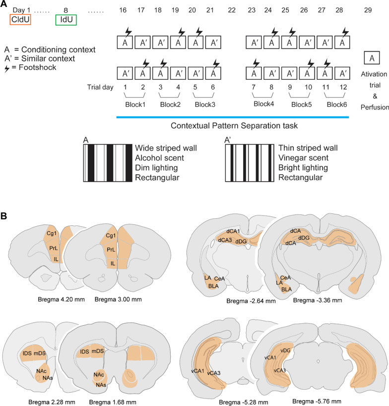
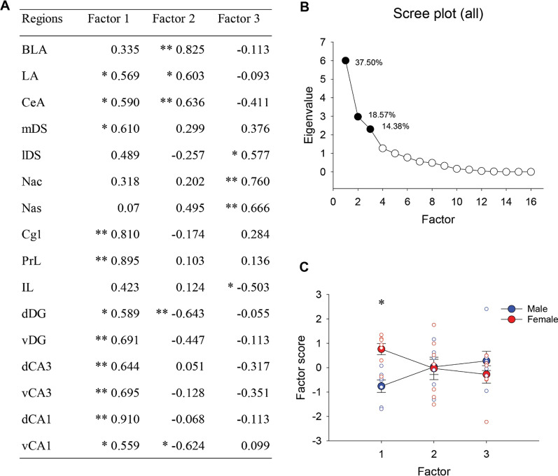
Methods: Two-month-old male and female Sprague-Dawley rats were injected with the DNA synthesis markers, iododeoxyuridine (IdU) and chlorodeoxyuridine (CldU) 3 weeks and 4 weeks before perfusion, respectively. One week after CldU injection, the rats underwent a context discrimination task in which rats were placed in context A (shock) and context A' (no shock) every day for 12 days. On the test day, rats were placed in the shock context (context A) to measure fear memory and expression of zif268, an immediate early gene across 16 different limbic and reward regions. Repeated-measures or factorial analysis of variance was conducted on our variables of interest. Pearson product-moment calculations and principal component analyses on zif268 expression across regions were also performed.
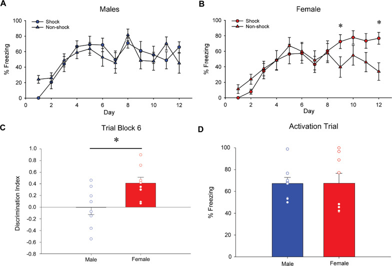
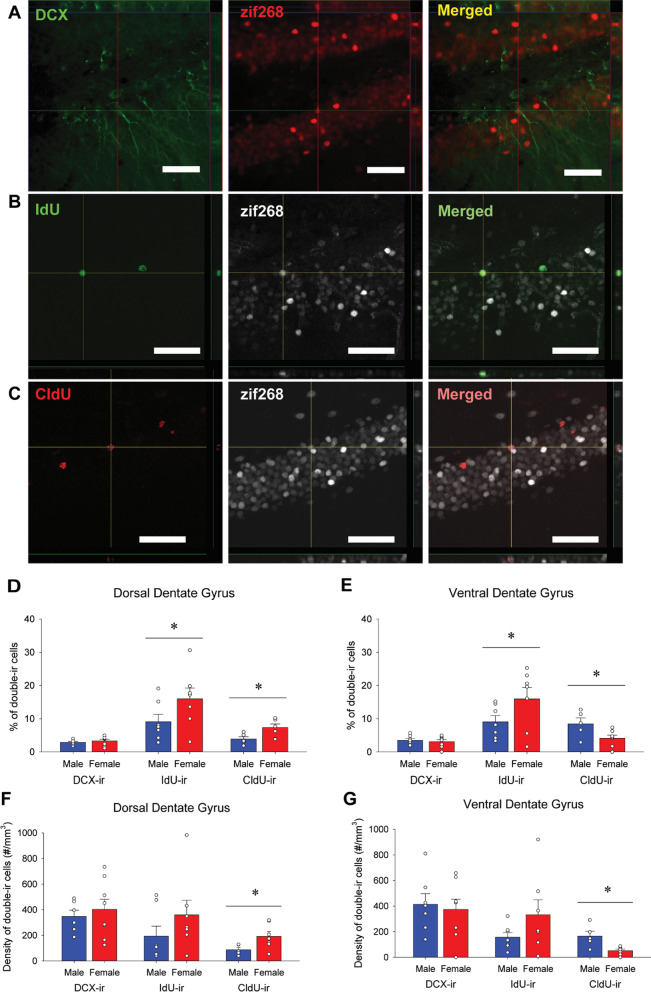
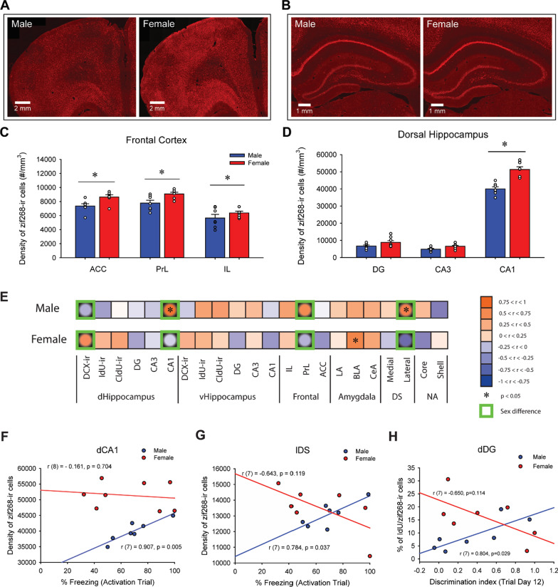
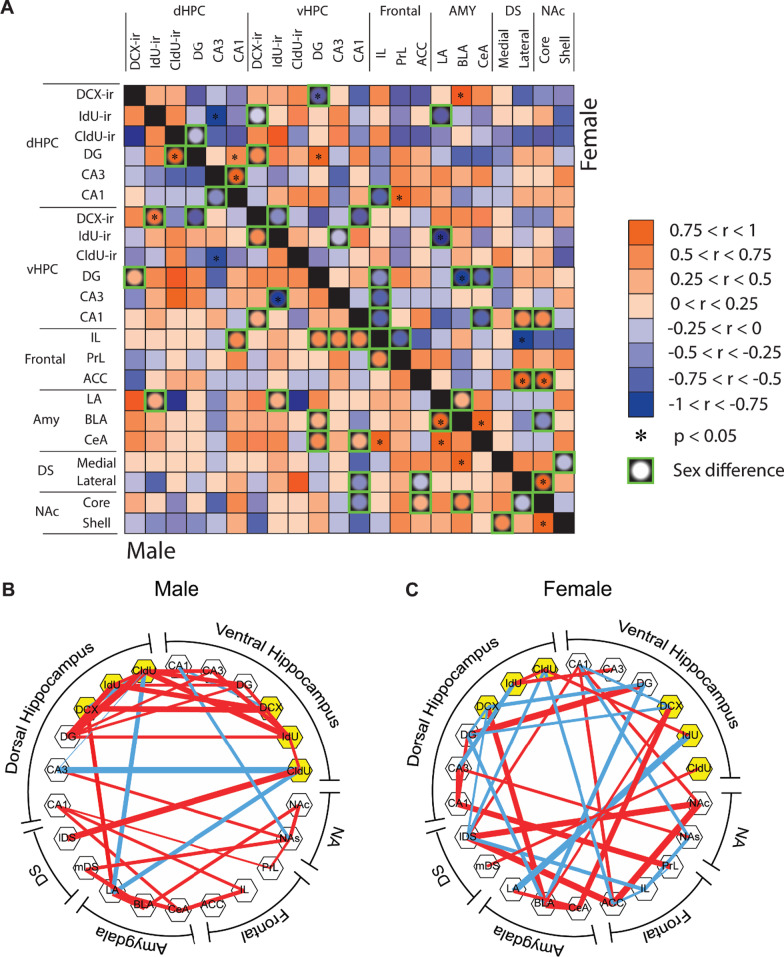
Results: We found that females, but not males, showed contextual discrimination during the last days of training. On the test day, both sexes displayed similar levels of freezing, indicating equivalent fear memory for context A. Despite similar fear memory, males showed more positive correlations of zif268 activation between the limbic regions and the striatum, whereas females showed more negative correlations among these regions. Females showed greater activation of the frontal cortex, dorsal CA1, and 3-week-old adult-born dentate granular cells compared to males.
Conclusions: These results highlight the importance of studying sex differences in fear memory and the contribution of adult neurogenesis to the neuronal network and may contribute to differences in susceptibility to fear-related disorders such as post-traumatic stress disorder. Highlights Female rats, but not male rats, show faster discrimination during a contextual pattern separation task. Three-week-old adult-born neurons are more active in response to fear memory in females compared to males. Females had greater neural activation compared to males in the frontal cortex and dorsal CA1 region of the hippocampus in response to fear memory. Males and females show distinct patterns in functional connectivity for fear memory across limbic regions. Males have many positive correlations between activated new neurons of different ages between the dorsal and ventral hippocampus, while females show more correlations between activated new neurons and other limbic regions.
© 2022. The Author(s).
Conflict of interest statement
The authors declare that they have no competing interests.
Figures
Similar articles
-
Sex and strategy use matters for pattern separation, adult neurogenesis, and immediate early gene expression in the hippocampus.Hippocampus. 2016 Jan;26(1):87-101. doi: 10.1002/hipo.22493. Epub 2015 Aug 4. Hippocampus. 2016. PMID: 26179150
-
Sex differences in neurogenesis and activation of new neurons in response to spatial learning and memory.Psychoneuroendocrinology. 2013 Aug;38(8):1236-50. doi: 10.1016/j.psyneuen.2012.11.007. Epub 2012 Dec 6. Psychoneuroendocrinology. 2013. PMID: 23219473
-
Dorsal and ventral hippocampal adult-born neurons contribute to context fear memory.Neuropsychopharmacology. 2018 Nov;43(12):2487-2496. doi: 10.1038/s41386-018-0109-6. Epub 2018 Jun 2. Neuropsychopharmacology. 2018. PMID: 29941977 Free PMC article.
-
Sex differences in hippocampal cognition and neurogenesis.Neuropsychopharmacology. 2019 Jan;44(1):200-213. doi: 10.1038/s41386-018-0208-4. Epub 2018 Sep 7. Neuropsychopharmacology. 2019. PMID: 30214058 Free PMC article. Review.
-
Acute stress yields a sex-dependent facilitation of signaled active avoidance in rats.Neurobiol Stress. 2024 Jun 13;31:100656. doi: 10.1016/j.ynstr.2024.100656. eCollection 2024 Jul. Neurobiol Stress. 2024. PMID: 38994219 Free PMC article. Review.
Cited by
-
Network Neuroscience Untethered: Brain-Wide Immediate Early Gene Expression for the Analysis of Functional Connectivity in Freely Behaving Animals.Biology (Basel). 2022 Dec 24;12(1):34. doi: 10.3390/biology12010034. Biology (Basel). 2022. PMID: 36671727 Free PMC article. Review.
-
Young adult and aged female rats are vulnerable to amygdala-dependent, but not hippocampus-dependent, memory impairment following short-term high-fat diet.Brain Res Bull. 2023 Apr;195:145-156. doi: 10.1016/j.brainresbull.2023.03.001. Epub 2023 Mar 2. Brain Res Bull. 2023. PMID: 36870621 Free PMC article. Review.
-
Morphine-context associative memory and locomotor sensitization in mice are modulated by sex and context in a dose-dependent manner.bioRxiv [Preprint]. 2023 Nov 5:2023.11.03.565492. doi: 10.1101/2023.11.03.565492. bioRxiv. 2023. PMID: 37961152 Free PMC article. Preprint.
-
ERPs in Children and Adolescents with Generalized Anxiety Disorder: Before and after an Intervention Program.Brain Sci. 2022 Sep 1;12(9):1174. doi: 10.3390/brainsci12091174. Brain Sci. 2022. PMID: 36138910 Free PMC article.
-
Sex differences in inflammation in the hippocampus and amygdala across the lifespan in rats: associations with cognitive bias.Immun Ageing. 2022 Oct 6;19(1):43. doi: 10.1186/s12979-022-00299-4. Immun Ageing. 2022. PMID: 36203171 Free PMC article.
References
-
- Marr D. Simple memory: a theory for archicortex. Philos Trans R Soc London Ser B, Biol Sci. 1971;262:24–80. - PubMed
Publication types
MeSH terms
LinkOut - more resources
Full Text Sources
Miscellaneous
