. 2022 Oct;127(7):1239-1253.
doi: 10.1038/s41416-022-01902-7. Epub 2022 Jul 21.

LHX2 facilitates the progression of nasopharyngeal carcinoma via activation of the FGF1/FGFR axis

Affiliations

LHX2 facilitates the progression of nasopharyngeal carcinoma via activation of the FGF1/FGFR axis

Tao Xie et al. Br J Cancer. 2022 Oct.

Abstract

Background: Distant metastasis and recurrence remain the main obstacle to nasopharyngeal carcinoma (NPC) treatment. However, the molecular mechanisms underlying NPC growth and metastasis are poorly understood.

Methods: LHX2 expression was examined in NPC cell lines and NPC tissues using quantitative reverse transcription-polymerase chain reaction, western blotting and Immunohistochemistry assay. NPC cells overexpressing or silencing LHX2 were used to perform CCK-8 assay, colony-formation assay, EdU assay, wound-healing and invasion assays in vitro. Xenograft tumour models and lung metastasis models were involved for the in vivo assays. The Gene Set Enrichment Analysis (GSEA), ELISA assay, western blot, chromatin immunoprecipitation (ChIP) assay and Luciferase reporter assay were applied for the downstream target mechanism investigation.

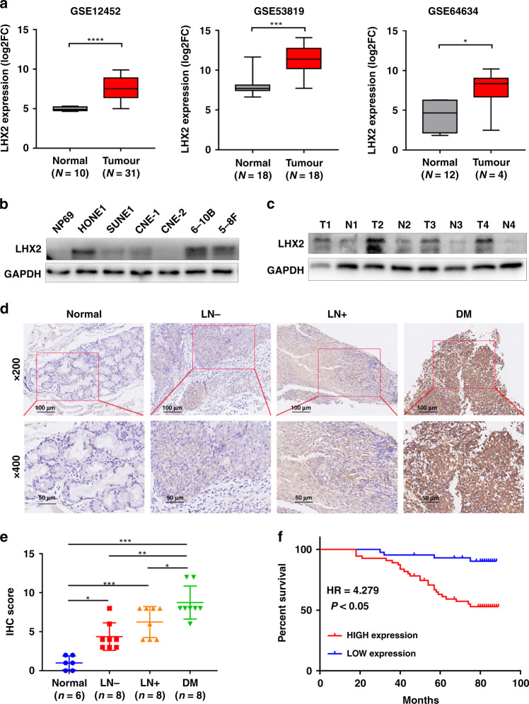
Results: LIM-homeodomain transcription factor 2 (LHX2) was upregulated in NPC tissues and cell lines. Elevated LHX2 was closely associated with poor survival in NPC patients. Ectopic LHX2 overexpression dramatically promoted the growth, migration and invasion of NPC cells both in vitro and in vivo. Mechanistically, LHX2 transcriptionally increased the fibroblast growth factor 1 (FGF1) expression, which in turn activated the phosphorylation of STAT3 (signal transducer and activator of transcription 3), ERK1/2 (extracellular regulated protein kinases 1/2) and AKT signalling pathways in an autocrine and paracrine manner, thereby promoting the growth and metastasis of NPC. Inhibition of FGF1 with siRNA or FGFR inhibitor blocked LHX2-induced nasopharyngeal carcinoma cell growth, migration and invasion.

Conclusions: Our study identifies the LHX2-FGF1-FGFR axis plays a key role in NPC progression and provides a potential target for NPC therapy.


Conflict of interest statement

The authors declare no competing interests.

Figures

Fig. 1
Fig. 1. LHX2 is highly expressed in NPC and predicts poor survival.
a Box plots showing LHX2 mRNA expression level is higher in nasopharyngeal carcinoma tissues than in normal tissues in GSE12452 (left), GSE53819 (middle) and GSE64634 (right). b Western blot analysis shows LHX2 expression level is higher in NPC cell lines than in normal nasopharyngeal epithelial cell lines. Each experiment was independently repeated at least three times. c Western blot analysis shows LHX2 expression level is higher in nasopharyngeal carcinoma tissues than in normal tissues. Each experiment was independently repeated at least three times. d Immunohistochemical staining and (e) statistical analysis of LHX2 in normal tissues, primary NPC tissues with (LN + ) or without (LN-) lymph node metastasis and primary NPC tissues with distant metastasis. f Kaplan–Meier survival curves indicate high expression of LHX2 is associated with poor survival in NPC patients. Data shown as mean ± SD. *P < 0.05, **P < 0.01, ***P < 0.001.
Fig. 2
Fig. 2. LHX2 promotes NPC cell growth in vitro and in vivo.
a Western blot analysis of LHX2 protein expression in HONE1 and SUNE1 cells stably overexpressing LHX2. Each experiment was independently repeated at least three times. b The CCK-8 assay and c the colony-formation assay of HONE1 and SUNE1 cells stably overexpressing LHX2. Each experiment was independently repeated at least three times. d Western blot analysis of LHX2 protein expression in HONE1 and SUNE1 cells transfected with LHX2 shRNAs or control cells. Each experiment was independently repeated at least three times. e The CCK-8 assay and f the colony-formation assay of HONE1 and SUNE1 cells transfected with LHX2 shRNAs or control. Each experiment was independently repeated at least three times. g, h Images of Xenograft tumours of nude mice 30 days after injecting with HONE1 and SUNE1 cells stably overexpressing LHX2 or vector (n = 5 in each group). i Tumour volume growth curves. j Average xenograft tumour weights. k Immunohistochemistry assay of LHX2 and Ki67 protein expression in xenograft tumours. Data shown as mean ± SD. *P < 0.05, **P < 0.01, ***P < 0.001.
Fig. 3
Fig. 3. LHX2 strengthens NPC cells migration, invasion and metastasis.
a GSEA enrichment plots demonstrated that metastasis was associated with upregulation of LHX2 in GSE12452 (left and middle) and GSE53819 (right). b Wound healing and (c) transwell migration and invasion assays in HONE1 cells stably overexpressing LHX2 or empty vector. Each experiment was independently repeated at least three times. d Wound healing and (e) transwell migration and invasion assays in HONE1 cells transfected with LHX2 shRNAs or control. Each experiment was independently repeated at least three times. fh Representative images of metastatic lungs (f), representative HE images (g) and numbers of metastatic foci per lung (h) injected with HONE1 and SUNE1 cells stably overexpressing LHX2 or empty vector (n = 5 in each group). i Western blot analysis of EMT-related protein expression in HONE1 and SUNE1 cells stably overexpressing LHX2 or empty vector. Each experiment was independently repeated at least three times. j IHC analysis of E-cadherin and vimentin protein expression in xenograft tumours. Data shown as mean ± SD. *P < 0.05, **P < 0.01, ***P < 0.001.
Fig. 4
Fig. 4. LHX2 activates pro-survival and β-catenin signalling pathways via transcriptionally regulating FGF1 expression.
a LHX2 motif. b, c Western blot analysis of FGF1 expression in HONE1 and SUNE1 cells with LHX2 overexpression (b) or knockdown (c). Each experiment was independently repeated at least three times. d, e ELISA assay of FGF1 expression in the supernatant derived from HONE1 and SUNE1 cells with LHX2 overexpression (d) or knockdown (e). Each experiment was independently repeated at least three times. f Luciferase activity of the reporter gene driven by serially truncated/mutated FGF1 promoters. Each experiment was independently repeated at least three times. g ChIP assay confirms the direct binding of LHX2 to the FGF1 promoters in HONE1 and SUNE1 cells. Each experiment was independently repeated at least three times. h Western blot assay reveals the p-STAT3, pERK1/2 and p-AKT were activated by LHX2. Each experiment was independently repeated at least three times. i GSEA enrichment plots demonstrates that β-catenin signalling is associated with upregulation of LHX2 in GSE53819. j Western blot assay of β-catenin and Ser9-GSK3β in HONE1 and SUNE1 cells stably overexpressing LHX2 or containing the empty vector. Each experiment was independently repeated at least three times. k RT-qPCR analysis of β-catenin response genes in HONE1 and SUNE1 cells stably overexpressing LHX2 or containing the empty vector. Each experiment was independently repeated at least three times. l Representative images of immunofluorescent staining of β-catenin in HONE1 and SUNE1 cells stably overexpressing LHX2 or containing the empty vector. Each experiment was independently repeated at least three times. Data shown as mean ± SD. *P < 0.05, **P < 0.01, ***P < 0.001.
Fig. 5
Fig. 5. FGF1 mediates LHX2-induced proliferation, migration and invasion in an autocrine or paracrine manner.
ae FGF1 siRNA was transfected into HONE1 cells stably overexpressing LHX2 or the empty vector. Colony-formation assay (a), wound-healing assay (b) and transwell migration and invasion assays (c). Western blot analysis of LHX2, FGF1 and pro-survival signalling pathways (d). Western blot analysis of β-catenin signalling pathway and EMT-related proteins (e). Each experiment was independently repeated at least three times. f, g HONE1 and SUNE1 cells were treated with FGF1-CM or EV-CM for the indicated time. Western blot analysis of pro-survival signalling pathways (f). Western blot analysis of β-catenin signalling pathway and EMT-related proteins (g). Each experiment was independently repeated at least three times. Data shown as mean ± SD. *P < 0.05, **P < 0.01. FGF1-CM FGF1-condition medium, EV empty vector.
Fig. 6
Fig. 6. Blocking FGF1/FGFR signalling suppresses the proliferation, migration and invasion of NPC cells.
a HONE1 cells were incubated for 4 h of AZD4547 at different concentrations and then lysed and immunoblotted for the indicated proteins. b, c HONE1 cells stably overexpressing LHX2 or the empty vector were treated with AZD4547 (100 nM) or DMSO. Whole-cell lysates were subjected to western blot analysis. Each experiment was independently repeated at least three times. df HONE1 cells stably overexpressing LHX2 or the empty vector were treated with AZD4547 (100 nM) or DMSO for 48 h. The cells were harvested and subjected to (d) colony-formation assay, (e) wound-healing assay and (f) transwell migration and invasion assays. Each experiment was independently repeated at least three times. gj LHX2-overexpressed or vector HONE1 cells (2 × 106) were injected into the dorsal flank of mice. One week after injection, the mice were treated with AZD4547 (12.5 mg/kg/d) or vehicle orally for 3 weeks. Images of Xenograft tumours (g). Tumour volume growth curves (h). The volume of xenograft tumours (i). Average xenograft tumour weights (j). LHX2-overexpressed or vector HONE1 cells (1 × 106) were injected into the tail vein of nude mice and the mice were treated with AZD4547 (12.5 mg/kg/d) or vehicle orally for 3 weeks. km Representative images of metastatic lungs (k), representative HE images (l) and numbers of metastatic foci per lung (m) (n = 5 in each group). Data are shown as means ± SD. *P < 0.05, **P < 0.01, ***P < 0.001.
Fig. 7
Fig. 7. LHX2 is positively correlated with FGF1 in NPC.
a Correlations between LHX2 and FGF1 mRNA expression in NPC tissues. b Representative immunostaining of LHX2 and FGF1 in consecutive paraffin sections of NPC tissues. c Representative immunostaining of FGF1, p-STAT3, p-ERK, p-AKT in LHX2 overexpression and control xenograft tumours.
Fig. 8
Fig. 8. A working model of LHX2-induced NPC progression.
LHX2 transcriptionally activates FGF1 expression. Tumour cells produce FGF1, which binds to FGFR, and the subsequent downstream signalling occurs through the intracellular receptor substrates FGFR substrate 2 (FRS2), resulting in the phosphorylation and activation of MAPK/ERK, JAK2/STAT3, and PI3K/AKT signalling pathways. (i) These pro-survival pathways are responsible for the promotion of tumour proliferation and growth. (ii) Phosphorylated AKT stimulates the Ser9-GSK3β/β-catenin signalling, leading to the EMT of NPC via the β-catenin targeted ZEB1 and TWIST1 genes and promotes tumour cell migration and invasion. (iii) FGF/FGFR trapping by AZD4547 could block the LHX2/FGF1-induced promotion on tumour growth and metastasis.

Similar articles

Cited by

References

    1. Bray F, Ferlay J, Soerjomataram I, Siegel RL, Torre LA, Jemal A. Global cancer statistics 2018: GLOBOCAN estimates of incidence and mortality worldwide for 36 cancers in 185 countries. CA Cancer J Clin. 2018;68:394–424. doi: 10.3322/caac.21492. - DOI - PubMed
    1. Lee AW, Ng WT, Chan LL, Hung WM, Chan CC, Sze HC, et al. Evolution of treatment for nasopharyngeal cancer-success and setback in the intensity-modulated radiotherapy era. Radiother Oncol. 2014;110:377–84. doi: 10.1016/j.radonc.2014.02.003. - DOI - PubMed
    1. Sun X, Su S, Chen C, Han F, Zhao C, Xiao W, et al. Long-term outcomes of intensity-modulated radiotherapy for 868 patients with nasopharyngeal carcinoma: an analysis of survival and treatment toxicities. Radiother Oncol. 2014;110:398–403. doi: 10.1016/j.radonc.2013.10.020. - DOI - PubMed
    1. Li Y, Tang LQ, Liu LT, Guo SS, Liang YJ, Sun XS, et al. Induction chemotherapy plus concurrent chemoradiotherapy versus concurrent chemoradiotherapy alone in locoregionally advanced nasopharyngeal carcinoma in children and adolescents: a matched cohort analysis. Cancer Res Treat. 2018;50:1304–15. doi: 10.4143/crt.2017.463. - DOI - PMC - PubMed
    1. Peter IS, Davidson EH. Evolution of gene regulatory networks controlling body plan development. Cell. 2011;144:970–85. doi: 10.1016/j.cell.2011.02.017. - DOI - PMC - PubMed

MeSH terms