. 2022 Aug 30;13(4):e0163922.
doi: 10.1128/mbio.01639-22. Epub 2022 Jul 12.

Extracellular Loops of the Treponema pallidum FadL Orthologs TP0856 and TP0858 Elicit IgG Antibodies and IgG+-Specific B-Cells in the Rabbit Model of Experimental Syphilis

Affiliations

Extracellular Loops of the Treponema pallidum FadL Orthologs TP0856 and TP0858 Elicit IgG Antibodies and IgG+-Specific B-Cells in the Rabbit Model of Experimental Syphilis

Kristina N Delgado et al. mBio. .

Abstract

The resurgence of syphilis in the new millennium has called attention to the importance of a vaccine for global containment strategies. Studies with immune rabbit serum (IRS) indicate that a syphilis vaccine should elicit antibodies (Abs) that promote opsonophagocytosis of treponemes by activated macrophages. The availability of three-dimensional models for Treponema pallidum's (Tp) repertoire of outer membrane proteins (OMPs) provides an architectural framework for identification of candidate vaccinogens with extracellular loops (ECLs) as the targets for protective Abs. Herein, we used Pyrococcus furiosus thioredoxin (PfTrx) as a scaffold to display Tp OMP ECLs to interrogate sera and peripheral blood mononuclear cells (PBMCs) from immune rabbits for ECL-specific Abs and B cells. We validated this approach using a PfTrx scaffold presenting ECL4 from BamA, a known opsonic target. Using scaffolds displaying ECLs of the FadL orthologs TP0856 and TP0858, we determined that ECL2 and ECL4 of both proteins are strongly antigenic. Comparison of ELISA and immunoblot results suggested that the PfTrx scaffolds present conformational and linear epitopes. We then used the FadL ECL2 and ECL4 PfTrx constructs as "hooks" to confirm the presence of ECL-specific B cells in PBMCs from immune rabbits. Our results pinpoint immunogenic ECLs of two newly discovered OMPs, while advancing the utility of the rabbit model for circumventing bottlenecks in vaccine development associated with large-scale production of folded OMPs. They also lay the groundwork for production of rabbit monoclonal Abs (MAbs) to characterize potentially protective ECL epitopes at the atomic level. IMPORTANCE Recent identification and structural modeling of Treponema pallidum's (Tp) repertoire of outer membrane proteins (OMPs) represent a critical breakthrough in the decades long quest for a syphilis vaccine. However, little is known about the antigenic nature of these β-barrel-forming OMPs and, more specifically, their surface exposed regions, the extracellular loops (ECLs). In this study, using Pyrococcus furiosus thioredoxin (PfTrx) as a scaffold to display Tp OMP ECLs, we interrogated immune rabbit sera and peripheral blood mononuclear cells for the presence of antibodies (Abs) and circulating rare antigen-specific B cells. Our results pinpoint immunogenic ECLs of two newly discovered OMPs, while advancing the utility of the rabbit model for surveying the entire Tp OMPeome for promising OMP vaccinogens. This work represents a major advancement toward characterizing potentially protective OMP ECLs and future vaccine studies. Additionally, this strategy could be applied to OMPs of nonspirochetal bacterial pathogens.

Keywords: B cells; FadL; Treponema pallidum; extracellular loop; outer membrane protein; syphilis; vaccine.


Conflict of interest statement

The authors declare no conflict of interest.

Figures
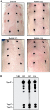
FIG 1
FIG 1
Immune rabbits. (A) Three rabbits inoculated intratesticularly with Tp Nichols were challenged 60 days later, along with an uninfected control, on their shaved backs at each of 8 sites with 1 × 103 freshly harvested Tp. Black marks indicate location of initial intradermal injections and used to monitoring animals for syphilitic lesion development (representative images 27 days postchallenge). (B) Immunoblot reactivity of sera from the three immune rabbits with Tp lysate strips.
FIG 2
FIG 2
Pyrococcus furiosus thioredoxin (PfTrx) as a scaffold for presentation of extracellular loops (ECLs). (A) ModWeb prediction of the β-barrel of Tp BamA (TP0326) (12) with ECL4 shown in red. (B) Phyre2 prediction of the PfTrx structure showing the insertion site (green) for ECLs (here BamA ECL4) and a linear map of the PfTrxBamA/ECL4 construct with N-His- and C-Avi- tags shown in pink and yellow, respectively. (C) Reactivity of PfTrxBamA/ECL4 and PfTrx with IRS, HSS, NRS, NHS, mouse anti-Avi-Tag, and rat anti-BamA ECL4 antiserum. The highly antigenic lipoprotein, Tpp17, was used as a positive control. (D) Supernatant (Sup) and elution (Elu) fractions from Protein G pull-downs of PfTrxBamA/ECL4 and PfTrx with IRS or NRS immunoblotted with Avi-Tag Abs.
FIG 3
FIG 3
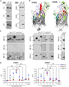
Reactivity of FadL orthologs and extracellular regions of TP0856 and TP0858. (A) Immunoblots of TP0856 and TP0858 against IRS and HSS. (B) trRosetta (89) predictions for the structures of TP0856 and TP0858 (6). The seven ECLs and hatches of each protein are identified; cysteine residues in ECL4 are shown in black. (C) Immunoblots of PfTrxTP0856/ECL1-ECL7 and PfTrxTP0856/Hatch (left); PfTrxTP0858/ECL1-ECL7 and PfTrxTP0858/Hatch (middle); and PfTrx scaffold and Tpp17 controls (right) against IRS. (D) Reactivity of PfTrxTP0856/ECL1-ECL7 and PfTrxTP0856/Hatch (left) and PfTrxTP0858/ECL1-ECL7 and PfTrxTP0858/Hatch (right) with IRS, measured as area under the curve (AUC) from ELISA dilutions corrected for PfTrx background. *, P ≤ 0.05 or **, P ≤ 0.01, significant differences between the means of the groups (respectively) determined by one-way ANOVA with Bonferroni's correction for multiple comparisons.
FIG 4
FIG 4
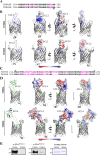
Comparison of sequences, structures and electrostatics of ECL2 and ECL4 of TP0856 and TP858 (Nichols). (A) Alignment of TP0856 and TP0858 ECL2 sequences with substitutions shown in magenta. (B) trRosetta models of TP0856 and TP0858 with ECL2 shown in blue (left). Electrostatics of ECL2s are shown in the same and opposite orientations (middle and right, respectively). (C) Alignment of TP0856 and TP0858 ECL4 sequences with substitutions and deletions shown in magenta. (D) trRosetta models of TP0856 and TP0858 with ECL4 shown in blue (left). Electrostatics of ECL4s are shown in the same and opposite orientations (middle and right, respectively). Some ECLs and the hatches are masked for optimal viewing of electrostatics. (E) Immunoblot reactivity of rabbit anti-PfTrxTP0856/ECL2 and anti-PfTrxTP0856/ECL4 against full-length TP0856 and TP0858.
FIG 5
FIG 5
B-cell epitope predictions of TP0856 and TP0858. (A) Cartoon schematics of TP0856 and TP0858 showing the positions of discontinuous (D) and linear (L) BCE predictions using ElliPro (32), DiscoTope 2.0 (31), IEDB (33), and BC pred (34) algorithms. (B) Ribbon diagrams (gray) of TP0856 and TP0858 with discontinuous BCEs predicted by ElliPro (threshold: 0.5) shown as transparent surfaces. Numeric values indicate the average prediction scores for corresponding epitopes. (C) Epitopes with scores ≥0.8 and ≥0.9 are shown as pink and magenta transparent surfaces, respectively.
FIG 6
FIG 6
Identification of Tpp17-specific rabbit IgG+ B cells. (A) Gating strategy: After gating lymphocytes, doublets were excluded in FSC-H and -W, and SSC-H and -W plots. IgG+ cells were identified from the IgM and IgA double-negative cells. (B) Tpp17-specific B cells were gated from IgG+ cells as double-positive cells for Tpp17-SP-AF405 and Tpp17-SP-AF647. B. burgdorferi OspC conjugated to SP-AF405 and SP-AF647 was used as a negative control. The mean frequencies of antigen-specific IgG+ B cells are shown in the accompanying bar graphs. (C) Identification of IgG+ B cells specific for PfTrxBamA/ECL4 using the gating strategy shown in panel A. IgG+ PfTrx+ cells were excluded using PfTrx-SP-APC-Cy7. From each immune rabbit, B cells specific for PfTrxBamA/ECL4 were identified within the IgG+ PfTrxNeg gate as cells double-positive for SP-AF405 and SP-AF647; PBMCs from a normal rabbit were used as a control. The mean frequencies of IgG+ B cells specific BamA ECL4 are shown in the accompanying bar graphs.
FIG 7
FIG 7
Identification of circulating B cells specific for ECL2 and ECL4 of TP0856 and TP0858. IgG+ B cells specific for ECL2 and ECL4 in TP0856 (A) and TP0858 (B) were identified within the IgG+ PfTrxNeg gate as cells double positive for SP-AF405 and SP-AF647; PBMCs from a normal rabbit were used as a control. The mean frequencies of the individual ECL-specific IgG+ B cells are shown in the accompanying bar graphs.

References

    1. Gottlieb SL, Deal CD, Giersing B, Rees H, Bolan G, Johnston C, Timms P, Gray-Owen SD, Jerse AE, Cameron CE, Moorthy VS, Kiarie J, Broutet N. 2016. The global roadmap for advancing development of vaccines against sexually transmitted infections: update and next steps. Vaccine 34:2939–2947. doi:10.1016/j.vaccine.2016.03.111. - DOI - PMC - PubMed
    1. Lithgow KV, Cameron CE. 2017. Vaccine development for syphilis. Expert Rev Vaccines 16:37–44. doi:10.1080/14760584.2016.1203262. - DOI - PMC - PubMed
    1. Liu J, Howell JK, Bradley SD, Zheng Y, Zhou ZH, Norris SJ. 2010. Cellular architecture of Treponema pallidum: novel flagellum, periplasmic cone, and cell envelope as revealed by cryo electron tomography. J Mol Biol 403:546–561. doi:10.1016/j.jmb.2010.09.020. - DOI - PMC - PubMed
    1. Cox DL, Luthra A, Dunham-Ems S, Desrosiers DC, Salazar JC, Caimano MJ, Radolf JD. 2010. Surface immunolabeling and consensus computational framework to identify candidate rare outer membrane proteins of Treponema pallidum. Infect Immun 78:5178–5194. doi:10.1128/IAI.00834-10. - DOI - PMC - PubMed
    1. Luthra A, Montezuma-Rusca JM, La Vake CJ, LeDoyt M, Delgado KN, Davenport TC, Fiel-Gan M, Caimano MJ, Radolf JD, Hawley KL. 2020. Evidence that immunization with TP0751, a bipartite Treponema pallidum lipoprotein with an intrinsically disordered region and lipocalin fold, fails to protect in the rabbit model of experimental syphilis. PLoS Pathog 16:e1008871. doi:10.1371/journal.ppat.1008871. - DOI - PMC - PubMed

Publication types