. 2022 Jul 20;13(1):4198.
doi: 10.1038/s41467-022-31794-3.

Kinesin-8-specific loop-2 controls the dual activities of the motor domain according to tubulin protofilament shape

Affiliations

Kinesin-8-specific loop-2 controls the dual activities of the motor domain according to tubulin protofilament shape

Byron Hunter et al. Nat Commun. .

Abstract

Kinesin-8s are dual-activity motor proteins that can move processively on microtubules and depolymerize microtubule plus-ends, but their mechanism of combining these distinct activities remains unclear. We addressed this by obtaining cryo-EM structures (2.6-3.9 Å) of Candida albicans Kip3 in different catalytic states on the microtubule lattice and on a curved microtubule end mimic. We also determined a crystal structure of microtubule-unbound CaKip3-ADP (2.0 Å) and analyzed the biochemical activity of CaKip3 and kinesin-1 mutants. These data reveal that the microtubule depolymerization activity of kinesin-8 originates from conformational changes of its motor core that are amplified by dynamic contacts between its extended loop-2 and tubulin. On curved microtubule ends, loop-1 inserts into preceding motor domains, forming head-to-tail arrays of kinesin-8s that complement loop-2 contacts with curved tubulin and assist depolymerization. On straight tubulin protofilaments in the microtubule lattice, loop-2-tubulin contacts inhibit conformational changes in the motor core, but in the ADP-Pi state these contacts are relaxed, allowing neck-linker docking for motility. We propose that these tubulin shape-induced alternations between pro-microtubule-depolymerization and pro-motility kinesin states, regulated by loop-2, are the key to the dual activity of kinesin-8 motors.


Conflict of interest statement

The authors declare no competing interests.

Figures
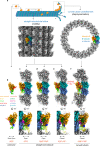
Fig. 1
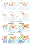
Fig. 1. Structures of microtubule-unbound, microtubule-bound, and curved tubulin-bound CaKip3.
a Top—Cartoon representation of catalytic intermediates of CaKip3’s motility and microtubule depolymerization cycles. Bottom—Example of a CaKip3-decorated microtubule (MT-CaKip3-MDC-ANP) cryo-EM map and the full CaKip3-decorated dolastatin-tubulin-ring cryo-EM map. b X-ray crystallographic density of CaKip3-MDN in the ADP state. ce Cryo-EM maps of microtubule-bound CaKip3-MDC in the APO, AMP-PNP, and ADP-AlFx nucleotide states. f Cryo-EM maps of curved tubulin-bound CaKip3-MDC in the AMP-PNP state. Map surfaces are colored regionally according to the segment of the fitted protein model they enclose: α-tubulin (cornflower blue), β-tubulin (sky blue), kinesin motor core (orange), Switch I loop (forest green), loop-11 of Switch II (magenta), neck-linker (red), nucleotide (tomato), loop-1 (yellow), loop-2 (lime green), helix-0 (dark blue). The figure was prepared with UCSF ChimeraX.
Fig. 2
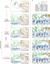
Fig. 2. Conformational changes in CaKip3 and tubulin.
a Displacement vectors for Cα atoms in CaKip3 when comparing the microtubule-unbound CaKip3-MDN-ADP structure and the MT-CaKip3-MDC-APO complex. The models were aligned to microtubule interacting regions H4, loop-11 and loop-8 of the MT-CaKip3-MDC-APO structure, specifically, residues 249-257 and 350-379. Displacement vectors for Cα atoms in CaKip3 and tubulin when comparing b MT-CaKip3-MDC-APO and MT-CaKip3-MDC-ANP, c MT-CaKip3-MDC-ANP and MT-CaKip3-MDC-AAF, and d MT-CaKip3-MDC-ANP and CT-CaKip3-MDC-ANP. All structures in bd were aligned to the β-tubulin chain of the MT-CaKip3-MDC-APO complex. Displacement vectors for Cα atoms are colored regionally to match the segment of the protein model that is compared, using the color scheme in Fig. 1. Views are from the minus end, down the long axis of the protofilament (left), side view (middle) and close-up of the tubulin interface (right). In each panel, the structure indicated on the left side of the arrow is shown as semi-transparent colored ribbons using the same regional color scheme as in Fig. 1. Only vectors with a magnitude of 1.0 Å or higher are shown.
Fig. 3
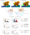
Fig. 3. Loop-2 electron and cryo-EM densities and tubulin contacts.
a 2mFobs – dFcalc electron density map (contoured at 1.0 σ) and cartoon model of loop-2 and the motor domain of the CaKip3-MDN crystal structure. b Sequence alignment of the tubulin contact region of CaKip3’s loop-2 with other kinesin-8s. Conserved tubulin-bonding residues are highlighted. Cryo-EM map and cartoon model of loop-2 of the c MT-CaKip3-MDC-APO complex, d MT-CaKip3-MDC-ANP complex, e MT-CaKip3-MDC-AAF complex, and f CT-CaKip3-MDC-ANP complex. Right panels show close-up view of loop-2-tubulin interactions for each complex. Green mesh maps were low-pass filtered at 7 Å (MT-CaKip3-MDC-AAF at 9 Å) to better display the noisier density of loop-2. Interacting residues are shown in stick representation. Pseudo-bonds between interacting atoms were determined and displayed using the “find clashes and contacts” routine in UCSF ChimeraX and are represented as yellow dashed lines.
Fig. 4
Fig. 4. Structures and activity of loop-2 swap mutants on microtubules.
Cryo-EM map of a MT-CaKip3-MDNL2-HsKHC in the APO state, b MT-CaKip3-MDNL2-HsKHC in the AMP-PNP state, and c MT-CaKip3-MDN in the AMP-PNP state. d Displacement vectors for Cα atoms in CaKip3 and tubulin when comparing the MT-CaKip3-MDN-ANP and MT-CaKip3-MDNL2-HsKHC-ANP complexes. Structures were aligned to the β-tubulin chain of MT-CaKip3-MDC-ANP. Displacement vectors for Cα atoms are colored regionally to match the segment of the protein model that is compared, using the color scheme in Fig. 1. MT-CaKip3-MDN-ANP structure is shown as semi-transparent colored ribbons using the same regional color scheme as in Fig. 1. Only vectors with a magnitude of 1.0 Å or higher are shown. e, f Microtubule-gliding assay velocity distributions for CaKip3-MDN, CaKip3-MDNL2-HsKHC, HsKHC-MDN, and HsKHC-MDNL2-CaKip3 using taxol-stabilized microtubules. n = 3 independent experiments for each protein. Asterisks indicate two-tailed unpaired t-test significance: p = 1.3e-135, p = 3.8e-122 for e and f, respectively. g Summary of motor velocities. Values are presented as mean ± SEM. h Microtubule-stimulated ATPase kinetics of CaKip3-MDN and CaKip3-MDNL2-HsKHC at steady state (n = 3 independent experiments). The basal rate was 0.13 ± 0.01 s−1 for both motors. Mean values (±SD) are shown. i Microtubule-stimulated ATPase kinetics of HsKHC-MDN and HsKHC-MDNL2-CaKip3 at steady state (n = 3 independent experiments). Basal rates were 0.012 ± 0.001 s−1 and 0.010 ± 0.001 s−1, respectively. Mean values (±SD) are shown. j Summary of the microtubule-stimulated ATPase activities of the motors. Values are presented as mean ± SEM. Data was fit to the Michaelis-Menten equation using GraphPad Prism to obtain K0.5, MT and kcat values. k Microtubule-co-sedimentation data for CaKip3-MDN and CaKip3-MDNL2-HsKHC (1 μM each) in the presence of 2 mM MgAMP-PNP (n = 3 independent experiments). Mean values (±SD) are shown. l Microtubule-co-sedimentation data for HsKHC-MDN and HsKHC-MDNL2-CaKip3 (1 μM each) in the presence of 2 mM MgAMP-PNP (n = 3 independent experiments). Mean values (±SD) are shown. Taxol-stabilized microtubules were pelleted by centrifugation to separate the free kinesin and microtubule-bound kinesin. SDS-PAGE and Coomassie brilliant blue staining were used to determine the fraction of microtubule-bound kinesin. m Microtubule-binding affinities (Kd values) of the motors were calculated using the quadratic equation given in Materials and Methods. Values are presented as mean ± SEM for three independent experiments. Source data are provided in the Source Data file.
Fig. 5
Fig. 5. Activity of loop-2 swap mutants on curved tubulin.
a Dolastatin-induced tubulin-ring (D-ring)-stimulated ATPase kinetics of CaKip3-MDN and CaKip3-MDNL2-HsKHC at steady state (n = 3 independent experiments). Mean values (±SD) are shown. b Free tubulin dimer-stimulated ATPase kinetics of CaKip3-MDN and CaKip3-MDNL2-HsKHC at steady state (n = 3 independent experiments). Mean values (±SD) are shown. c Free tubulin dimer-stimulated ATPase kinetics of HsKHC-MDN and HsKHC-MDNL2-CaKip3 at steady state (n = 3 independent experiments). Mean values (±SD) are shown. d Summary of the D-ring-stimulated ATPase activities of the motors. e Summary of the tubulin-stimulated ATPase activities of the motors. Values are presented as mean ± SEM. Data were fit to the Michaelis-Menten equation using GraphPad Prism to obtain K0.5, MT and kcat values. Source data are provided in the Source Data file.
Fig. 6
Fig. 6. Loop-1 and loop-2 are major contributors to microtubule depolymerization activity.
a Cartoon representation of CaKip3 in the CT-CaKip3-MDC-ANP complex showing the polyasparagine section of loop-1 (magenta) inserted into the preceding motor domain (shown in surface representation). The insert shows “close up” view of the polyasparagine section in order to visualize structural elements involved in complex formation. Sequence alignment shows polyasparagine section of loop-1 in CaKip3 compared to ScKip3. b Cryo-EM map of the CT-CaKip3-MDC-ANP complex. A low-pass filtered map (5 Å) is shown as a black mesh around the polyasparagine track of loop-1. c Microtubule depolymerization by sedimentation dose-response curves for the indicated CaKip3 proteins. 2 µM GMP-CPP-stabilized microtubules were incubated with increasing concentrations of CaKip3-MDN, CaKip3-MDNΔL1, CaKip3-MDNL2-HsKHC, or CaKip3-MDNL2-HsKHC+ΔL1 in the presence of ATP. The data from three independent experiments were analyzed and fit to the four-parameter logistic equation. Mean values (±SD) are shown. EC50 values are presented as the mean ± SEM. d Depolymerization of 2 µM GMP-CPP-stabilized microtubules by 3 µM of HsKHC-MDN, 3 µM HsKHC-MDNL2-CaKip3, and 1 µM CaKip3-MDN assessed by sedimentation. Reactions were incubated for 20 min in the presence of 20 mM MgATP, then free tubulin and microtubule polymers were separated into supernatant (S) and pellet (P) fractions by ultra-centrifugation. Equal portions of (S) and (P) were loaded and analyzed on a 10% Coomassie blue-stained SDS-PAGE gel. Similar results were obtained from two independent experiments. Source data are provided in the Source Data file.
Fig. 7
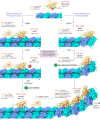
Fig. 7. Model for tubulin shape-induced alternations between pro-motility and pro-depolymerization states of kinesin-8.
When kinesin-8 binds tubulin protofilaments that are constrained to a straight conformation by the lateral protofilament interactions of the microtubule lattice (left), the interactions between loop-2 and α-tubulin restrict the motor domain from transitioning to the closed conformation with a docked neck-linker. Unable to stably interact with the preceding motor via loop-1, or to curve tubulin enough to disrupt protofilament contacts, some of the loop-2-tubulin bonds eventually break, allowing the motor domain to form the pro-motility state, in which the neck-linker docks and the tethered head is moved to the next αβ-tubulin subunit. Alternatively, when kinesin-8 encounters curved tubulin protofilaments, or protofilaments that can become curved, which are found at the microtubule plus-end (right), its motor domain readily forms a pro-depolymerization state. In this state, the elongated loop-2 region moves toward the motor core to accommodate the position of α-tubulin in curved αβ-tubulin subunits. This displacement of loop-2 is accompanied by closing of the ATP pocket to form a nucleotide-hydrolysis-competent active site and a docked neck-linker. In addition, loop-1 becomes more structurally ordered and inserts into the deep groove between loop-8 and the core β-sheet of the preceding motor domain. By maintaining loop-2 contacts with tubulin, and the loop-1 linkage between motors, the conformational transition of multiple motor domains to a closed conformation could increase tubulin protofilament curvature enough to trigger microtubule depolymerization.

Similar articles

Cited by

References

    1. Gupta ML, Jr., Carvalho P, Roof DM, Pellman D. Plus end-specific depolymerase activity of Kip3, a kinesin-8 protein, explains its role in positioning the yeast mitotic spindle. Nat. Cell Biol. 2006;8:913–923. doi: 10.1038/ncb1457. - DOI - PubMed
    1. Varga V, et al. Yeast kinesin-8 depolymerizes microtubules in a length-dependent manner. Nat. Cell Biol. 2006;8:957–962. doi: 10.1038/ncb1462. - DOI - PubMed
    1. Mayr MI, et al. The human kinesin Kif18A is a motile microtubule depolymerase essential for chromosome congression. Curr. Biol. 2007;17:488–498. doi: 10.1016/j.cub.2007.02.036. - DOI - PubMed
    1. Varga V, Leduc C, Bormuth V, Diez S, Howard J. Kinesin-8 motors act cooperatively to mediate length-dependent microtubule depolymerization. Cell. 2009;138:1174–1183. doi: 10.1016/j.cell.2009.07.032. - DOI - PubMed
    1. McHugh T, Gluszek AA, Welburn JP. Microtubule end tethering of a processive kinesin-8 motor Kif18b is required for spindle positioning. J. Cell Biol. 2018;217:2403–2416. doi: 10.1083/jcb.201705209. - DOI - PMC - PubMed

Publication types