Effects of Electroacupuncture on Gastrointestinal Motility Function, Pain, and Inflammation via Transient Receptor Potential Vanilloid 1 in a Rat Model after Colonic Anastomoses
- PMID: 35845135
- PMCID: PMC9277154
- DOI: 10.1155/2022/5113473
Effects of Electroacupuncture on Gastrointestinal Motility Function, Pain, and Inflammation via Transient Receptor Potential Vanilloid 1 in a Rat Model after Colonic Anastomoses
Abstract
Background: Complications after colon surgery are a major obstacle to postoperative recovery. The purpose of this study was to investigate the effect of electroacupuncture (EA) at Zusanli (ST36) on gastrointestinal motility in rats after colonic anastomosis and the mechanism of transient receptor potential vanillin 1 (TRPV1) channel in regulating gastrointestinal motility, pain, and inflammation.
Methods: The rats were randomly divided into six groups, including the control, model, EA, sham-EA, capsaicin, and capsaicin+EA groups, with preoperative capsaicin pretreatment and EA treatment at ST36 acupoint after surgery. Rats were treated using EA at ST36 or sham acupoints after surgery for 5 days. Capsaicin was intraperitoneally injected into rats 3 hours before surgery. Gastrointestinal motility was assessed by measuring the gastric residue, small intestinal propulsion in vivo, contractile tension, and frequency of isolated muscle strips in vitro. The mechanical withdrawal threshold (MWT) of abdominal incision skin and spontaneous nociceptive scores were observed and recorded in rats after colon anastomosis. The expressions of TRPV1, substance P (SP), neurokinin 1 (NK1) receptor, nuclear factor kappa-B (NF-κB), interleukin- (IL-) 6, L-1β, and tumor necrosis factor- (TNF-) α were determined.
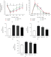
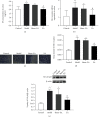
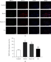
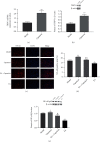
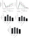
Results: Compared with the model group, electroacupuncture at ST36 point could significantly reduce the residual rate of stomach in rats after operation and increase the propulsive force of the small intestine and the contraction tension of the isolated smooth muscle. Electroacupuncture also increased postoperative day 3 MWT values and decreased postoperative spontaneous nociception scores. In addition, electroacupuncture treatment downregulated the expressions of IL-6, IL-1β, TNF-α, TRPV1, NF-κB, SP, and NK1 receptors in the colon tissue of rats after colonic anastomosis.
Conclusions: Our study showed that electroacupuncture at ST36 acupoint could improve gastrointestinal motility in rats after colonic anastomosis and relieve intestinal inflammation and pain. The mechanism may be to inhibit the activation of NF-κB and SP/NK1 receptor signaling pathways by inhibiting TRPV1.
Copyright © 2022 Xuelai Zhong et al.
Conflict of interest statement
No potential conflicts of interest were disclosed.
Figures


Similar articles
-
Downregulation of electroacupuncture at ST36 on TNF-alpha in rats with ulcerative colitis.World J Gastroenterol. 2003 May;9(5):1028-33. doi: 10.3748/wjg.v9.i5.1028. World J Gastroenterol. 2003. PMID: 12717850 Free PMC article.
-
[Effect of electroacupuncture pretreatment on transient receptor potential vanilloid 1(TRPV1)/calcitonin gene-related peptide(CGRP) signal and NF-κB p65 protein expression in rats with acute myocardial ischemia].Zhen Ci Yan Jiu. 2021 Jan 25;46(1):58-63. doi: 10.13702/j.1000-0607.200905. Zhen Ci Yan Jiu. 2021. PMID: 33559427 Chinese.
-
[Effect of electroacupuncture of "Hegu" (LI4) and "Zusanli" (ST36) on intestinal sensitivity and motility in irritable bowel syndrome rats].Zhen Ci Yan Jiu. 2020 Apr 25;45(4):293-8. doi: 10.13702/j.1000-0607.190743. Zhen Ci Yan Jiu. 2020. PMID: 32333534 Chinese.
-
Electroacupuncture in Regulating Gastrointestinal Symptoms of COVID-19: A Mini-review.Curr Pharm Des. 2023 Jun 6;29(15):1163-1165. doi: 10.2174/1381612829666230516164527. Curr Pharm Des. 2023. PMID: 37194937 Review.
-
Clinical Effect of Electroacupuncture on Acute Pancreatitis: Efficacies and Mechanisms.J Inflamm Res. 2023 Jul 28;16:3197-3203. doi: 10.2147/JIR.S410618. eCollection 2023. J Inflamm Res. 2023. PMID: 37534302 Free PMC article. Review.
Cited by
-
TRP (transient receptor potential) ion channel family: structures, biological functions and therapeutic interventions for diseases.Signal Transduct Target Ther. 2023 Jul 5;8(1):261. doi: 10.1038/s41392-023-01464-x. Signal Transduct Target Ther. 2023. PMID: 37402746 Free PMC article. Review.
-
Bibliometric analysis of recent research on the association between TRPV1 and inflammation.Channels (Austin). 2023 Dec;17(1):2189038. doi: 10.1080/19336950.2023.2189038. Channels (Austin). 2023. PMID: 36919561 Free PMC article.
References
MeSH terms
Substances
LinkOut - more resources
Full Text Sources

