

Convection-Enhanced Arborizing Catheter System Improves Local/Regional Delivery of Infusates Versus a Single-Port Catheter in Ex Vivo Porcine Brain Tissue
- PMID: 35832263
- PMCID: PMC8597548
- DOI: 10.1115/1.4048935
Convection-Enhanced Arborizing Catheter System Improves Local/Regional Delivery of Infusates Versus a Single-Port Catheter in Ex Vivo Porcine Brain Tissue
Abstract
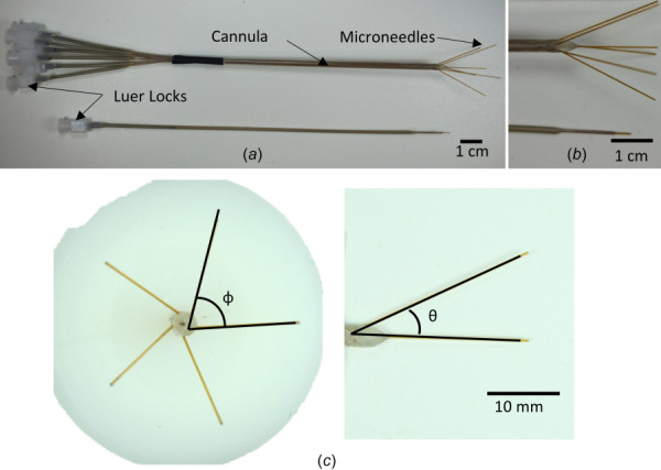
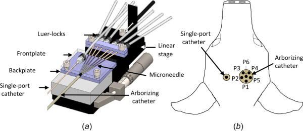
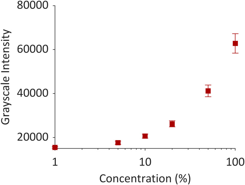
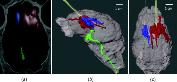
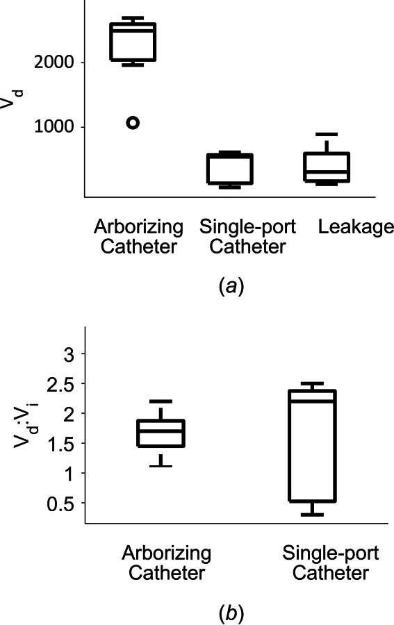
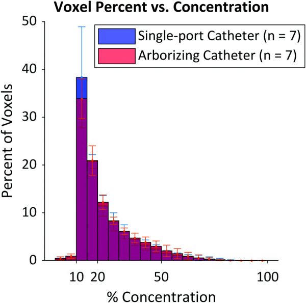
Standard treatment for glioblastoma is noncurative and only partially effective. Convection-enhanced delivery (CED) was developed as an alternative approach for effective loco-regional delivery of drugs via a small catheter inserted into the diseased brain. However, previous CED clinical trials revealed the need for improved catheters for controlled and satisfactory distribution of therapeutics. In this study, the arborizing catheter, consisting of six infusion ports, was compared to a reflux-preventing single-port catheter. Infusions of iohexol at a flow rate of 1 μL/min/microneedle were performed, using the arborizing catheter on one hemisphere and a single-port catheter on the contralateral hemisphere of excised pig brains. The volume dispersed (Vd) of the contrast agent was quantified for each catheter. Vd for the arborizing catheter was significantly higher than for the single-port catheter, 2235.8 ± 569.7 mm3 and 382.2 ± 243.0 mm3, respectively (n = 7). Minimal reflux was observed; however, high Vd values were achieved with the arborizing catheter. With simultaneous infusion using multiple ports of the arborizing catheter, high Vd was achieved at a low infusion rate. Thus, the arborizing catheter promises a highly desirable large volume of distribution of drugs delivered to the brain for the purpose of treating brain tumors.
Keywords: convection-enhanced delivery; drug delivery; glioblastoma; intracranial infusion; volume dispersed.
Copyright © 2021 by ASME.
Figures
Similar articles
-
Maximizing Local Access to Therapeutic Deliveries in Glioblastoma. Part II: Arborizing Catheter for Convection-Enhanced Delivery in Tissue Phantoms.In: De Vleeschouwer S, editor. Glioblastoma [Internet]. Brisbane (AU): Codon Publications; 2017 Sep 27. Chapter 18. In: De Vleeschouwer S, editor. Glioblastoma [Internet]. Brisbane (AU): Codon Publications; 2017 Sep 27. Chapter 18. PMID: 29251864 Free Books & Documents. Review.
-
Convection Enhanced Delivery: A Comparison of infusion characteristics in ex vivo and in vivo non-human primate brain tissue.Ann Neurosci. 2013 Jul;20(3):108-14. doi: 10.5214/ans.0972.7531.200306. Ann Neurosci. 2013. PMID: 25206026 Free PMC article.
-
In-vitro and in-vivo performance studies of a porous infusion catheter designed for intraparenchymal delivery of therapeutic agents of varying size.J Neurosci Methods. 2022 Aug 1;378:109643. doi: 10.1016/j.jneumeth.2022.109643. Epub 2022 Jun 9. J Neurosci Methods. 2022. PMID: 35691412
-
Parametric Study of the Design Variables of an Arborizing Catheter on Dispersal Volume Using a Biphasic Computational Model.J Eng Sci Med Diagn Ther. 2019 Aug;2(3):0310021-310029. doi: 10.1115/1.4042874. Epub 2019 Apr 1. J Eng Sci Med Diagn Ther. 2019. PMID: 35833170 Free PMC article.
-
Convection-enhanced delivery for the treatment of glioblastoma.Neuro Oncol. 2015 Mar;17 Suppl 2(Suppl 2):ii3-ii8. doi: 10.1093/neuonc/nou354. Neuro Oncol. 2015. PMID: 25746090 Free PMC article. Review.
Cited by
-
Biocompatibility of the fiberoptic microneedle device chronically implanted in the rat brain.Res Vet Sci. 2022 Mar;143:74-80. doi: 10.1016/j.rvsc.2021.12.018. Epub 2021 Dec 31. Res Vet Sci. 2022. PMID: 34995824 Free PMC article.
-
Advancements in drug delivery methods for the treatment of brain disease.Front Vet Sci. 2022 Oct 18;9:1039745. doi: 10.3389/fvets.2022.1039745. eCollection 2022. Front Vet Sci. 2022. PMID: 36330152 Free PMC article. Review.
-
Convection-Enhanced Drug Delivery: Experimental and Analytical Studies of Infusion Behavior in an In Vitro Brain Surrogate.Ann Biomed Eng. 2024 Jun;52(6):1693-1705. doi: 10.1007/s10439-024-03482-4. Epub 2024 Mar 19. Ann Biomed Eng. 2024. PMID: 38502430
-
Solid Fiber Inside of Capillary and Modified Fusion-Spliced Fiber Optic Microneedle Devices for Improved Light Transmission Efficiency.J Med Device. 2022 Dec 1;16(4):041014. doi: 10.1115/1.4055607. Epub 2022 Sep 27. J Med Device. 2022. PMID: 36353365 Free PMC article.
-
Delivery strategies for cell-based therapies in the brain: overcoming multiple barriers.Drug Deliv Transl Res. 2021 Dec;11(6):2448-2467. doi: 10.1007/s13346-021-01079-1. Epub 2021 Oct 30. Drug Deliv Transl Res. 2021. PMID: 34718958 Free PMC article.
References
-
- Ostrom, Q. T. , Gittleman, H. , Xu, J. , Kromer, C. , Wolinsky, Y. , Kruchko, C. , and Barnholtz-Sloan, J. S. , 2016, “ CBTRUS Statistical Report: Primary Brain and Other Central Nervous System Tumors Diagnosed in the United States in 2009-2013,” Neuro Oncol., 18(suppl_5), pp. v1–v75.10.1093/neuonc/now207 - DOI - PMC - PubMed
-
- Debinski, W. , Priebe, W. , and Tatter, S. B. , 2017, Maximizing Local Access to Therapeutic Deliveries in Glioblastoma. Part I: Targeted Cytotoxic Therapy, De Vleeschouwer Glioblastoma S., ed., Codon Publications, Brisbane, Australia, pp. 341–358. - PubMed
-
- Sanai, N. , and Berger, M. S. , 2012, “ Recent Surgical Management of Gliomas,” Glioma: Immunotherapeutic Approaches, Yamanaka R., ed., Springer, New York, pp. 12–25. - PubMed
Grants and funding
LinkOut - more resources
Full Text Sources