2.7 Å cryo-EM structure of ex vivo RML prion fibrils
- PMID: 35831275
- PMCID: PMC9279362
- DOI: 10.1038/s41467-022-30457-7
2.7 Å cryo-EM structure of ex vivo RML prion fibrils
Abstract
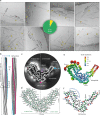
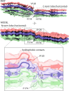
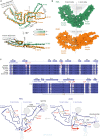
Mammalian prions propagate as distinct strains and are composed of multichain assemblies of misfolded host-encoded prion protein (PrP). Here, we present a near-atomic resolution cryo-EM structure of PrP fibrils present in highly infectious prion rod preparations isolated from the brains of RML prion-infected mice. We found that prion rods comprise single-protofilament helical amyloid fibrils that coexist with twisted pairs of the same protofilaments. Each rung of the protofilament is formed by a single PrP monomer with the ordered core comprising PrP residues 94-225, which folds to create two asymmetric lobes with the N-linked glycans and the glycosylphosphatidylinositol anchor projecting from the C-terminal lobe. The overall architecture is comparable to that of recently reported PrP fibrils isolated from the brain of hamsters infected with the 263K prion strain. However, there are marked conformational variations that could result from differences in PrP sequence and/or represent distinguishing features of the distinct prion strains.
© 2022. The Author(s).
Conflict of interest statement
J.C. is a Director and J.C. and J.D.F.W. are shareholders of D-Gen Limited, an academic spin-out company working in the field of prion disease diagnosis, decontamination, and therapeutics. D-Gen supplied the ICSM35 and ICSM18 antibodies used for western blot and ELISA performed in this study. The other authors declare no competing interests.
Figures


Comment in
-
The shape of things to come: structural insights into how prion proteins encipher heritable information.Nat Commun. 2022 Jul 13;13(1):4003. doi: 10.1038/s41467-022-31460-8. Nat Commun. 2022. PMID: 35831278 Free PMC article.
Similar articles
-
Prion strains viewed through the lens of cryo-EM.Cell Tissue Res. 2023 Apr;392(1):167-178. doi: 10.1007/s00441-022-03676-z. Epub 2022 Aug 27. Cell Tissue Res. 2023. PMID: 36028585 Free PMC article. Review.
-
Structural biology of ex vivo mammalian prions.J Biol Chem. 2022 Aug;298(8):102181. doi: 10.1016/j.jbc.2022.102181. Epub 2022 Jun 23. J Biol Chem. 2022. PMID: 35752366 Free PMC article. Review.
-
Ex vivo mammalian prions are formed of paired double helical prion protein fibrils.Open Biol. 2016 May;6(5):160035. doi: 10.1098/rsob.160035. Epub 2016 May 4. Open Biol. 2016. PMID: 27249641 Free PMC article.
-
Cryo-EM structure of anchorless RML prion reveals variations in shared motifs between distinct strains.Nat Commun. 2022 Jul 13;13(1):4005. doi: 10.1038/s41467-022-30458-6. Nat Commun. 2022. PMID: 35831291 Free PMC article.
-
High-resolution structure and strain comparison of infectious mammalian prions.Mol Cell. 2021 Nov 4;81(21):4540-4551.e6. doi: 10.1016/j.molcel.2021.08.011. Epub 2021 Aug 25. Mol Cell. 2021. PMID: 34433091
Cited by
-
Evidence of Orientation-Dependent Early States of Prion Protein Misfolded Structures from Single Molecule Force Spectroscopy.Biology (Basel). 2022 Sep 16;11(9):1358. doi: 10.3390/biology11091358. Biology (Basel). 2022. PMID: 36138837 Free PMC article.
-
Characterisation and prion transmission study in mice with genetic reduction of sporadic Creutzfeldt-Jakob disease risk gene Stx6.Neurobiol Dis. 2024 Jan;190:106363. doi: 10.1016/j.nbd.2023.106363. Epub 2023 Nov 22. Neurobiol Dis. 2024. PMID: 37996040 Free PMC article.
-
Sporadic Creutzfeldt-Jakob disease infected human cerebral organoids retain the original human brain subtype features following transmission to humanized transgenic mice.Acta Neuropathol Commun. 2023 Feb 14;11(1):28. doi: 10.1186/s40478-023-01512-1. Acta Neuropathol Commun. 2023. PMID: 36788566 Free PMC article.
-
High-throughput cryo-EM structure determination of amyloids.Faraday Discuss. 2022 Nov 8;240(0):243-260. doi: 10.1039/d2fd00034b. Faraday Discuss. 2022. PMID: 35913272 Free PMC article.
-
Phospholipid cofactor solubilization inhibits formation of native prions.J Neurochem. 2023 Sep;166(5):875-884. doi: 10.1111/jnc.15930. Epub 2023 Aug 7. J Neurochem. 2023. PMID: 37551010 Free PMC article.
References
Publication types
MeSH terms
Substances
Grants and funding
LinkOut - more resources
Full Text Sources
Molecular Biology Databases
Research Materials

