. 2022 Jun 24;23(13):7042.
doi: 10.3390/ijms23137042.

Evidence for Dual Activation of IK(M) and IK(Ca) Caused by QO-58 (5-(2,6-Dichloro-5-fluoropyridin-3-yl)-3-phenyl-2-(trifluoromethyl)-1H-pyrazolol[1,5-a]pyrimidin-7-one)

Affiliations

Evidence for Dual Activation of IK(M) and IK(Ca) Caused by QO-58 (5-(2,6-Dichloro-5-fluoropyridin-3-yl)-3-phenyl-2-(trifluoromethyl)-1H-pyrazolol[1,5-a]pyrimidin-7-one)

Chao-Liang Wu et al. Int J Mol Sci. .

Abstract

QO-58 (5-(2,6-dichloro-5-fluoropyridin-3-yl)-3-phenyl-2-(trifluoromethyl)-1H-pyrazolol[1,5-a]pyrimidin-7-one) has been regarded to be an activator of KV7 channels with analgesic properties. However, whether and how the presence of this compound can result in any modifications of other types of membrane ion channels in native cells are not thoroughly investigated. In this study, we investigated its perturbations on M-type K+ current (IK(M)), Ca2+-activated K+ current (IK(Ca)), large-conductance Ca2+-activated K+ (BKCa) channels, and erg-mediated K+ current (IK(erg)) identified from pituitary tumor (GH3) cells. Addition of QO-58 can increase the amplitude of IK(M) and IK(Ca) in a concentration-dependent fashion, with effective EC50 of 3.1 and 4.2 μM, respectively. This compound could shift the activation curve of IK(M) toward a leftward direction with being void of changes in the gating charge. The strength in voltage-dependent hysteresis (Vhys) of IK(M) evoked by upright triangular ramp pulse (Vramp) was enhanced by adding QO-58. The probabilities of M-type K+ (KM) channels that will be open increased upon the exposure to QO-58, although no modification in single-channel conductance was seen. Furthermore, GH3-cell exposure to QO-58 effectively increased the amplitude of IK(Ca) as well as enhanced the activity of BKCa channels. Under inside-out configuration, QO-58, applied at the cytosolic leaflet of the channel, activated BKCa-channel activity, and its increase could be attenuated by further addition of verruculogen, but not by linopirdine (10 μM). The application of QO-58 could lead to a leftward shift in the activation curve of BKCa channels with neither change in the gating charge nor in single-channel conductance. Moreover, cell exposure of QO-58 (10 μM) resulted in a minor suppression of IK(erg) amplitude in response to membrane hyperpolarization. The docking results also revealed that there are possible interactions of the QO-58 molecule with the KCNQ or KCa1.1 channel. Overall, dual activation of IK(M) and IK(Ca) caused by the presence of QO-58 eventually may have high impacts on the functional activity (e.g., anti-nociceptive effect) residing in electrically excitable cells. Care must be exercised when interpreting data generated with QO-58 as it is not entirely KCNQ/KV7 selective.

Keywords: 5-(2,6-dichloro-5-fluoro-3-pyridinyl)-3-phenyl-2-(trifluoromethyl)-pyrazolo[1,5-a]pyrimidin-7(4H)-one); Ca2+-activated K+ current; M-type K+ channel; M-type K+ current; QO-58 (5-(2,6-dichloro-5-fluoropyridin-3-yl)-3-phenyl-2-(trifluoromethyl)-1H-pyrazolol[1,5-a]pyrimidin-7-one; large-conductance Ca2+-activated K+ channel; voltage-dependent hysteresis.


Conflict of interest statement

The authors declare no conflict of interests that are directly relevant to the present study. The content and writing of this paper are solely the responsibility of the authors.

Figures

Figure 1
Figure 1
QO-58-induced stimulation of M-type K+ current (IK(M)) recorded from pituitary GH3 cells. In these experiments, we placed cells in high-K+ (145 mM), Ca2+-free solution that contained 1 μM tetrodotoxin (TTX) and 0.5 mM CdCl2 and the electrodes that we used were filled up with a K+-containing solution. (A) Superimposed current traces obtained in the control period (a), during cell exposure of 1 μM QO-58 (b) or 3 μM QO-58 (c), and washout of QO-58 (d). The top part indicates the voltage-clamp protocol applied, while the lower part is an expanded record from purple dash box in the upper part. (B) Concentration-dependent stimulation of QO-58 effect on the amplitude of IK(M) (mean ± SEM; n = 8 for each point). Current amplitudes during cell exposure to different QO-58 concentrations were measured at the end of depolarizing pulse from −50 to −10 mV with a duration of 1 s. Sigmoid smooth curve indicates best fit to a modified Hill function described in Section 4.
Figure 2
Figure 2
Effect of QO-58 on the current versus voltage (I-V) relationship (A) and the activation curve (B) of IK(M). The experimental protocol applied is the same as that used in Figure 1. (A) Average I-V relationship of IK(M) amplitude acquired in the control period (i.e., absence of QO-58, solid black squares) and during cell exposure to 3 μM QO-58 (open red circles). (B) Steady-state activation curve of IK(M) in the absence (solid black squares) and presence (open red circles) of 3 μM QO-58 (mean ± SEM; n = 8). Smooth curves drawn were optimally fitted to a modified Boltzmann function as elaborated in Section 4. Of note, there is a leftward shift along the voltage axis in the quasi-steady-state activation curve of IK(M) in QO-58 presence, despite being void of changes in the gating charge of the curve.
Figure 3
Figure 3
Effect of QO-58 on IK(M) in response to an upright isosceles-triangular ramp pulse (Vramp) observed in GH3 cells. The Vramp with different durations (0.4–3.2 s) (or with ramp speed between ±31.25 and ±250 mV/s) was designed to mimic different depolarizing and repolarizing slopes in bursting patterns of neuronal firing. (A) Representative IK(M) traces in response to the double-pulse Vramp in the absence (upper) and presence (lower) of 3 μM QO-58. The voltage protocol used is indicated in the uppermost part, and voltage waveforms appearing in different colors are indicated to correspond with current traces evoked by the waveforms. (B) Effect of QO-58 (3 μM) on voltage-dependent hysteresis (Vhys) (i.e., the relationship of forward and backward current versus membrane voltage) of IK(M) evoked by triangular Vramp with a duration of 3.2 s. Black or red current trajectory indicates the absence or presence of 3 μM QO-58, respectively. The direction of IK(M) in which time passes during the elicitation by 3.2 s triangular Vramp is indicated by dashed arrows. (C) Summary scatter graph showing the effect of QO-58 on the Vhys’s ∆area (mean ± SEM; n = 8 for each point). ∆area means the area encircling the forward (upsloping) and backward (downsloping) limbs of current trajectory evoked by Vramp. * Significantly different from control (p < 0.05), and ** significantly different from 3 μM QO-58 alone group (p < 0.05).
Figure 4
Figure 4
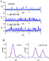
Stimulatory effect of QO-58 on the activity of M-type K+ (KM) channels recorded from GH3 cells. In this series of cell-attached current recordings, cells were bathed in high-K+, Ca2+-free solution, and we filled up the recording pipette with low-K+ (5.4 mM) solution. (A) Representative single KM channels in the control period (upper) and during cell exposure to 1 μM QO-58 (middle) or 3 μM QO-58 (lower). The channel activity in the absence or presence of QO-58 was taken at the level of +20 mV relative to the bath. The upward deflection indicates the opening event of the channel. (B) Amplitude histogram taken in the absence (left) and presence (right) of 3 mM QO-58. (C) Average I-V relationship of single KM channels in the absence (solid black squares) and presence (open red circles) of 3 μM QO-58 (mean ± SEM; n = 8 for each point). Of note, the linear relationship of KM channels versus ∆voltage was superimposable between the absence and presence of QO-58; hence, the single-channel conductance of the channel between the absence and presence of 3 μM QO-58 did not differ. (D) Summary scatter graph showing effect of QO-58 (1 and 3 μM) and QO-58 (3 μM) plus linopirdine (Lino, 10 μM) on the probability of KM channels that would be open (mean ± SEM; n = 8 for each point). Channel activity was measured at +20 mV relative to the bath. * Significantly different from control (p < 0.05) and ** significantly different from QO-58 (3 μM) alone group (p < 0.05).
Figure 4
Figure 4
Stimulatory effect of QO-58 on the activity of M-type K+ (KM) channels recorded from GH3 cells. In this series of cell-attached current recordings, cells were bathed in high-K+, Ca2+-free solution, and we filled up the recording pipette with low-K+ (5.4 mM) solution. (A) Representative single KM channels in the control period (upper) and during cell exposure to 1 μM QO-58 (middle) or 3 μM QO-58 (lower). The channel activity in the absence or presence of QO-58 was taken at the level of +20 mV relative to the bath. The upward deflection indicates the opening event of the channel. (B) Amplitude histogram taken in the absence (left) and presence (right) of 3 mM QO-58. (C) Average I-V relationship of single KM channels in the absence (solid black squares) and presence (open red circles) of 3 μM QO-58 (mean ± SEM; n = 8 for each point). Of note, the linear relationship of KM channels versus ∆voltage was superimposable between the absence and presence of QO-58; hence, the single-channel conductance of the channel between the absence and presence of 3 μM QO-58 did not differ. (D) Summary scatter graph showing effect of QO-58 (1 and 3 μM) and QO-58 (3 μM) plus linopirdine (Lino, 10 μM) on the probability of KM channels that would be open (mean ± SEM; n = 8 for each point). Channel activity was measured at +20 mV relative to the bath. * Significantly different from control (p < 0.05) and ** significantly different from QO-58 (3 μM) alone group (p < 0.05).
Figure 5
Figure 5
Stimulatory effect of QO-58 on the amplitude of whole-cell (i.e., macroscopic) Ca2+-activated K+ current (IK(Ca)) measured from GH3 cells. In this series of voltage-clamp current recordings on these cells, we used normal Tyrode’s solution containing 1.8 mM CaCl2 as a bathing medium, and the recording pipette used was backfilled with K+-enriched solution. As whole-cell configuration was established, we evoked IK(Ca) from a holding potential of 0 mV to test potentials in the range of 0 and +70 mV (10-mV in increments) at a rate of 0.1 Hz. (A) Superimposed current traces activated in response to a series of voltage steps (indicated in the uppermost part). Current traces in the upper part are controls (i.e., absence of QO-58), those in the middle part were recorded during cell exposure to 3 μM QO-58, while those in lower part were taken after washout of the QO-58. The duration of rectangular voltage commands applied was set in the range of 280 and 160 ms (20-ms decrements), indicating a progressive increase with membrane depolarization (i.e., an outwardly-rectifying property). (B) Average I-V relationship of IK(Ca) obtained in the control period, during the exposure to 3 μM QO-58, and after washout of QO-58. Current amplitude was measured at the end of each depolarizing pulse. Each point represents the mean ± SEM (n = 8). (C) Concentration-response relationship for QO-58-induced stimulation of IK(Ca) (mean ± SEM; n = 8 for each point). The gray continuous line drawn is reliably fitted to the Hill equation. The values for EC50 or nH were yielded to be 4.2 μM or 1.2, respectively.
Figure 6
Figure 6
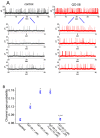
Stimulatory effect of QO-58 on the activity of BKCa channels recorded from GH3 cells. We conducted these inside-out current recordings in cells which were bathed in high-K+ solution containing 0.1 μM Ca2+, and the recording pipette was then filled up with K+-containing solution. (A) Original BKCa-channel currents obtained in the control period (left, black color) and after bath application of 3 μM QO-58 (right, red color). The detached patch was voltage-clamped at +60 mV. The lower part indicates the expanded records from the uppermost part. The opening event of the channel is indicated by the upward deflection. (B) Summary scatter graph showing effect of QO-58, QO-58 plus linopirdine (Lino), or QO-58 plus verruculogen (Ver) (mean ± SEM; n = 8 for each point). Under inside-out configuration, the channel open probability was measured at the level of +60 mV. * Significantly different from control (p < 0.05) and ** significantly different from QO-58 (3 μM) alone group (p < 0.05).
Figure 7
Figure 7
Effect of QO-58 on single-channel conductance (A) and activation curve (B) of BKCa channels in GH3 cells. In this series of inside-out current recordings, we voltage-clamped the excised patched at different levels of membrane potentials, and the recording pipette was filled with K+-enriched solution. (A) Average I-V relationship of single BKCa-channel currents (i.e., linear regression between membrane potential and mean single-channel amplitude) obtained in the absence (black filled squares) and presence (orange open circles) of 3 μM QO-58 (mean ± SEM; n = 8 for each point). Of note, the linear relationship of BKCa channels between the absence and presence of QO-58 is superimposed, and the value of reversal potential with or without the QO-58 addition was pointed toward zero mV. (B) Steady-state activation curve of BKCa channels obtained in the control period (black filled squares) and during exposure to 3 μM QO-58 (orange open circles) (mean ± SEM; n = 8 for each point). Sigmoid lines indicate the best fit to a Boltzmann function as stated in Section 4. Of note, there is a leftward shift of the activation curve of the channel, although neither single-channel conductance of the channel nor gating charge of the curve was altered by QO-58 presence.
Figure 8
Figure 8
Minor inhibition of erg-mediated K+ current (IK(erg)) produced by the presence of QO-58 in GH3 cells. In these experiments, we used high-K+, Ca2+-free solution as bathing medium and the recording pipette was filled up with K+-containing (145 mM) solution. (A) Superimposed current traces obtained in the control period (upper) and during cell exposure to 10 μM QO-58 (lower). The top part indicates the voltage-clamp protocol imposed. (B) Average I-V relationship of peak (black squares, upper) or sustained (brown circles, lower) component of IK(erg) obtained in the absence (filled symbols) and presence (open symbols) of 10 μM QO-58 (mean ± SEM; n = 7 for each point). Peak or sustained IK(erg) obtained with or without the QO-58 addition was measured at the start or end-point of each hyperpolarizing step with a duration of 1 s. Of note, the presence of 10 μM QO-58 slightly inhibited IK(erg) in these cells.
Figure 9
Figure 9
Docking results of KCa1.1 channel and QO-58 (A) or KCNQ2 and QO-58 (B). Protein structure of KCa1.1 or KCNQ2 channel was acquired from PDB (PDB ID: 6V3G or 7CR1), respectively, while chemical structure of QO-58 was from PubChem (Compound CID: 51351551). The structure of KCa1.1 or KCNQ2 channel was docked by the QO-58 molecule through PyRx (https://pyrx.sourceforge.io/, accessed on 23 June 2022). The diagram of interactions between KCa1.1 or KCNQ2 channel and the QO-58 molecule was generated by LigPlot+ (https://www.ebi.ac.uk/thornton-srv/software/LIGPLOT/, accessed on 23 June 2022). Of note, the red arcs with spokes radiating toward the ligand (i.e., QO-58) in the lower part of each panel denote the hydrophobic contact, while green dot line indicates the hydrogen bond.

Similar articles

Cited by

References

    1. Qi J., Zhang F., Mi Y., Fu Y., Xu W., Zhang D., Wu Y., Du X., Jia Q., Wang K., et al. Design, synthesis and biological activity of pyrazolo[1,5-a]pyrimidin-7(4H)-ones as novel Kv7/KCNQ potassium channel activators. Eur. J. Med. Chem. 2011;46:934–943. doi: 10.1016/j.ejmech.2011.01.010. - DOI - PubMed
    1. Zhang F., Mi Y., Qi J.L., Li J.W., Si M., Guan B.C., Du X.N., An H.L., Zhang H.L. Modulation of K(v)7 potassium channels by a novel opener pyrazolo[1,5-a]pyrimidin-7(4H)-one compound QO-58. Br. J. Pharmacol. 2013;168:1030–1042. doi: 10.1111/j.1476-5381.2012.02232.x. - DOI - PMC - PubMed
    1. Zhang J., Jia Q., Qi J., Zhang H., Wu Y., Shi X. Exploring in vivo metabolism and excretion of QO-58L using ultra-high-performance liquid chromatography coupled with tandem mass spectrometry. Eur. J. Pharm. Sci. 2018;117:379–391. doi: 10.1016/j.ejps.2018.02.015. - DOI - PubMed
    1. Zhang F., Liu Y., Zhang D., Fan X., Shao D., Li H. Suppression of KCNQ/M Potassium Channel in Dorsal Root Ganglia Neurons Contributes to the Development of Osteoarthritic Pain. Pharmacology. 2019;103:257–262. doi: 10.1159/000496422. - DOI - PubMed
    1. Teng B.-C., Song Y., Zhang F., Ma T.-Y., Qi J.-L., Zhang H.-L., Li G., Wang K. Activation of neuronal Kv7/KCNQ/M-channels by the opener QO58-lysine and its anti-nociceptive effects on inflammatory pain in rodents. Acta Pharmacol. Sin. 2016;37:1054–1062. doi: 10.1038/aps.2016.33. - DOI - PMC - PubMed