. 2023 Jan;19(1):324-337.
doi: 10.1080/15548627.2022.2090727. Epub 2022 Jul 1.

Stimulating the autophagic-lysosomal axis enhances host defense against fungal infection in a zebrafish model of invasive Aspergillosis

Affiliations

Stimulating the autophagic-lysosomal axis enhances host defense against fungal infection in a zebrafish model of invasive Aspergillosis

G Forn-Cuní et al. Autophagy. 2023 Jan.

Abstract

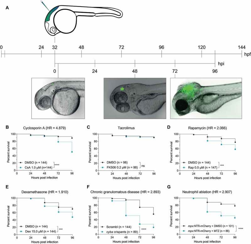
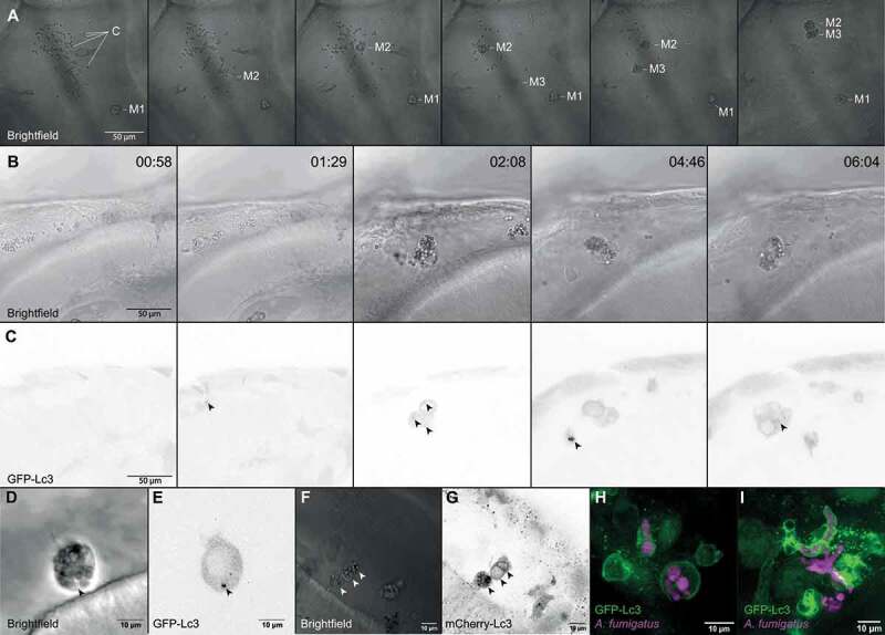
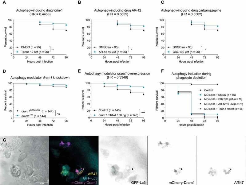
The increasing prevalence of antifungal-resistant human pathogenic fungi, particularly azole-resistant Aspergillus fumigatus, is a life-threatening challenge to the immunocompromised population. Autophagy-related processes such as LC3-associated phagocytosis have been shown to be activated in the host response against fungal infection, but their overall effect on host resistance remains uncertain. To analyze the relevance of these processes in vivo, we used a zebrafish animal model of invasive Aspergillosis. To confirm the validity of this model to test potential treatments for this disease, we confirmed that immunosuppressive treatments or neutropenia rendered zebrafish embryos more susceptible to A. fumigatus. We used GFP-Lc3 transgenic zebrafish to visualize the autophagy-related processes in innate immune phagocytes shortly after phagocytosis of A. fumigatus conidia, and found that both wild-type and melanin-deficient conidia elicited Lc3 recruitment. In macrophages, we observed GFP-Lc3 accumulation in puncta after phagocytosis, as well as short, rapid events of GFP-Lc3 decoration of single and multiple conidia-containing vesicles, while neutrophils covered single conidia-containing vesicles with bright and long-lasting GFP-Lc3 signal. Next, using genetic and pharmacological stimulation of three independent autophagy-inducing pathways, we showed that the antifungal autophagy response improves the host survival against A. fumigatus infection, but only in the presence of phagocytes. Therefore, we provide proof-of-concept that stimulating the (auto)phagolysosomal pathways is a promising approach to develop host-directed therapies against invasive Aspergillosis, and should be explored further either as adjunctive or stand-alone therapy for drug-resistant Aspergillus infections.Abbreviations: DMSO: dimethyl sulfoxide; HR: hazard ratio; HDT: host-directed therapy; Hpf: hours post fertilization; IA: invasive Aspergillosis; LAP: LC3-associated phagocytosis; MTZ: metronidazole; PTU: N-phenylthiourea; ROS: reactive oxygen species.

Keywords: Aspergillus; autophagic defense; fungal infection; host-pathogen interaction; immunomodulation; innate immunity; phagocytes; zebrafish.


Conflict of interest statement

No potential conflict of interest was reported by the author(s).

Figures

Figure 1.
Figure 1.
The zebrafish embryo model recapitulates susceptibilities of clinical IA. (a) Schematic representation of the zebrafish embryo – A. fumigatus infection model. Conidia of A. fumigatus D141 expressing an EGFP fluorescent plasmid were injected in the hindbrain at 32 hpf and representative images of infection at 2, 48 and 72 h post infection are shown. (B, C, D, and E) Survival curves of zebrafish embryos infected with A. fumigatusKu80 and exposed via waterborne treatment to 1 μM of cyclosporin A (b), 0.2 μM of FK506 (c), 0.5 μM of rapamycin (d), 15 μM of dexamethasone (e), or DMSO as a vehicle control at the same v/v. (f) Survival curves of cyba crispants or scramble control zebrafish embryos infected with A. fumigatus ∆Ku80. (g) Survival curves of zebrafish embryos with or without neutrophil ablation with metronidazole infected with A. fumigatus ∆Ku80. (b, d and f) are representative of at least 3 independent biological replicates, while (c) is representative of 2 independent biological replicates. The hazard ratio (HR) indicated is calculated vs. the control condition using the logrank method. Significance in the curve comparison is calculated using Log-rank (Mantel-Cox test): ns non-significant; * P ≤ 0.05; ** P ≤ 0.01; *** P ≤ 0.001; **** P ≤ 0.0001.
Figure 2.
Figure 2.
GFP-Lc3 dynamics in phagocytes after phagocytosing A. fumigatus conidia. (a) Representative sequence of events following injection of A. fumigatus conidia into the hindbrain of a zebrafish embryo. More than 100 A. fumigatusKu80 conidia are initially phagocytosed by the first 3 macrophages to arrive at the infection foci. Arrows indicate macrophage arrival and phagocytosis start. Images taken from Movie S1. (b and c) Confocal microscopy images showing the brightfield (b) or GFP-Lc3 fluorescence intensity (c) during the first 6 h and 30 min after infection. Most conidia are phagocytosed in the first 5 h post infection by a small number of phagocytes. Arrows indicate GFP-Lc3 levels in immune cells after phagocytosing A. fumigatus conidia, including individual GFP-Lc3 rings around the conidia. Images taken from Movie S2. (d and e) Brightfield (d) and GFP-Lc3 fluorescence intensity (e) of a macrophage with several conidia showing a GFP-Lc3 puncta. (f and g) Brightfield (f) and mCherry-Lc3 fluorescence intensity (g) confocal microscopy images showing mCherry-Lc3 accumulation across vesicles with individual and multiple spores. (h and i) Confocal images of GFP-Lc3 accumulation around germinating conidia of A. fumigatus D141 expressing dsRed fluorescent plasmid [40] in the hindbrain of zebrafish embryos fixed 15 h post infection.
Figure 3.
Figure 3.
Differential GFP-Lc3 dynamics in phagocytes after phagocytosing A. fumigatus conidia. (a, b, c, d, and e) Quantification of phagocytosis and Lc3 dynamics in 15 hours of timelapse imaging from 5 individual larvae infected with A. fumigatusKu80 conidia during the first hours post infection. (a) Most of the injected conidia in the zebrafish hindbrain are phagocytosed by macrophages, and (b) macrophages phagocytose more conidia per cell than neutrophils. (c, d, and e) In the events of conidial phagocytosis by neutrophils, we observed rapid bright Lc3 rings covering single-conidia vesicles, while Lc3 decoration of conidia-containing vesicles in macrophages was scarcer in the first hours post infection. (f) A non-labeled phagocyte (neutrophil, N) strongly mobilizes GFP-Lc3 to single conidia-containing vesicles shortly after phagocytosis (arrowheads), while labeled mCherryF-macrophages show high GFP-Lc3 fluorescence but not colocalizing with A. fumigatusKu80 conidia labeled with Alexa Fluor™ NHS 647 (Af647). Image taken from the Movie S3. (G, H, I, and J) Examples of GFP-Lc3 puncta (g), and GFP-Lc3 signal around single (h) or multiple conidia (h and i) in mCherryF labeled macrophages. Note that the membrane-bound mCherryF signal of macrophages colocalizes with GFP-Lc3 signal after conidia phagocytosis. Contrasting to the bright GFP-Lc3 signal in neutrophils (f), macrophages show weaker GFP-Lc3 targeting to phagocytosed conidia. (j) GFP-Lc3 targeting of single-conidia continues during the first hours after formation of a granuloma-like structure.
Figure 4.
Figure 4.
Suppression of autophagy pathways increases susceptibility to A. fumigatus infection. (a and b) Survival curves of atg13 mutation (a) or rubcn (b) knockdown or control zebrafish embryos infected with A. fumigatus ∆Ku80. (c) Survival curve of sqstm1 mutants or non-mutant siblings infected with A. fumigatus ∆Ku80. (d) Survival curve of zebrafish embryos infected with equal inoculum (150 conidia) of A. fumigatus ∆Ku80 or DHN-Melanin-deficient A. fumigatuspksP. (e and f) Survival curves of atg13 mutation (e) or rubcn (f) knockdown or control zebrafish embryos infected with A. fumigatuspksP. (G, H, I, J, and K) Quantification of phagocytosis and Lc3 dynamics in 21 hours of timelapse imaging from 5 individual larvae infected with A. fumigatuspksP conidia labeled with Alexa Fluor™ NHS 647 during the first hours post infection. (g) Most of the injected conidia in the zebrafish hindbrain are phagocytosed by macrophages, and (h) macrophages phagocytose more conidia per cell than neutrophils. (i, j, and k) In the events of conidial phagocytosis by neutrophils, we observed rapid bright Lc3 rings covering single-conidia vesicles, while Lc3 decoration of conidia-containing vesicles in macrophages was scarcer and shorter in the first hours post infection. Examples of GFP-Lc3 signal around single A. fumigatuspksP conidia labeled with Alexa Fluor™ NHS 647 (Af∆pksP) in mCherryF negative phagocytes (l, m), or mCherryF labeled macrophage (n). Images taken from Movie S4. All survival curves are representative of at least 3 independent biological replicates. The hazard ratio (HR) indicated is calculated vs. the control condition using the logrank method. Significance in the curve comparison is calculated using Log-rank (Mantel-Cox test): ns non-significant; * P ≤ 0.05; ** P ≤ 0.01; *** P ≤ 0.001; **** P ≤ 0.0001.
Figure 5.
Figure 5.
Stimulating the (auto)phagolysosomal axis increases the host survival against IA. (a, b and c) Survival curves of zebrafish embryos infected with A. fumigatus ∆Ku80 and exposed via waterborne treatment to 10 nM of torin 1 (a), 10 μM of AR-12 (b), 100 μM or carbamazepine (c), or DMSO as a vehicle control at the same v:v. (a, b, and c) infections were performed together but showed in split graphs for visualization purposes. (d) Survival curve of dram1 mutants or non-mutant siblings infected with A. fumigatus ∆Ku80. (e) Survival curve of zebrafish embryos overexpressing Dram1 after injection of 100 pg of dram1 mRNA or their control infected with A. fumigatus ∆Ku80. (e) Confocal image demonstrating colocalization (arrowheads) of mCherry-Dram1 with Alexa Fluor™ NHS 647-labeled A. fumigatus conidia (Af647) inside a phagocyte in the zebrafish hindbrain shortly after infection. All survival curves are representative of at least 3 independent biological replicates. The hazard ratio (HR) indicated is calculated vs. the control condition using the logrank method. Significance in the curve comparison is calculated using Log-rank (Mantel-Cox test): ns non-significant; * P ≤ 0.05; ** P ≤ 0.01; *** P ≤ 0.001; **** P ≤ 0.0001.

Similar articles

Cited by

References

    1. Aimanianda V, Bayry J, Bozza S, et al. Surface hydrophobin prevents immune recognition of airborne fungal spores. Nature. 2009;460:1117–1121. - PubMed
    1. Stop neglecting fungi. Nat Microbiol. 2017;2:17120. - PubMed
    1. Kousha M, Tadi R, Soubani AO.. Pulmonary aspergillosis: a clinical review. Eur Respir Rev Off J Eur Respir Soc. 2011;20:156–174. - PMC - PubMed
    1. Burgos A, Zaoutis TE, Dvorak CC, et al. Pediatric invasive aspergillosis: a multicenter retrospective analysis of 139 contemporary cases. Pediatrics. 2008;121:e1286–1294. - PubMed
    1. van Paassen J, Russcher A, and In ’t Veld-van Wingerden AW, et al. Emerging aspergillosis by azole-resistant Aspergillus fumigatus at an intensive care unit in the Netherlands, 2010 to 2013. Euro Surveill Bull Eur Sur Mal Transm Eur Commun Dis Bull. 2016;21(30):30300. https://www.nature.com/articles/nmicrobiol2017120 - PubMed

Publication types

Substances

Grants and funding

This work was supported by the H2020 Marie Skłodowska-Curie Actions [H2020-COFUND-2015-FP707404].

LinkOut - more resources