. 2022 Jul 1;11(1):34.
doi: 10.1186/s40035-022-00309-x.

Loss of tau expression attenuates neurodegeneration associated with α-synucleinopathy

Affiliations

Loss of tau expression attenuates neurodegeneration associated with α-synucleinopathy

Scott C Vermilyea et al. Transl Neurodegener. .

Abstract

Background: Neuronal dysfunction and degeneration linked to α-synuclein (αS) pathology is thought to be responsible for the progressive nature of Parkinson's disease and related dementia with Lewy bodies. Studies have indicated bidirectional pathological relationships between αS pathology and tau abnormalities. We recently showed that A53T mutant human αS (HuαS) can cause post-synaptic and cognitive deficits that require microtubule-associated protein tau expression. However, the role of tau in the development of αS pathology and subsequent neuronal dysfunction has been controversial. Herein, we set out to determine the role of tau in the onset and progression of αS pathology (α-synucleinopathy) using a transgenic mouse model of α-synucleinopathy lacking mouse tau expression.

Methods: Transgenic mice expressing A53T mutant HuαS (TgA53T) were crossed with mTau-/- mice to generate TgA53T/mTau-/-. To achieve more uniform induction of α-synucleinopathy in mice, we used intramuscular injections of αS preformed fibrils (PFF) in non-transgenic (nTg), TgA53T, TgA53T/mTau-/-, and mTau-/- mice. Motor behavior was analyzed at 70 days post inoculation (dpi) of PFF and tissues for biochemical and neuropathological analysis were collected at 40 dpi, 70 dpi, and end stage.

Results: Loss of tau expression significantly delayed the onset of motor deficits in the TgA53T model and the progression of α-synucleinopathy disease, as evidenced by a significant reduction in histopathological and behavioral markers of neurodegeneration and disease, and a significant improvement in survival. In vitro application of PFF to primary mouse hippocampal neurons demonstrated no changes in PFF uptake and processing or pS129 αS aggregation as a function of tau expression. However, PFF-induced neurotoxicity, including morphological deficits in nTg neurons, was prevented with tau removal.

Conclusions: Collectively, our data suggest that tau is likely acting downstream of αS pathology to affect neuronal homeostasis and survival. This work further supports the investigation of tau in α-synucleinopathies to identify novel disease-modifying therapeutic strategies.

Keywords: Lewy body disease; Neurodegeneration; Parkinson’s disease; Tau; α-Synuclein.


Conflict of interest statement

All experimental protocols involving mice were in strict adherence to the NIH Animal Care and Guidelines and were approved by the Institutional Animal Care and Use Committee (IACUC) at the University of Minnesota. All applicable ethical standards required by University of Minnesota IACUC was followed. All authors declare no conflict of interest.

The authors declare no conflict of interest.

Figures
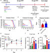
Fig. 1
Fig. 1
Loss of tau in TgA53T mice delays disease onset and progression following αS PFF inoculation. a Schematic of study paradigm, including genotypic groups, collection time points, behavior, and disease staging analysis. b, c Kaplan–Meier plots of all groups showing disease onset (onset of motor abnormalities) (b; P = 0.0122; Log-rank (Mantel Cox) test) and time to reach end stage (hind limb paralysis) (c; P = 0.0022; Log-rank (Mantel Cox) test). Both onset and time to end stage were delayed in TgA53T mice lacking tau (TgA53T/mTau−/−). d Average duration from disease onset to end stage was also significantly extended with loss of tau (P = 0.0247; t-test). e Performance on rotarod at 70 dpi showed that TgA53T mice exhibited decreased latency to fall compared to TgA53T/mTau−/− subjects. One-way ANOVA with Tukey’s post-hoc analysis; Day 3: F(3,18) = 4.925, P = 0.0114; Day 4: F(3,18) = 3.150, P = 0.0505. f, g TgA53T mice required more time to descend the pole (f) and complete (turn and descend) the pole test (g) compared to nTg and mTau−/− controls, while performance of the TgA53T/mTau−/− mice was not significantly different from all other groups, indicating that loss of tau expression results in partial protection from impaired performance. Abbreviations: preformed fibril, PFF; days post inoculation, dpi; immunohistochemistry, IHC; biochemistry, BC; seconds, s. One-way ANOVA with Tukey’s post-hoc analysis; time to descend: F(3,20) = 4.186, P = 0.0188; time to perform: F(3,20) = 4.220; P = 0.0182. n = 8–11 animals/genotype (survival), n = 57 animals/genotype (behavior). *P < 0.05 and **P < 0.01; error bars represent mean ± SEM
Fig. 2
Fig. 2
Biochemical abundance of pS129 αS in spinal cord is not affected by tau expression in end-stage TgA53T mice. a Representative immunoblots of total αS (a) and pS129 αS (b) in spinal cords of end-stage mice. c, d Quantitation of immunoblots shown in a and b. The level of pS129 αS was normalized to total αS. The levels of total αS and pS129 αS/total αS were not different between TgA53T and TgA53T/mTau−/− mice in end-stage spinal cord lysates (t-test: P = 0.0518 and P = 0.4267, respectively). All quantified bands were normalized to the respective ponceau S total protein. Abbreviations: age-matched, A.M.; arbitrary units, A.U.; ponceau S, ponc; n = 5 animals/genotype; error bars represent mean ± SEM
Fig. 3
Fig. 3
Loss of tau does not affect end-stage neuropathology in TgA53T mice. Spinal cord sections from end-stage nTg, mTau−/−, TgA53T and TgA53T/mTau−/− mice were stained for pS129 αS (a–e), Iba1 (microglia) (f–j), and GFAP (astrocytes) (k–o). Shown are representative images and corresponding quantitative analysis of % area covered by immunoreactivity in the lumbar region. In both TgA53T and TgA53T/mTau−/− mice the indices of neuropathology were significantly increased to a similar extent. One-way ANOVA with Tukey’s post-hoc analysis. pS129 αS: F(3,40) = 30.64; P < 0.0001. Iba1: F(3,40) = 54.81; P < 0.0001. GFAP: F(3,20) = 88.16; P < 0.0001. n = 6–15 sections from 3 to 5 animals/genotype; scale bars = 100 μm; error bars represent mean ± SEM
Fig. 4
Fig. 4
Bulk tissue level of pS129 αS in spinal cord is not affected by tau expression in TgA53T mice at 70 dpi. a Representative immunoblots of total αS (a) and pS129 αS (b) in spinal cord of 70-dpi mice. c, d Quantitation of immunoblots shown in a and b. Total αS (c), and pS129 αS normalized to total αS (d) were not different between TgA53T and TgA53T/mTau−/− mice in 70-dpi spinal cord lysates (t test, P = 0.1575 and P = 0.7993, respectively). All quantified bands were normalized to the respective ponceau S total protein. Abbreviations: ponceau S, ponc; arbitrary units, A.U.; n = 4 animals/genotype; error bars represent mean ± SEM
Fig. 5
Fig. 5
Loss of tau attenuates intermediate neuropathology in TgA53T mice. Spinal cord sections from 70-dpi nTg, mTau−/−, TgA53T, and TgA53T/mTau−/− mice were stained for pS129 αS (a–e), Iba1 (f–j), and GFAP (k–o). Shown are representative images and corresponding quantitative analysis of % area covered by immunoreactivity in the lumbar region. The indices of neuropathology were all significantly reduced in TgA53T/mTau−/− compared to TgA53T mice at 70 dpi. One-way ANOVA with Tukey’s post-hoc analysis. pS129 αS: F(3,23) = 82.46; P < 0.0001. Iba1: F(3,26) = 25.79; P < 0.0001. GFAP: F(3,18) = 69.86; P < 0.0001. n = 5–9 sections from 3 animals/genotype; scale bars = 100 μm; error bars represent mean ± SEM
Fig. 6
Fig. 6
Loss of tau delays α-synucleinopathy-associated spinal motor neuron degeneration in TgA53T mice. Spinal cord sections from  end-stage (a–g) and 70-dpi (h–n) TgA53T mice and controls (nTg, mTau−/−) were stained for NeuN (neuronal marker). Shown are representative images of lumbar ventral horn region. In end-stage mice (a–g), TgA53T and TgA53T/mTau−/− mice show significant loss of ventral horn (V. Horn) motor neurons (a–e; F(3,26) = 82.45, P < 0.0001). NeuN quantification further demonstrated significant neurodegeneration within the ventral horn (f; F(3,20) = 21.75, P < 0.0001), but not dorsal horn (D. Horn) (g; F(3,20) = 1.257; P = 0.3160). At 70 dpi (h–n), ventral horn motor neuron degeneration (l; F(3,20) = 14.15, P < 0.0001) as well as ventral NeuN+ neuronal loss (m—Ventral: F(3,20) = 6.490, P = 0.0030; n—Dorsal: F(3,20) = 0.4447, P = 0.7237) was delayed with tau loss. Abbreviation: days post inoculation, dpi; One-way ANOVA with Tukey’s post-hoc analysis. n = 5–10 sections from 3 to 5 animals/genotype; scale bars = 100 μm; error bars represent mean ± SEM
Fig. 7
Fig. 7
Soluble αS oligomers detected by Syn33 are not affected by tau expression. a Dot blot detection of human αS (LB509), αS off-pathway oligomers (Syn33), and total αS (4D6) in buffer-soluble spinal cord fractions of 70-dpi and end-stage mice. b No differences in LB509 or Syn33 were observed as a result of tau expression. c, d Syn33+ αS oligomers were not detected in the detergent-insoluble spinal cord fractions of both 70-dpi and end-stage mice. Abbreviations: days post inoculation, dpi; end stage, ES; arbitrary units, A.U.; n = 4–5 animals/genotype; error bars represent mean ± SEM
Fig. 8
Fig. 8
Neurons lacking tau expression are protected from αS PFF-induced loss of dendrites without impacting the development of pS129 αS+ aggregates. a, b Primary mouse hippocampal neurons from nTg (a) and mTau−/− (b) were induced to develop αS pathology by αS PFF treatment. Same levels of pS129 αS+ aggregates, normalized to MAP2 area,  were seen in nTg (c; P = 0.0001, PBS vs PFF) and mTau−/− (c; P = 0.0002, PBS vs PFF) neurons 14 days after PFF treatment. pS129 αS+ aggregates normalized to NeuN demonstrated a significant difference between nTg and mTau−/− (d; P < 0.0001, PBS vs PFF for both nTg and mTau−/−, and nTg PFF vs mTau−/− PFF). Analysis of MAP2+ neurites/dendrites at 14 days post-PBS or αS PFF addition to nTg (e) and mTau−/− (f) neurons. αS PFF treatment led to significant loss of MAP2 area in nTg neurons (e, g; P = 0.0010, PBS vs PFF; P = 0.0004, nTg PFF vs mTau−/− PFF; P = 0.0230, nTg PFF vs mTau−/− PBS) but not mTau−/− primary hippocampal neurons (f, g; P = 0.8297, PBS vs PFF), compared to PBS controls. Abbreviations: phosphate buffered saline, PBS; preformed fibril, PFF; Two-way ANOVA with Sidak’s multiple comparisons test; *P < 0.05, **P < 0.01, and ***P < 0.001. Scale bars = 100 μm;  n = 6–16 randomly selected areas from 3 to 5 independent cultures; error bars represent mean ± SEM

Similar articles

Cited by

References

    1. Braak H, Tredici KD, Rüb U, de Vos RAI, Jansen Steur ENH, Braak E. Staging of brain pathology related to sporadic Parkinson’s disease. Neurobiol Aging. 2003;24:197–211. doi: 10.1016/S0197-4580(02)00065-9. - DOI - PubMed
    1. Del Tredici K, Rüb U, de Vos RAI, Bohl JRE, Braak H. Where does Parkinson disease pathology begin in the brain? J Neuropathol Exp Neurol. 2002;61:413–426. doi: 10.1093/jnen/61.5.413. - DOI - PubMed
    1. Lee MK, Stirling W, Xu Y, Xu X, Qui D, Mandir AS, et al. Human α-synuclein-harboring familial Parkinson’s disease-linked Ala-53 → Thr mutation causes neurodegenerative disease with α-synuclein aggregation in transgenic mice. Proc Natl Acad Sci USA. 2002;99:8968–8973. doi: 10.1073/pnas.132197599. - DOI - PMC - PubMed
    1. Singh B, Covelo A, Martell-Martínez H, Nanclares C, Sherman MA, Okematti E, et al. Tau is required for progressive synaptic and memory deficits in a transgenic mouse model of α-synucleinopathy. Acta Neuropathol. 2019;138(4):551–574. doi: 10.1007/s00401-019-02032-w. - DOI - PMC - PubMed
    1. Teravskis PJ, Covelo A, Miller EC, Singh B, Martell-Martínez HA, Benneyworth MA, et al. A53T mutant alpha-synuclein induces tau-dependent postsynaptic impairment independently of neurodegenerative changes. J Neurosci. 2018;38:9754–9767. doi: 10.1523/JNEUROSCI.0344-18.2018. - DOI - PMC - PubMed

Publication types