AnatomySketch: An Extensible Open-Source Software Platform for Medical Image Analysis Algorithm Development
- PMID: 35768752
- PMCID: PMC9712876
- DOI: 10.1007/s10278-022-00660-5
AnatomySketch: An Extensible Open-Source Software Platform for Medical Image Analysis Algorithm Development
Abstract
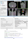
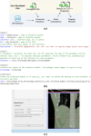
The development of medical image analysis algorithm is a complex process including the multiple sub-steps of model training, data visualization, human-computer interaction and graphical user interface (GUI) construction. To accelerate the development process, algorithm developers need a software tool to assist with all the sub-steps so that they can focus on the core function implementation. Especially, for the development of deep learning (DL) algorithms, a software tool supporting training data annotation and GUI construction is highly desired. In this work, we constructed AnatomySketch, an extensible open-source software platform with a friendly GUI and a flexible plugin interface for integrating user-developed algorithm modules. Through the plugin interface, algorithm developers can quickly create a GUI-based software prototype for clinical validation. AnatomySketch supports image annotation using the stylus and multi-touch screen. It also provides efficient tools to facilitate the collaboration between human experts and artificial intelligent (AI) algorithms. We demonstrate four exemplar applications including customized MRI image diagnosis, interactive lung lobe segmentation, human-AI collaborated spine disc segmentation and Annotation-by-iterative-Deep-Learning (AID) for DL model training. Using AnatomySketch, the gap between laboratory prototyping and clinical testing is bridged and the development of MIA algorithms is accelerated. The software is opened at https://github.com/DlutMedimgGroup/AnatomySketch-Software .
Keywords: Algorithm development; Deep learning; Image annotation; Medical image analysis; User interaction.
© 2022. The Author(s).
Conflict of interest statement
The authors declare no competing interests.
Figures


Similar articles
-
ATOMMIC: An Advanced Toolbox for Multitask Medical Imaging Consistency to facilitate Artificial Intelligence applications from acquisition to analysis in Magnetic Resonance Imaging.Comput Methods Programs Biomed. 2024 Nov;256:108377. doi: 10.1016/j.cmpb.2024.108377. Epub 2024 Aug 22. Comput Methods Programs Biomed. 2024. PMID: 39180913
-
An extensible software platform for interdisciplinary cardiovascular imaging research.Comput Methods Programs Biomed. 2020 Feb;184:105277. doi: 10.1016/j.cmpb.2019.105277. Epub 2019 Dec 12. Comput Methods Programs Biomed. 2020. PMID: 31891904
-
MaD GUI: An Open-Source Python Package for Annotation and Analysis of Time-Series Data.Sensors (Basel). 2022 Aug 5;22(15):5849. doi: 10.3390/s22155849. Sensors (Basel). 2022. PMID: 35957406 Free PMC article.
-
RIL-Contour: a Medical Imaging Dataset Annotation Tool for and with Deep Learning.J Digit Imaging. 2019 Aug;32(4):571-581. doi: 10.1007/s10278-019-00232-0. J Digit Imaging. 2019. PMID: 31089974 Free PMC article. Review.
-
GADMA2: more efficient and flexible demographic inference from genetic data.Gigascience. 2022 Dec 28;12:giad059. doi: 10.1093/gigascience/giad059. Epub 2023 Aug 23. Gigascience. 2022. PMID: 37609916 Free PMC article. Review.
Cited by
-
Two-stage multi-task deep learning framework for simultaneous pelvic bone segmentation and landmark detection from CT images.Int J Comput Assist Radiol Surg. 2024 Jan;19(1):97-108. doi: 10.1007/s11548-023-02976-1. Epub 2023 Jun 15. Int J Comput Assist Radiol Surg. 2024. PMID: 37322299
References
-
- Yoo TS, Ackerman MJ, Lorensen WE, Schroeder W, Chalana V, Aylward S, Metaxas D, Whitaker R. Engineering and algorithm design for an image processing Api: a technical report on ITK–the Insight Toolkit. Stud Health Technol Inform. 2002;85:586–592. - PubMed
-
- Schroeder, W.; Martin, K.; Lorensen, B. The Visualization Toolkit (4th ed.); Kitware: 2006.
Publication types
MeSH terms
LinkOut - more resources
Full Text Sources
Miscellaneous

