Review
. 2022 Jul 29;42(7):BSR20221212.
doi: 10.1042/BSR20221212.

The insulin receptor family in the heart: new light on old insights

Affiliations
Review

The insulin receptor family in the heart: new light on old insights

Angela Clerk et al. Biosci Rep. .

Abstract

Insulin was discovered over 100 years ago. Whilst the first half century defined many of the physiological effects of insulin, the second emphasised the mechanisms by which it elicits these effects, implicating a vast array of G proteins and their regulators, lipid and protein kinases and counteracting phosphatases, and more. Potential growth-promoting and protective effects of insulin on the heart emerged from studies of carbohydrate metabolism in the 1960s, but the insulin receptors (and the related receptor for insulin-like growth factors 1 and 2) were not defined until the 1980s. A related third receptor, the insulin receptor-related receptor remained an orphan receptor for many years until it was identified as an alkali-sensor. The mechanisms by which these receptors and the plethora of downstream signalling molecules confer cardioprotection remain elusive. Here, we review important aspects of the effects of the three insulin receptor family members in the heart. Metabolic studies are set in the context of what is now known of insulin receptor family signalling and the role of protein kinase B (PKB or Akt), and the relationship between this and cardiomyocyte survival versus death is discussed. PKB/Akt phosphorylates numerous substrates with potential for cardioprotection in the contractile cardiomyocytes and cardiac non-myocytes. Our overall conclusion is that the effects of insulin on glucose metabolism that were initially identified remain highly pertinent in managing cardiomyocyte energetics and preservation of function. This alone provides a high level of cardioprotection in the face of pathophysiological stressors such as ischaemia and myocardial infarction.

Keywords: insulin receptors; insulin signalling; intracellular signaling; regulation of metabolism.


Conflict of interest statement

The authors declare that there are no competing interests associated with the manuscript.

Figures
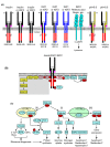
Figure 1
Figure 1. Insulin receptor family signalling
(A) Insulin receptors (INSRs, black) and the related receptor family members, IGF1 receptors (IGF1Rs, blue) and insulin receptor-related receptors (INSRRs, yellow) are expressed at the cell surface as preformed heterotetramers, each comprising two hemi-receptors composed of one α- and one β-subunit. Each α- and β-subunit and the two α-subunits are cross-linked by disulphide bridges. The extracellular α subunits bind insulin or IGFs as indicated or are responsive to alkaline pHo. The β-subunits have a tyrosine kinase domain (red). Alternative splicing generates two isoforms of INSR, INSR-A and INSR-B, without or with (respectively) exon 11 (pink). Hybrid receptors can form between the family members. IGF2 also binds to the cation-independent mannose 6-phosphate receptor (CI-M6PR) that directs IGF2 and other molecules to the lysosome for degradation. (B) Insulin or IGFs bind to INSRs/IGF1Rs causing receptor phosphorylation and full activation of the tyrosine kinase, with additional phosphorylation of tyrosine residues (pY) on the β-subunit. These recruit adapter proteins including insulin receptor substrates (IRS) that become phosphorylated. IRS proteins recruit phosphoinositide 3′ kinase (PI3K) that stimulates production of phosphatidylinositol 3,4,5 tris phosphate in the membrane (red lipids), recruiting PDK1 and protein kinase B (PKB, also known as Akt). PDK1 phosphorylates PKB/Akt on Thr308. In some cells, INSRs/IGF1Rs also signal via Grb2 and the exchange factor Sos to the small G protein Ras. Activated Ras recruits Raf kinases that phosphorylate and activate mitogen-activated protein kinase kinases 1/2 (MKK1/2) that phosphorylate extracellular signal-regulated kinases 1 and 2 (ERK1/2). However, this pathway is not thought to be activated significantly in the heart and is shaded. (C) (i) PKB/Akt(T308) phosphorylates AS160 (Tbc1d4) to increase translocation of the glucose transporter GLUT4 to the membrane for glucose uptake. Phosphorylation and inhibition of glycogen synthase kinase 3 (GSK3) results in activation of glycogen synthase (GS) to increase glycogen synthesis. Other GSK3 substrates influence apoptosis, metabolism and cardiac hypertrophy. (ii) PKB/Akt(T308) activates mammalian target of rapamycin (mTOR) in mTOR complex 1 (mTORC1). Signalling via p70 ribosomal S6 kinase (p70S6K) increases ribosome biogenesis by increasing translation of RNAs with 5′ terminal oligopyrimidine (TOP) sequences), phosphorylation of Rps6 and regulation of other eukaryotic initiation factors (eIFs). mTORC1-dependent phosphorylation of 4E-BP causes it to dissociate from eIF4E that is then available to bind to the 5′ cap structure of mRNAs to increase translation and enhance protein synthesis. (iii) PKB/Akt(T308) is phosphorylated on Ser473 in mTORC2. Dually phosphorylated PKB/Akt(T308/S473) has greater activity and additional target specificity for forkhead transcription factors of the FoxO family. Phosphorylation of FoxO transcription factors inhibits their effects on gene expression, to influence apoptosis and metabolism.
Figure 2
Figure 2. Glucose metabolism through the glycolytic and pentose phosphate pathways: regulation by PKB/Akt
PKB/Akt phosphorylates RabGAPs (AS160/Tbc1d4, Tbc1d1) to prevent their inhibitory action (red type), increasing translocation of vesicles containing the glucose transporter GLUT4 to the membrane (blue arrow). This increases glucose uptake into the cell. Hexokinase II (HKII) converts glucose to glucose 6-phosphate (G6P). It is tethered to the mitochondrial membrane, interacting with the voltage-dependent anion channel (VDAC), allowing it to access ATP directly from the mitochondria, and facilitating direct transfer of ADP back into the mitochondria. HKII expression and activity is increased by PKB/Akt signalling (blue type) increasing production of G6P. Glycolysis (centre). G6P is converted to fructose 6-phosphate which is then converted to fructose 1,6-bisphosphate by phosphofructokinase (PFK) in the rate-limiting step of the pathway. Fructose 6-phosphate interconversion with fructose 2,6-bisphosphate is performed by the cardiac isoform of phosphofructokinase 2 (PFKB2), an enzyme with dual kinase (K) and phosphatase (P) activity. PKB/Akt phosphorylates and inhibits the phosphatase activity, resulting in accumulation of fructose 2,6-bisphosphate, an allosteric activator of PFK. This increases glycolytic flux to produce pyruvate for oxidative phosphorylation in the mitochondria. If oxidative phosphorylation is insufficient to manage additional pyruvate, pyruvate is converted to lactate by lactate dehydrogenase (LDH). This causes acidification and excess lactate is exported from the cell. Pentose phosphate pathway (left section). G6P is converted to 6-phospho-gluconolactone by G6P dehydrogenase (G6PDH), relying on production of G6P that is increased by PKB/Akt signalling. This reaction produces NADPH, required to reduce oxidised glutathione (GSSG) to GSH. GSH is a critical antioxidant in the cell, scavenging reactive oxygen species (ROS). 6-phospho-gluconolactone can be returned to the glycolytic pathway via further oxidative and non-oxidative reactions.
Figure 3
Figure 3. Cross-talk from α1-adrenergic receptors (α1-ARs) to INSR/INSRR receptors in the heart: possible mechanisms
(i) α1-ARs are G-protein-coupled receptors that couple through Gαq to activate phospholipase C (PLC) β and possibly PLCε. PLCβ/ε hydrolyses phosphatidylinositol 4,5 bisphosphate (PIP2) to produce diacylglycerol (DAG) which activates protein kinase C (PKC) family members and guanine nucleotide exchange factors for Ras (e.g. RasGRP). This results in activation of the ERK1/2 cascade that affects downstream signalling and gene expression. (ii) Activation of the small G protein Rac1 results in an increase in production of reactive oxygen species (ROS) via NADPH oxidases (NOX). ROS inhibit protein tyrosine phosphatases (PTPs), and this may lead to increased tyrosine phosphorylation of INSRs/INSRRs, with receptor activation and downstream signalling. (iii) Src family kinases (SFKs) may be activated, possibly by phosphatidic acid produced by phospholipase D (activated by PKC; pathway not shown). SFKs may phosphorylate INSRs/INSRRs directly to cause receptor activation. (iv) α1-ARs increase intracellular pHi via an unknown mechanism and this needs to be neutralised. SFKs phosphorylate anion exchanger 3 (AE3) to increase exchange of Cl and HCO3 delivering HCO3 to the extracellular space. Intracellular HCO3 is probably generated by carbonic anhydrase II (CAII). Simultaneous production of H+ reduces intracellular pHi. Extracellular HCO3 may increase extracellular pHo to activate INSRRs via a conformational change of the extracellular α-subunit.
Figure 4
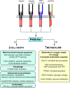
Figure 4. Possible mechanisms of cardioprotection resulting from stimulation of insulin receptor family members
Activation of insulin receptor family members results in activation of protein kinase B (PKB, also known as Akt). (i) PKB/Akt signalling influences cell death by reducing mitochondrial-dependent apoptosis, increasing hexokinase II (HKII) and inhibiting glycogen synthase kinase 3 (GSK3) activities. This prevents opening of the mitochondrial permeability transition pore (mPTP). Inhibition of FoxO transcription factors reduces expression of pro-apoptotic proteins. Signalling via mTORC1 inhibits autophagy via Ulk1. Activation of eNOS may reduce cell death, acting to promote vasodilation and, thus, reduce workload. It also reduces vascular inflammation. Phosphorylation of cGAS and STING inhibits interferon production. Increased flux through the pentose phosphate pathway (PPP) increases antioxidant capacity in the cell by increasing production of NADPH and glutathione (GSH). (ii) PKB/Akt increases metabolism, providing the cell with options for reducing oxygen needs whilst sustaining energy production, and maintaining antioxidant capability via the NADPH/glutathione system. Glucose uptake is increased by translocation of GLUT4 to the membrane. HKII increases availability of glucose 6-phosphate (G6P) for both glycolysis and the pentose phosphate pathway (PPP). Phosphorylation of the cardiac isoform of phosphofructokinase 2 (PFKB2) inhibits the phosphatase activity, increasing production of fructose 2,6-bisphosphate, an allosteric activator of glycolysis. Glycogen storage is increased by GSK3 inhibition, providing a source of glucose during ischaemia. FoxO inhibition increases glucose metabolism.

Similar articles

Cited by

References

    1. Ahuja P., Sdek P. and MacLellan W.R. (2007) Cardiac myocyte cell cycle control in development, disease, and regeneration. Physiol. Rev. 87, 521–544 10.1152/physrev.00032.2006 - DOI - PMC - PubMed
    1. Broughton K.M. and Sussman M.A. (2019) Adult cardiomyocyte cell cycle detour: off-ramp to quiescent destinations. Trends Endocrinol. Metab. 30, 557–567 10.1016/j.tem.2019.05.006 - DOI - PMC - PubMed
    1. Dorn G.W. II, Robbins J. and Sugden P.H. (2003) Phenotyping hypertrophy: eschew obfuscation. Circ. Res. 92, 1171–1175 10.1161/01.RES.0000077012.11088.BC - DOI - PubMed
    1. Gibb A.A. and Hill B.G. (2018) Metabolic coordination of physiological and pathological cardiac remodeling. Circ. Res. 123, 107–128 10.1161/CIRCRESAHA.118.312017 - DOI - PMC - PubMed
    1. Messerli F.H., Rimoldi S.F. and Bangalore S. (2017) The transition from hypertension to heart failure: contemporary update. JACC Heart Fail. 5, 543–551 10.1016/j.jchf.2017.04.012 - DOI - PubMed

MeSH terms