

Studies of neurodegenerative diseases using Drosophila and the development of novel approaches for their analysis
- PMID: 35765969
- PMCID: PMC9336468
- DOI: 10.1080/19336934.2022.2087484
Studies of neurodegenerative diseases using Drosophila and the development of novel approaches for their analysis
Abstract
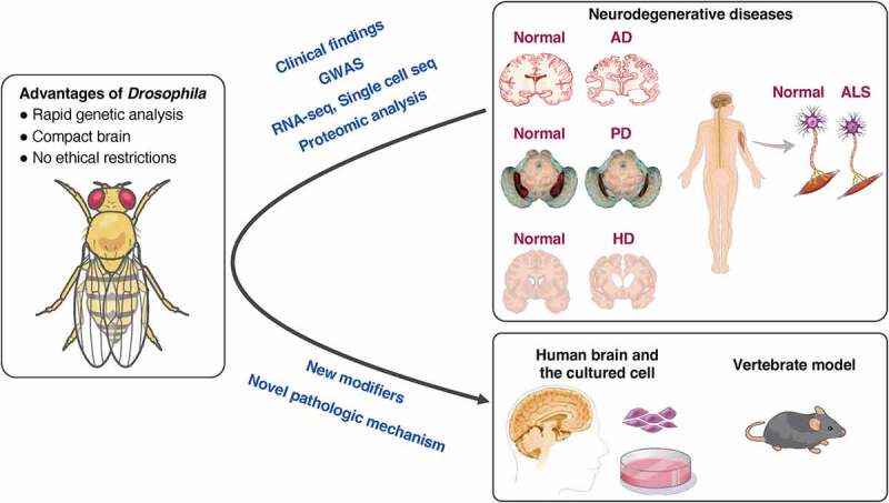
The use of Drosophila in neurodegenerative disease research has contributed to the identification of modifier genes for the pathology. The basis for neurodegenerative disease occurrence in Drosophila is the conservation of genes across species and the ability to perform rapid genetic analysis using a compact brain. Genetic findings previously discovered in Drosophila can reveal molecular pathologies involved in human neurological diseases in later years. Disease models using Drosophila began to be generated during the development of genetic engineering. In recent years, results of reverse translational research using Drosophila have been reported. In this review, we discuss research on neurodegenerative diseases; moreover, we introduce various methods for quantifying neurodegeneration in Drosophila.
Keywords: Drosophila; neurodegenerative diseases; reverse translational research.
Conflict of interest statement
No potential conflict of interest was reported by the author(s).
Figures
Similar articles
-
Recent advances in using Drosophila to model neurodegenerative diseases.Apoptosis. 2009 Aug;14(8):1008-20. doi: 10.1007/s10495-009-0347-5. Apoptosis. 2009. PMID: 19373559 Free PMC article. Review.
-
Neurodegenerative mutants in Drosophila: a means to identify genes and mechanisms involved in human diseases?Invert Neurosci. 2005 Nov;5(3-4):97-109. doi: 10.1007/s10158-005-0005-8. Epub 2005 Oct 24. Invert Neurosci. 2005. PMID: 16187075 Review.
-
From fruit fly to bedside: translating lessons from Drosophila models of neurodegenerative disease.Curr Opin Neurol. 2003 Aug;16(4):443-9. doi: 10.1097/01.wco.0000084220.82329.60. Curr Opin Neurol. 2003. PMID: 12869801 Review.
-
Drosophila models of neurodegenerative disease.NeuroRx. 2005 Jul;2(3):438-46. doi: 10.1602/neurorx.2.3.438. NeuroRx. 2005. PMID: 16389307 Free PMC article. Review.
-
Genetic strategies to tackle neurological diseases in fruit flies.Curr Opin Neurobiol. 2018 Jun;50:24-32. doi: 10.1016/j.conb.2017.10.017. Epub 2017 Nov 9. Curr Opin Neurobiol. 2018. PMID: 29128849 Free PMC article. Review.
Cited by
-
Brazilian kefir fraction mitigates the Alzheimer-like phenotype in Drosophila melanogaster with β-amyloid overexpression model.Sci Rep. 2024 Oct 26;14(1):25474. doi: 10.1038/s41598-024-76601-9. Sci Rep. 2024. PMID: 39461991 Free PMC article.
-
Ethnopharmacological Effects of Urtica dioica, Matricaria chamomilla, and Murraya koenigii on Rotenone-Exposed D. melanogaster: An Attenuation of Cellular, Biochemical, and Organismal Markers.Antioxidants (Basel). 2022 Aug 21;11(8):1623. doi: 10.3390/antiox11081623. Antioxidants (Basel). 2022. PMID: 36009342 Free PMC article.
-
A novel NONO variant that causes developmental delay and cardiac phenotypes.Sci Rep. 2023 Jan 18;13(1):975. doi: 10.1038/s41598-023-27770-6. Sci Rep. 2023. PMID: 36653413 Free PMC article.
-
The overexpression of DSP1 in neurons induces neuronal dysfunction and neurodegeneration phenotypes in Drosophila.Mol Brain. 2024 Jul 13;17(1):43. doi: 10.1186/s13041-024-01117-2. Mol Brain. 2024. PMID: 39003465 Free PMC article.
-
Saccharomyces cerevisiae as a Model for Studying Human Neurodegenerative Disorders: Viral Capsid Protein Expression.Int J Mol Sci. 2023 Dec 7;24(24):17213. doi: 10.3390/ijms242417213. Int J Mol Sci. 2023. PMID: 38139041 Free PMC article. Review.
References
Publication types
MeSH terms
Grants and funding
LinkOut - more resources
Full Text Sources
Other Literature Sources
Medical
Molecular Biology Databases