. 2022 Jun 26;23(1):39.
doi: 10.1186/s12868-022-00726-8.

Opposing actions of CRF-R1 and CB1 receptor on facial stimulation-induced MLI-PC plasticity in mouse cerebellar cortex

Affiliations

Opposing actions of CRF-R1 and CB1 receptor on facial stimulation-induced MLI-PC plasticity in mouse cerebellar cortex

Guang-Gao Li et al. BMC Neurosci. .

Abstract

Background: Corticotropin-releasing factor (CRF) is the major neuromodulator orchestrating the stress response, and is secreted by neurons in various regions of the brain. Cerebellar CRF is released by afferents from inferior olivary neurons and other brainstem nuclei in response to stressful challenges, and contributes to modulation of synaptic plasticity and motor learning behavior via its receptors. We recently found that CRF modulates facial stimulation-evoked molecular layer interneuron-Purkinje cell (MLI-PC) synaptic transmission via CRF type 1 receptor (CRF-R1) in vivo in mice, suggesting that CRF modulates sensory stimulation-evoked MLI-PC synaptic plasticity. However, the mechanism of how CRF modulates MLI-PC synaptic plasticity is unclear. We investigated the effect of CRF on facial stimulation-evoked MLI-PC long-term depression (LTD) in urethane-anesthetized mice by cell-attached recording technique and pharmacological methods.

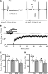
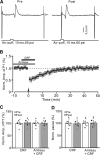
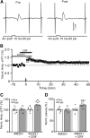
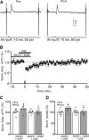
Results: Facial stimulation at 1 Hz induced LTD of MLI-PC synaptic transmission under control conditions, but not in the presence of CRF (100 nM). The CRF-abolished MLI-PC LTD was restored by application of a selective CRF-R1 antagonist, BMS-763,534 (200 nM), but it was not restored by application of a selective CRF-R2 antagonist, antisauvagine-30 (200 nM). Blocking cannabinoid type 1 (CB1) receptor abolished the facial stimulation-induced MLI-PC LTD, and revealed a CRF-triggered MLI-PC long-term potentiation (LTP) via CRF-R1. Notably, either inhibition of protein kinase C (PKC) with chelerythrine (5 µM) or depletion of intracellular Ca2+ with cyclopiazonic acid (100 µM), completely prevented CRF-triggered MLI-PC LTP in mouse cerebellar cortex in vivo.

Conclusions: The present results indicated that CRF blocked sensory stimulation-induced opioid-dependent MLI-PC LTD by triggering MLI-PC LTP through CRF-R1/PKC and intracellular Ca2+ signaling pathway in mouse cerebellar cortex. These results suggest that activation of CRF-R1 opposes opioid-mediated cerebellar MLI-PC plasticity in vivo in mice.

Keywords: Corticotropin-releasing factor (CRF); Long-term plasticity; Molecular layer interneuron (MLI); Mouse cerebellar cortex; Purkinje cell; Sensory stimulation; in vivo cell-attached recording.


Conflict of interest statement

We have no conflicts of interest in this manuscript.

Figures
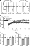
Fig. 1
Fig. 1
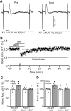
Effect of CRF on 1 Hz facial stimulation-evokedMLI-PC LTD in vivo in mice. A Upper: Representative cell-attached recording traces showing air-puff stimulation (10 ms, 60 psi; arrows)-evoked responses in cerebellar PCs before (Pre) and after (post) delivering 1 Hz (240 pulses) facial stimulation during treatments with ACSF (control) and CRF (100 nM). B Summary of data showing the time course of normalized P1 amplitude before and after delivery of 1 Hz facial stimulation (arrow head) in ACSF (control, filled circle) and CRF (open circles). C Bar graph with individual data showing the normalized amplitude of P1 before (Pre), after (Post) delivery of 1 Hz stimulation. D Mean (± S.E.M.) with individual data showing the normalized pause of simple spike firing before (Pre), after (Post) delivery of 1 Hz stimulation. #P < 0.05 versus Pre of control; *P < 0.05 versus post of control. n = 7 mice in each group
Fig. 2
Fig. 2
Blockade of CRF-R1, CRF failed to prevent facial stimulation-evoked MLI-PC LTD. A Upper: Representative cell-attached recording traces showing air-puff stimulation (10 ms, 60 psi; arrows)-evoked responses in a cerebellar PC before (Pre) and after (post) delivering 1 Hz (240 pulses) facial stimulation in the presence of a mixture of BSM (200 nM) and CRF (100 nM). B Summary of data showing the time course of normalized P1 amplitude before and after delivery of 1 Hz facial stimulation (arrow head) in the presence of the mixture. C Bar graph with individual data showing the normalized amplitude of P1 before (Pre), after (Post) delivery of 1 Hz stimulation. D Mean (± S.E.M.) with individual data showing the normalized pause of simple spike firing before (Pre), after (Post) delivery of 1 Hz stimulation. #P < 0.05 versus Pre of BSM + CRF; *P < 0.05 versus post of CRF. n = 8 mice in each group
Fig. 3
Fig. 3
Blockade of CRF-R2 did not prevent the effect of CRF on facial stimulation-evoked MLI-PC LTD. A Upper: Representative cell-attached recording traces showing air-puff stimulation (10 ms, 60 psi; arrows)-evoked responses in a cerebellar PC before (Pre) and after (post) delivering 1 Hz (240 pulses) stimulation in the presence of a mixture of antisauvagine-30 (Antisau; 200 nM)and CRF (100 nM). B Summary of data showing the time course of normalized P1 amplitude before and after delivery of 1 Hz facial stimulation (arrow head) in the presence of Antisau and CRF. C Mean (± S.E.M) with individual data showing the normalized amplitude of P1 before (Pre), after (Post) delivery of 1 Hz stimulation. D Mean (± S.E.M.) with individual data showing the normalized pause of simple spike firing before (Pre), after (Post) delivery of 1 Hz stimulation. n = 7 mice in each group
Fig. 4
Fig. 4
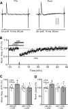
Blockade of CB1 receptor, CRF triggers MLI-PC LTP. A Upper: Representative cell-attached recording traces showing air-puff stimulation (10 ms, 60 psi; arrows)-evoked responses in a cerebellar PC before (Pre) and after (post) delivering 1 Hz (240 pulses) stimulation in the presence of a mixture of AM251(5 µM)and CRF (100 nM). B Summary of data showing the time course of normalized P1 amplitude before and after delivery of 1 Hz facial stimulation (arrow head) in the presence of AM-251 and CRF. C Bar graph with individual data showing the normalized amplitude of P1 before (Pre), after (Post) delivery of 1 Hz stimulation. D Mean (± S.E.M.) with individual data showing the normalized pause of simple spike firing before (Pre), after (Post) delivery of 1 Hz stimulation. #P < 0.05 versus baseline (Pre) of AM251 + CRF; *P < 0.05 versus post of AM251. n = 8 mice in each group
Fig. 5
Fig. 5
Blockade CB1 receptor and CRF-R1, CRF could not trigger MLI-PC LTP. A Upper: Representative cell-attached recording traces showing air-puff stimulation (10 ms, 60 psi; arrows)-evoked responses in a cerebellar PC before (Pre) and after (post) delivering 1 Hz (240 pulses) stimulation in the presence of a mixture of AM251(5 µM)+ BSM (200 nM) + CRF (100 nM). B Summary of data (n = 7) showing the time course of normalized P1 amplitude before and after delivery of 1 Hz facial stimulation (arrow head) in the presence of a mixture of AM251 + BSM + CRF. C Bar graph with individual data showing the normalized amplitude of P1 before (Pre), after (Post) delivery of 1 Hz stimulation. D Mean (± S.E.M.) with individual data showing the normalized pause of simple spike firing before (Pre), after (Post) delivery of 1 Hz stimulation. n = 8 mice in each group
Fig. 6
Fig. 6
Blockade of PKC and CB1 receptor, CRF could not trigger MLI-PC LTP. A Upper: Representative cell-attached recording traces showing air-puff stimulation (10 ms, 60 psi; arrows)-evoked responses in a cerebellar PC before (Pre) and after (post) delivering 1 Hz (240 pulses) stimulation in the presence of a mixture of AM251 (5 µM) + chelerythrine (5 µM)+ CRF (100 nM). B Summary of data showing the time course of normalized P1 amplitude before and after delivery of 1 Hz facial stimulation (arrow head) in the presence of AM251 + chelerythrine + CRF. C Bar graph with individual data showing the normalized amplitude of P1 before (Pre), after (Post) delivery of 1 Hz stimulation. D Mean (± S.E.M.) with individual data showing the normalized pause of simple spike firing before (Pre), after (Post) delivery of 1 Hz stimulation. n = 8 mice in each group
Fig. 7
Fig. 7
Depletion of intracellular Ca2+, CRF could not trigger MLI-PC LTP. A Upper: Representative cell-attached recording traces showing air-puff stimulation (10 ms, 60 psi; arrows)-evoked responses in a cerebellar PC before (Pre) and after (post) delivering 1 Hz (240 pulses) stimulation in the presence of a mixture of AM251 (5 µM) + cyclopiazonic acid (CPA, 100 µM) + CRF (100 nM). B Summary of data showing the time course of normalized P1 amplitude before and after delivery of 1 Hz facial stimulation (arrow head) in treatment with a mixture of AM251 + CPA + CRF. C Bar graph with individual data showing the normalized amplitude of P1 before (Pre), after (Post) delivery of 1 Hz stimulation. D Mean (± S.E.M.) with individual data showing the normalized pause of simple spike firing before (Pre), after (Post) delivery of 1 Hz stimulation. #P < 0.05 vs. baseline (Pre) of AM-251 + CRF; *P < 0.05 vs. post of AM-251 + CRF. n = 8 mice in each group

Similar articles

Cited by

References

    1. Barmack NH, Young WS., 3rd Optokinetic stimulation increases corticotropin-releasing factor mRNA in inferior olivary neurons of rabbits. J Neurosci. 1990;10(2):631–40. doi: 10.1523/JNEUROSCI.10-02-00631.1990. - DOI - PMC - PubMed
    1. Luo X, Kiss A, Makara G, Lolait SJ, Aguilera G. Stress-specific regulation of corticotropin releasing hormone receptor expression in the paraventricular and supraoptic nuclei of the hypothalamus in the rat. J Neuroendocrinol. 1994;6:689–696. doi: 10.1111/j.1365-2826.1994.tb00636.x. - DOI - PubMed
    1. Palkovits M, Leranth C, Gorcs T, Young WS., 3rd Corticotropin-releasing factor in the olivocerebellar tract of rats: demonstration by light- and electron-microscopic immunohistochemistry and in situ hybridization histochemistry. Proc Natl Acad Sci USA. 1987;84:3911–3915. doi: 10.1073/pnas.84.11.3911. - DOI - PMC - PubMed
    1. Tian JB, Bishop GA. Frequency-dependent expression of corticotropin releasing factor in the rat’s cerebellum. Neuroscience. 2003;121:363–377. doi: 10.1016/S0306-4522(03)00493-7. - DOI - PubMed
    1. Ezra-Nevo G, Prestori F, Locatelli F, Soda T, Ten Brinke MM, Engel M, Boele HJ, Botta L, Leshkowitz D, Ramot A, Tsoory M, Biton IE, Deussing J, D’Angelo E, De Zeeuw CI, Chen A. Cerebellar Learning Properties Are Modulated by the CRF Receptor. J Neurosci. 2018;38:6751–6765. doi: 10.1523/JNEUROSCI.3106-15.2018. - DOI - PMC - PubMed

Publication types

MeSH terms

Substances

LinkOut - more resources