. 2022 May 27;14(6):1163.
doi: 10.3390/v14061163.

The M13 Phage Assembly Machine Has a Membrane-Spanning Oligomeric Ring Structure

Affiliations

The M13 Phage Assembly Machine Has a Membrane-Spanning Oligomeric Ring Structure

Maximilian Haase et al. Viruses. .

Abstract

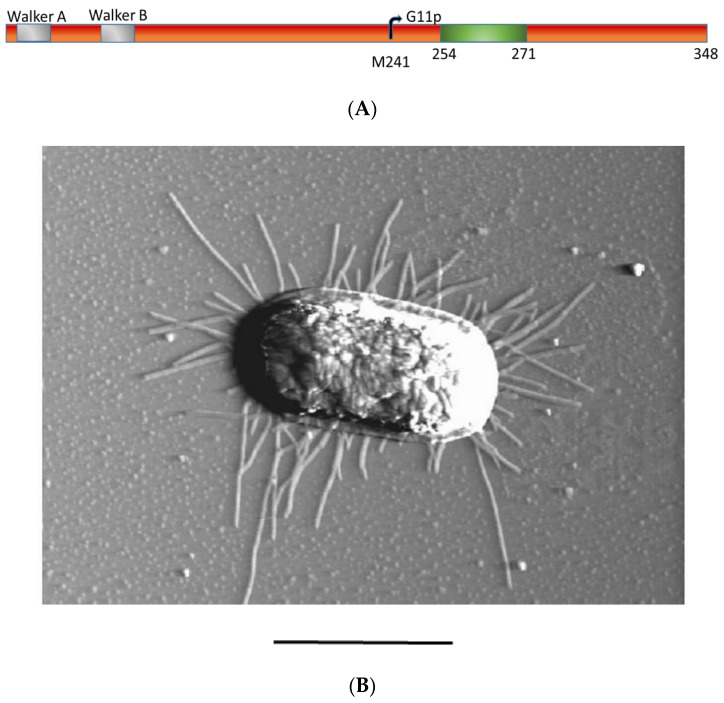
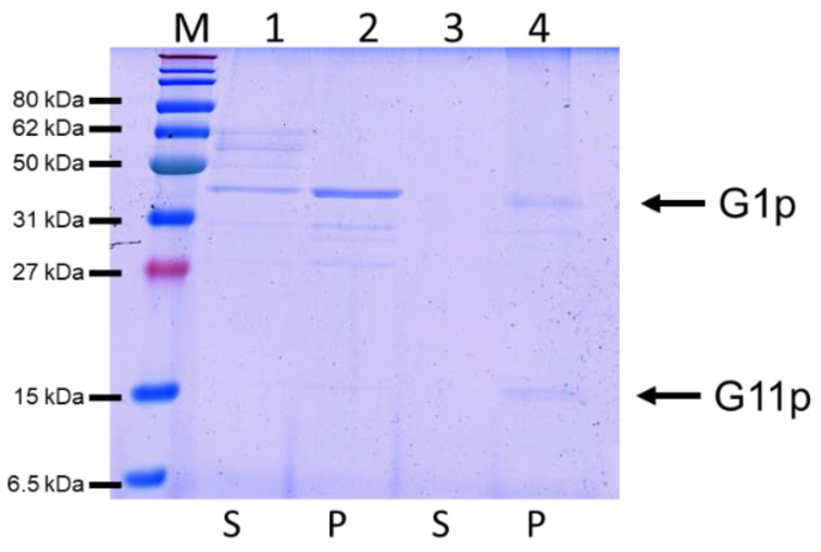
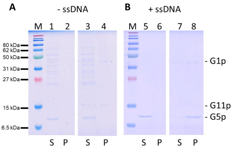
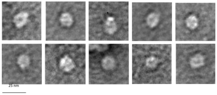
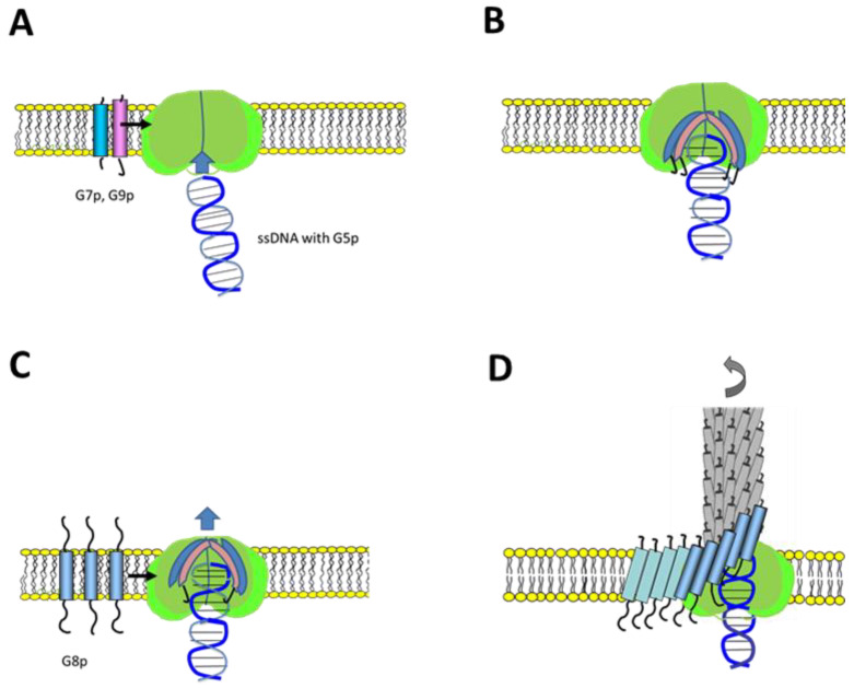
Bacteriophage M13 assembles its progeny particles in the inner membrane of the host. The major component of the assembly machine is G1p and together with G11p it generates an oligomeric structure with a pore-like inner cavity and an ATP hydrolysing domain. This allows the formation of the phage filament, which assembles multiple copies of the membrane-inserted major coat protein G8p around the extruding single-stranded circular DNA. The phage filament then passes through the G4p secretin that is localized in the outer membrane. Presumably, the inner membrane G1p/G11p and the outer G4p form a common complex. To unravel the structural details of the M13 assembly machine, we purified G1p from infected E. coli cells. The protein was overproduced together with G11p and solubilized from the membrane as a multimeric complex with a size of about 320 kDa. The complex revealed a pore-like structure with an outer diameter of about 12 nm, matching the dimensions of the outer membrane G4p secretin. The function of the M13 assembly machine for phage generation and secretion is discussed.

Keywords: affinity chromatography; bacteriophage M13; circular dichroism; membrane protein; phage assembly machine.


Conflict of interest statement

The authors declare no conflict of interest.

Figures

Figure 1
Figure 1
The secretion of M13 phage by E. coli is mediated by the M13 assembly machine. (A) Schematic representation of the G1p protein regions with the indicated Walker box sequences (grey, residues 8 to 15 and 84 to 89), the internal start codon M241 for G11p and the membrane anchor segment (green, 254 to 271) and the periplasmic domain (residues 272 to 348). (B) An M13-secreting E. coli cell 10 min post-infection was visualized by atomic force microscopy (AFM). The bar represents 1 µm.
Figure 2
Figure 2
Expression and functionality of plasmid-encoded G1p. (A) The expression of the plasmid-encoded G1p was analysed in M15F+ cells by Western blot. Lane 1: empty plasmid, lane 2: G1p/G11p, lane 3: HisG1p/G11p and lane 4: G1pHis/G11pHis were expressed from the respective plasmids after induction with 0.5 mM IPTG for 1 h and processed for immunoprecipitation. (B) The plasmids encoding G1p and G11p were transformed into M15F+ cells and analysed for complementation of M13am1 phage propagation, either with 0.025 IPTG (left column) or no IPTG (right column). For a control, E. coli K37 (sup+) was included in the analysis.
Figure 3
Figure 3
Purification of G1p/G11p by Ni2+-affinity chromatography. The HisG1p and G11p elute in the same fractions (A), similarly to the C-his construct (B) as the Coomassie stained gels show. Size exclusion chromatography with a Superdex 200 10/30 column was used to further purify the N-his complex (C) and the C-his complex (D). Both purification profiles show a single peak at an elution volume of about 10 mL, corresponding to a molecular weight of 320 kDa. M refers to Marker, F to flow through, W to wash and E to elution fraction.
Figure 4
Figure 4
The G1p/G11p contains a high α-helical content and shows thermal stability with a transition point of Tm = 65 °C. (A) The purified G1p was analysed for its secondary structure with circular dichroism (CD). (B) The unfolding of the α-helical content was followed at 220 nm with increasing temperature, showing a transition point at 65 °C. (C) Refolding of the unfolded protein was measured at 220 nm from 90 °C to 25 °C.
Figure 5
Figure 5
Reconstitution of HisG1p/G11p (lanes 1, 2) and G1p/G11pHis (lanes 3, 4) into PC liposomes to generate proteoliposomes. After sedimentation of the proteoliposomes, G1p and G11p were mainly found in the pellet (P) fraction and not in the supernatant (S).
Figure 6
Figure 6
Affinity purification of the DNA-binding protein G5p. The elution fractions (lanes 1 to 4) were visualized with Coomassie staining (A) and on a Western blot with a his tag antibody (B). The purified G5p was incubated with purified M13-ssDNA and analysed with EMSA (C). Lanes 1 to 8 contained 200 ng of M13-ssDNA. Increasing amounts of G5p were added; in lane 2: 15 ng, lane 3: 80 ng, lane 4: 130 ng, lane 5: 160 ng, lane 6: 480 ng and lane 7: 800 ng.
Figure 7
Figure 7
Binding of G5p-ssDNA to proteoliposomes containing G1p/G11pHis. (A) Purified G5p was added to liposomes (lanes 1, 2) or to proteoliposomes containing G1p/G11p (lanes 3, 4). (B) G5p-ssDNA complexes were added to liposomes (lanes 5, 6) or proteoliposomes (lanes 7, 8). After the vesicles were spun down, the supernatant (S, odd lanes) and pellet (P, even lanes) were analysed for their protein content with PAGE and Coomassie staining.
Figure 8
Figure 8
Electron micrographs of G1p/G11p complexes. The purified G1p/G11p was applied to a 400-mesh grid, negatively stained with UAc and analysed by electron microscopy. Note the white dots that appear in some of the complexes.
Figure 9
Figure 9
Model of M13 phage assembly steps. (A) Binding of the single-stranded DNA to the G1p/G11p complex. (B) Lateral entry of the transmembrane G7p and G9p proteins into the G1p/G11p complex (green). (C) Lateral entry of multiple G8p proteins into the assembly complex. (D) Movement of the nascent phage out of the pore structure.

Similar articles

Cited by

References

    1. Kuhn A., Leptihn S. Encyclopedia of Virology. Elsevier; Amsterdam, The Netherlands: 2021. Helical and Filamentous Phages; pp. 53–60.
    1. Ploss M., Kuhn A. Kinetics of filamentous phage assembly. Phys. Biol. 2010;7:45002. doi: 10.1088/1478-3975/7/4/045002. - DOI - PubMed
    1. Loh B., Haase M., Mueller L., Kuhn A., Leptihn S. The Transmembrane Morphogenesis Protein gp1 of Filamentous Phages Contains Walker A and Walker B Motifs Essential for Phage Assembly. Viruses. 2017;9:73. doi: 10.3390/v9040073. - DOI - PMC - PubMed
    1. Rapoza M.P., Webster R.E. The products of gene I and the overlapping in-frame gene XI are required for filamentous phage assembly. J. Mol. Biol. 1995;248:627–638. doi: 10.1006/jmbi.1995.0247. - DOI - PubMed
    1. Ikoku A.S., Hearst J.E. Identification of a structural hairpin in the filamentous chimeric phage M13Gori1. J. Mol. Biol. 1981;151:245–259. doi: 10.1016/0022-2836(81)90514-3. - DOI - PubMed

Publication types

MeSH terms

Grants and funding

This work was supported by a grant from the Deutsche Forschungsgemeinschaft KU 749/7-1 to AK.

LinkOut - more resources