Synthetic Thymidine Analog Labeling without Misconceptions
- PMID: 35741018
- PMCID: PMC9220989
- DOI: 10.3390/cells11121888
Synthetic Thymidine Analog Labeling without Misconceptions
Abstract
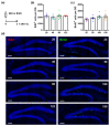
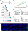
Tagging proliferating cells with thymidine analogs is an indispensable research tool; however, the issue of the potential in vivo cytotoxicity of these compounds remains unresolved. Here, we address these concerns by examining the effects of BrdU and EdU on adult hippocampal neurogenesis and EdU on the perinatal somatic development of mice. We show that, in a wide range of doses, EdU and BrdU label similar numbers of cells in the dentate gyrus shortly after administration. Furthermore, whereas the administration of EdU does not affect the division and survival of neural progenitor within 48 h after injection, it does affect cell survival, as evaluated 6 weeks later. We also show that a single injection of various doses of EdU on the first postnatal day does not lead to noticeable changes in a panel of morphometric criteria within the first week; however, higher doses of EdU adversely affect the subsequent somatic maturation and brain growth of the mouse pups. Our results indicate the potential caveats in labeling the replicating DNA using thymidine analogs and suggest guidelines for applying this approach.
Keywords: BrdU; DNA labeling; EdU; cell division; neural stem cell; neurogenesis; proliferation; thymidine analog.
Conflict of interest statement
The authors declare no conflict of interest. The funders had no role in the design of the study; in the collection, analyses, or interpretation of data; in the writing of the manuscript, or in the decision to publish the results.
Figures


Similar articles
-
Thymidine analog methods for studies of adult neurogenesis are not equally sensitive.J Comp Neurol. 2009 Nov 10;517(2):123-33. doi: 10.1002/cne.22107. J Comp Neurol. 2009. PMID: 19731267 Free PMC article.
-
Measurement of chemically induced cell proliferation in rodent liver and kidney: a comparison of 5-bromo-2'-deoxyuridine and [3H]thymidine administered by injection or osmotic pump.Carcinogenesis. 1990 Dec;11(12):2245-51. doi: 10.1093/carcin/11.12.2245. Carcinogenesis. 1990. PMID: 2265476
-
Evaluation of 5-ethynyl-2'-deoxyuridine staining as a sensitive and reliable method for studying cell proliferation in the adult nervous system.Brain Res. 2010 Mar 10;1319:21-32. doi: 10.1016/j.brainres.2009.12.092. Epub 2010 Jan 11. Brain Res. 2010. PMID: 20064490 Free PMC article.
-
BrdU immunohistochemistry for studying adult neurogenesis: paradigms, pitfalls, limitations, and validation.Brain Res Rev. 2007 Jan;53(1):198-214. doi: 10.1016/j.brainresrev.2006.08.002. Epub 2006 Oct 3. Brain Res Rev. 2007. PMID: 17020783 Review.
-
Thymidine analogues for tracking DNA synthesis.Molecules. 2011 Sep 15;16(9):7980-93. doi: 10.3390/molecules16097980. Molecules. 2011. PMID: 21921870 Free PMC article. Review.
Cited by
-
Neoblast-like stem cells of Fasciola hepatica.PLoS Pathog. 2024 May 28;20(5):e1011903. doi: 10.1371/journal.ppat.1011903. eCollection 2024 May. PLoS Pathog. 2024. PMID: 38805551 Free PMC article.
-
Detection of De Novo Dividing Stem Cells In Situ through Double Nucleotide Analogue Labeling.Cells. 2022 Dec 10;11(24):4001. doi: 10.3390/cells11244001. Cells. 2022. PMID: 36552766 Free PMC article.
-
Age-related decline in cognitive flexibility is associated with the levels of hippocampal neurogenesis.Front Neurosci. 2023 Aug 14;17:1232670. doi: 10.3389/fnins.2023.1232670. eCollection 2023. Front Neurosci. 2023. PMID: 37645372 Free PMC article.
-
Methods to Assess Proliferation of Stimulated Human Lymphocytes In Vitro: A Narrative Review.Cells. 2023 Jan 20;12(3):386. doi: 10.3390/cells12030386. Cells. 2023. PMID: 36766728 Free PMC article. Review.
-
Perfect duet: Dual recombinases improve genetic resolution.Cell Prolif. 2023 May;56(5):e13446. doi: 10.1111/cpr.13446. Epub 2023 Apr 14. Cell Prolif. 2023. PMID: 37060165 Free PMC article. Review.
References
Publication types
MeSH terms
Substances
Grants and funding
LinkOut - more resources
Full Text Sources

