

Eight years' advances on Bourbon virus, a tick-born Thogotovirus of the Orthomyxovirus family
- PMID: 35727718
- PMCID: PMC9206863
- DOI: 10.15212/zoonoses-2022-0012
Eight years' advances on Bourbon virus, a tick-born Thogotovirus of the Orthomyxovirus family
Abstract
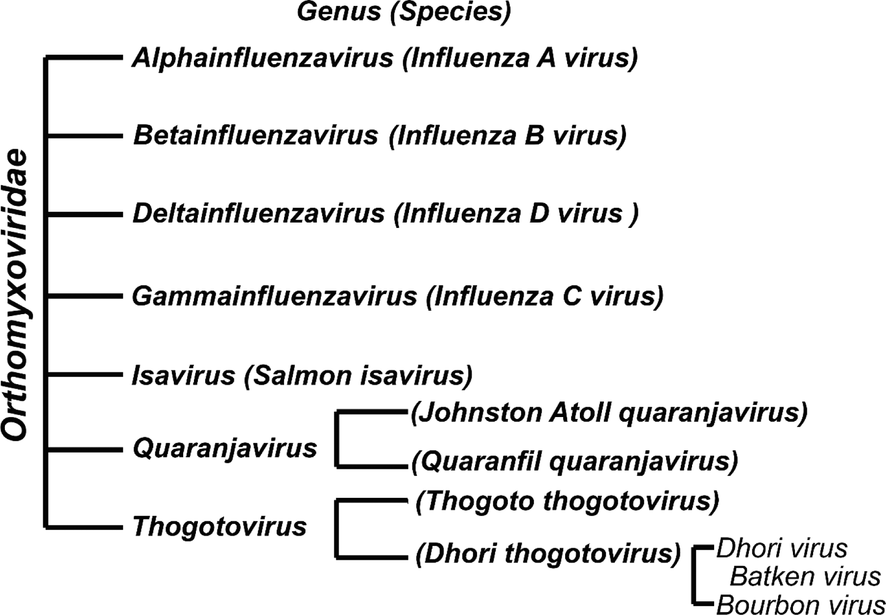
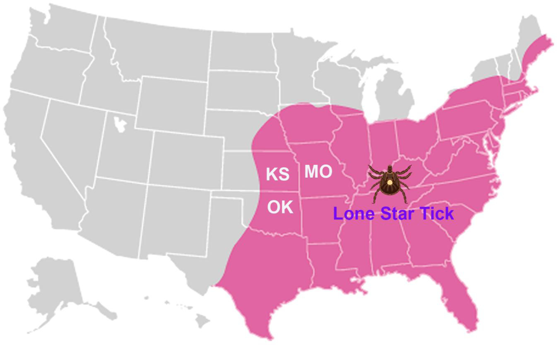
Bourbon virus (BRBV) was first isolated from a blood sample collected from a male patient living in Bourbon county, Kansas, during the spring of 2014. The patient later died due to complications associated with multiorgan failure. Currently, several BRBV infection-caused deaths have been reported in the United States, and misdiagnosed cases are often undercounted. BRBV is a member of the genus Thogotovirus of the Orthomyxoviridae family, and is transmitted through the Lone Star tick, Amblyomma Americanum, in North America. Currently, there are no specific antivirals or vaccinations available to treat or prevent BRBV infection. Several small molecular compounds have been identified to effectively inhibit BRBV infection of in vitro cell cultures at a single- or sub-micromolar level. Favipiravir, an RNA-dependent RNA polymerase inhibitor, prevented the death of Type I interferon receptor knockout mice infected with BRBV infection.
Keywords: Bourbon virus; Lone Star tick; antivirals; infection.
Conflict of interest statement
Conflicts of Interest The authors declared no conflicts of interest exist.
Figures
Similar articles
-
Bourbon virus, a newly discovered zoonotic thogotovirus.J Gen Virol. 2023 Aug;104(8). doi: 10.1099/jgv.0.001887. J Gen Virol. 2023. PMID: 37643129
-
Establishment of a Replicon Reporter of the Emerging Tick-Borne Bourbon Virus and Use It for Evaluation of Antivirals.Front Microbiol. 2020 Sep 8;11:572631. doi: 10.3389/fmicb.2020.572631. eCollection 2020. Front Microbiol. 2020. PMID: 33013808 Free PMC article.
-
Detection of Bourbon Virus-Specific Serum Neutralizing Antibodies in Human Serum in Missouri, USA.mSphere. 2022 Jun 29;7(3):e0016422. doi: 10.1128/msphere.00164-22. Epub 2022 May 24. mSphere. 2022. PMID: 35607948 Free PMC article.
-
Emerging tickborne viruses vectored by Amblyomma americanum (Ixodida: Ixodidae): Heartland and Bourbon viruses.J Med Entomol. 2023 Nov 14;60(6):1183-1196. doi: 10.1093/jme/tjad060. J Med Entomol. 2023. PMID: 37862097 Review.
-
The expanding spectrum of disease caused by the Lone Star Tick, Amblyomma americanum.Infez Med. 2021 Sep 10;29(3):378-385. doi: 10.53854/liim-2903-8. eCollection 2021. Infez Med. 2021. PMID: 35146342 Free PMC article. Review.
Cited by
-
Natural killer cells and their exosomes in viral infections and related therapeutic approaches: where are we?Cell Commun Signal. 2023 Sep 25;21(1):261. doi: 10.1186/s12964-023-01266-2. Cell Commun Signal. 2023. PMID: 37749597 Free PMC article. Review.
-
Diversification of Bourbon Virus in New York State.Microorganisms. 2023 Jun 15;11(6):1590. doi: 10.3390/microorganisms11061590. Microorganisms. 2023. PMID: 37375092 Free PMC article.
-
Widespread Circulation of Tick-Borne Viruses in Virginia-Evidence of Exposure to Heartland, Bourbon, and Powassan Viruses in Wildlife and Livestock.Microorganisms. 2024 Apr 30;12(5):899. doi: 10.3390/microorganisms12050899. Microorganisms. 2024. PMID: 38792729 Free PMC article.
-
Serosurveillance and the first detection of Bourbon virus RNA in a wildlife host.bioRxiv [Preprint]. 2024 Jun 4:2024.06.04.597417. doi: 10.1101/2024.06.04.597417. bioRxiv. 2024. PMID: 38895490 Free PMC article. Preprint.
References
-
- Kawaoka Y, Palese P (2006) Family Orthomyxoviridae. In: Fauquet CM, Mayo MA, Maniloff J, Desselberger U, Ball L, editors. Virus Taxonomy: Eighth Report of the International Committee on Taxonomy of Viruses. San Diego: Elsevier Academic. pp. 681–693.
-
- HAIG DA, WOODALL JP, DANSKIN D (1965) Thogoto virus: A hitherto underscribed agent isolated from tocks in kenya. J Gen Microbiol 38: 389–394. - PubMed
-
- Anderson CR, Casals J (1973) Dhori virus, a new agent isolated from Hyalomma dromedarii in India. Indian J Med Res 61: 1416–1420. - PubMed
-
- Butenko AM, Leshchinskaia EV, Semashko IV, Donets MA, Mart’ianova LI (1987) [Dhori virus--a causative agent of human disease. 5 cases of laboratory infection]. Vopr Virusol 32: 724–729. - PubMed
Grants and funding
LinkOut - more resources
Full Text Sources