Review
. 2022 Nov;23(11):732-749.
doi: 10.1038/s41580-022-00498-3. Epub 2022 Jun 20.

The Mediator complex as a master regulator of transcription by RNA polymerase II

Affiliations
Review

The Mediator complex as a master regulator of transcription by RNA polymerase II

William F Richter et al. Nat Rev Mol Cell Biol. 2022 Nov.

Abstract

The Mediator complex, which in humans is 1.4 MDa in size and includes 26 subunits, controls many aspects of RNA polymerase II (Pol II) function. Apart from its size, a defining feature of Mediator is its intrinsic disorder and conformational flexibility, which contributes to its ability to undergo phase separation and to interact with a myriad of regulatory factors. In this Review, we discuss Mediator structure and function, with emphasis on recent cryogenic electron microscopy data of the 4.0-MDa transcription preinitiation complex. We further discuss how Mediator and sequence-specific DNA-binding transcription factors enable enhancer-dependent regulation of Pol II function at distal gene promoters, through the formation of molecular condensates (or transcription hubs) and chromatin loops. Mediator regulation of Pol II reinitiation is also discussed, in the context of transcription bursting. We propose a working model for Mediator function that combines experimental results and theoretical considerations related to enhancer-promoter interactions, which reconciles contradictory data regarding whether enhancer-promoter communication is direct or indirect. We conclude with a discussion of Mediator's potential as a therapeutic target and of future research directions.


Conflict of interest statement

D.J.T. is a member of the scientific advisory board of Dewpoint Therapeutics. All the other authors declare no competing interests.

Figures

Fig. 1
Fig. 1. Structures of human Mediator and Mediator–PIC.
a | Structural features of Mediator in the tail-bent conformation. In this conformation (compared with the tail-extended conformation), MED23 and MED24 separate in combination with a shift in the MED16 propeller domain. The scaffold subunits MED17 and MED14 run through the head and middle modules and contact the tail. For the ‘top view’ shown on the right, structural disorder among Mediator subunits is indicated as semi-transparent ovals. The size of the oval correlates with the size of the disordered region unresolved by cryogenic electron microscopy. The location of MED6 is denoted with an asterisk. The middle module includes MED1, MED4, MED7, MED9, MED10, MED19, MED21, MED26 and MED31; the head module includes MED6, MED8, MED11, MED14, MED17, MED18, MED20, MED22, MED27, MED28 and MED30; the tail module includes MED15, MED16, MED23, MED24, MED25 and MED29. See also Supplementary Movie 1. b | Surface renderings of two views of the Mediator–preinitiation complex (PIC) structure. The Mediator complex is shown in the tail-bent conformation, suggesting it contains MED16 isoform 1 (ref.). Mediator sits atop the PIC, with the cyclin-dependent kinase (CDK)-activating kinase (CAK) module of transcription factor IIH (TFIIH) sandwiched between the Mediator hook and MED6. The CAK module is tethered to core TFIIH through its flexible MAT1 subunit. Consistent with its role in promoting phosphorylation of the RNA polymerase II (Pol II) caboxy-terminal domain (CTD), Mediator positions the CTD near the CAK, which contains CDK7. The TFIID subunit TATA box-binding protein (TBP) binds DNA upstream of the transcription start site. The TAF2 subunit of TFIID contacts the TFIIH subunits p52 and p8 (not shown) to help position the DNA translocase XPB, thereby promoting its interaction with downstream DNA, the Pol II jaw (not shown) and the Mediator hook domain. Finally, the Pol II stalk is an interaction hub, making contact with several Mediator subunits and with TFIIE, TFIIH and TFIIB in the PIC. See also Supplementary Movie 2.
Fig. 2
Fig. 2. Models for Mediator function at enhancers.
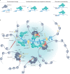
a | Potential mechanisms of reciprocal Mediator–transcription factor (TF) regulation: TFs can induce conformational changes in Mediator upon binding (left), which may influence Mediator function. Different molecules of a given TF could exchange with each other on DNA over time, especially if their local concentration is high, but this may not alter Mediator occupancy if it is bound by multiple TFs (middle). Also, intrinsically disordered activation domains of TFs may displace each other on Mediator, in a process called ‘competitive substitution’, which is favoured at high local concentrations (middle). Finally, TFs may transiently dissociate from DNA, but if Mediator-bound they would remain positioned to rebind DNA (right). b | Mediator function at a super-enhancer, which is shown at the centre, densely bound by TFs, Mediator complexes and other preinitiation complex (PIC) factors (not shown). This clustering of factors favours a high local concentration of intrinsically disordered regions (not shown), which are present in TFs, Mediator and other PIC components. Mutual weak attraction forces among intrinsically disordered regions will enforce a high local concentration of these factors (blue shading). Multiple Mediator complexes can bind the super-enhancer and can be oriented in all directions, to enable a single super-enhancer to activate multiple promoters at once if they are in spatial proximity. Enhancer RNAs (eRNAs), which are transcribed bidirectionally from the enhancers, may contribute to gene activation through the formation of additional multivalent weak interactions or through binding TFs or other regulatory factors. Over time, many promoters may colocalize with a super-enhancer, but activation will not occur unless a PIC can be fully assembled for activation. CAK, cyclin-dependent kinase-activating kinase module of transcription factor IIH; CTD, carboxy-terminal domain; H3K27ac, acetylated histone H3 Lys27; Pol II, RNA polymerase II; TBP, TATA box-binding protein; TFIIA, transcription factor IIA; TFIIB, transcription factor IIB; TFIID, transcription factor IID; TFIIE, transcription factor IIE; TFIIF, transcription factor IIF, TFIIH, transcription factor IIH.
Fig. 3
Fig. 3. A working model for Mediator function.
An enhancer–promoter interaction (loop) is shown on the left, within a larger topologically associating domain formed by CTCF and cohesin. Mediator is bound to one or more transcription factors (TFs) that occupy the enhancer, and the preinitiation complex (PIC) at the promoter is fully assembled and active. Such local architecture of enhancer–promoter chromatin looping could be further stabilized by Mediator-associated cohesin, but this association would be transient (dashed circle) relative to topologically associating domain boundaries (solid circle). Following a brief, direct enhancer–promoter interaction, the enhancer detaches from the promoter (for example, through dissociation of TFs from enhancer DNA); however, if one or more TFs remain bound to Mediator, the complex could remain in an active conformational state. This state could allow continued transcription reinitiation (bursting) from the PIC scaffold complex, provided RNA polymerase II (Pol II) and other PIC factors continue to associate for reinitiation (right). Ultimately, reinitiation may stop (not shown), because of TF–Mediator dissociation, binding of the kinase module to Mediator (which would block Mediator–Pol II interaction) or PIC disassembly. The light blue shading represents a hub or condensate that establishes a high local concentration of PIC components that promotes transcription initiation and bursting. TFIIH, transcription factor IIH.
Fig. 4
Fig. 4. Mediator as a therapeutic target.
a | Structure of the Mediator kinase module of Saccharomyces cerevisiae, in which a MED12 interaction with β-catenin in human cells is indicated. b | The human Mediator structure is shown, with a subset of identified transcription factor (TF)-binding sites highlighted. A potential strategy to manipulate TF function is to block specific TF–Mediator interactions, or to degrade a Mediator subunit targeted by a specific TF. Because different TFs bind Mediator at different sites, this strategy may allow gene activation by other TFs to occur normally. AR, androgen receptor; CDK8, cyclin-dependent kinase 8; ER, oestrogen receptor; ETS, E26 transformation-specific; GR, glucocorticoid receptor; SREBP1A, sterol regulatory element-binding protein 1A.

Similar articles

Cited by

References

    1. Schier AC, Taatjes DJ. Structure and mechanism of the RNA polymerase II transcription machinery. Genes Dev. 2020;34:465–488. doi: 10.1101/gad.335679.119. - DOI - PMC - PubMed
    1. Chen FX, Smith ER, Shilatifard A. Born to run: control of transcription elongation by RNA polymerase II. Nat. Rev. Mol. Cell Biol. 2018;19:464–478. doi: 10.1038/s41580-018-0010-5. - DOI - PubMed
    1. Cramer P. Organization and regulation of gene transcription. Nature. 2019;573:45–54. doi: 10.1038/s41586-019-1517-4. - DOI - PubMed
    1. Furlong EEM, Levine M. Developmental enhancers and chromosome topology. Science. 2018;361:1341–1345. doi: 10.1126/science.aau0320. - DOI - PMC - PubMed
    1. Hnisz D, Shrinivas K, Young RA, Chakraborty AK, Sharp PA. A phase separation model for transcriptional control. Cell. 2017;169:13–23. doi: 10.1016/j.cell.2017.02.007. - DOI - PMC - PubMed

Publication types

LinkOut - more resources