. 2022 Jun 16;18(6):e1010207.
doi: 10.1371/journal.ppat.1010207. eCollection 2022 Jun.

p166 links membrane and intramitochondrial modules of the trypanosomal tripartite attachment complex

Affiliations

p166 links membrane and intramitochondrial modules of the trypanosomal tripartite attachment complex

Bernd Schimanski et al. PLoS Pathog. .

Abstract

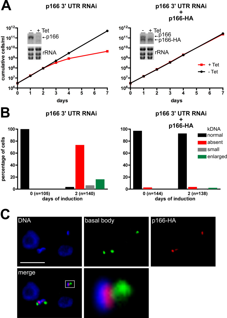
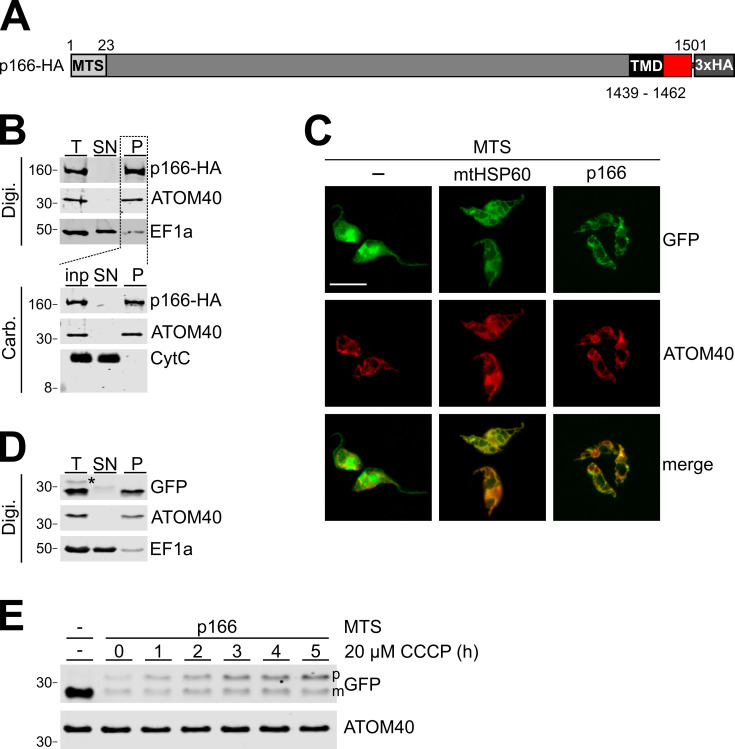
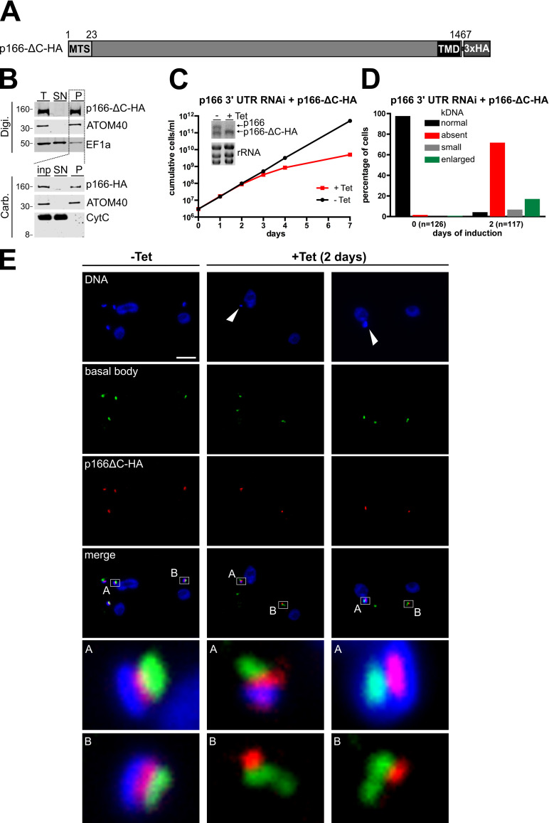
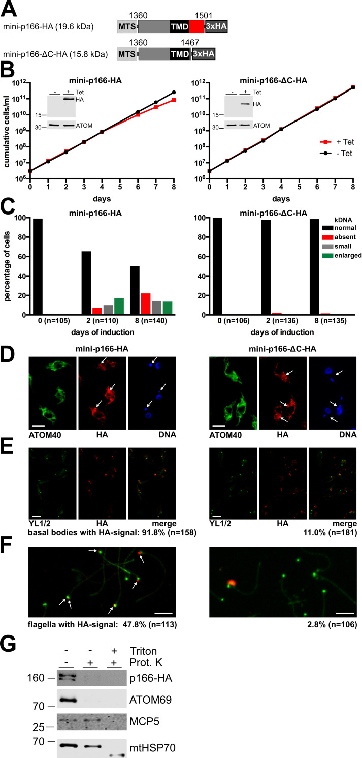
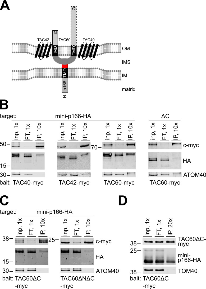
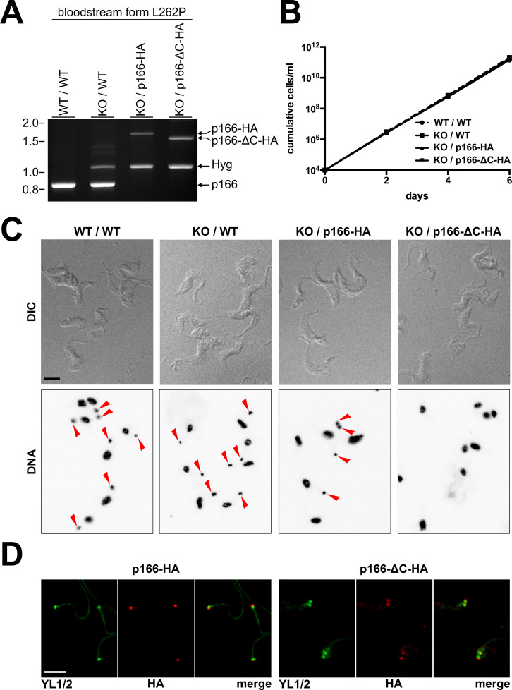
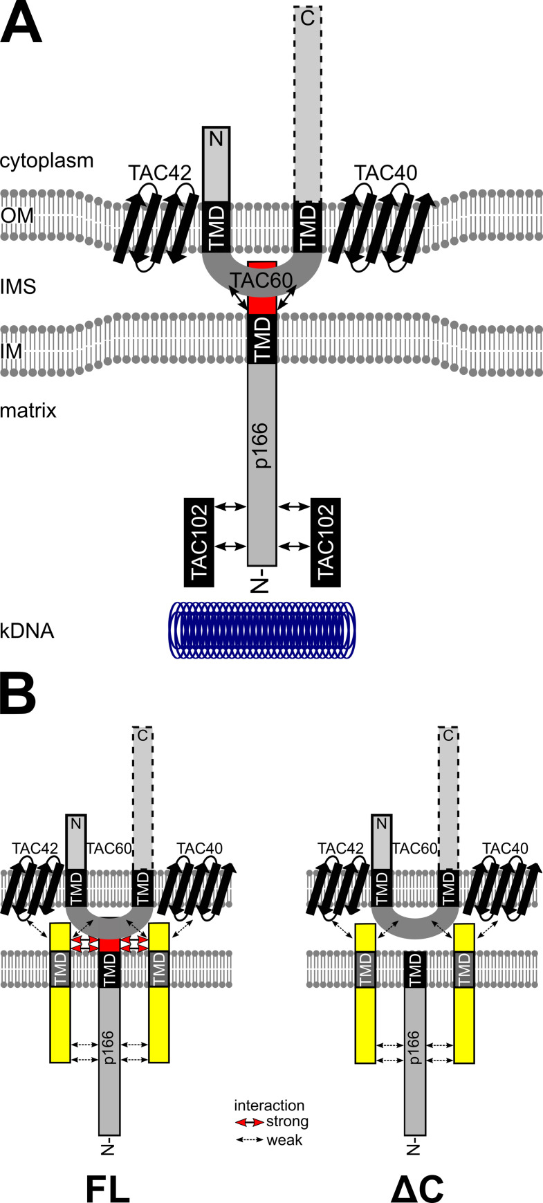
The protist parasite Trypanosoma brucei has a single mitochondrion with a single unit genome termed kinetoplast DNA (kDNA). Faithfull segregation of replicated kDNA is ensured by a complicated structure termed tripartite attachment complex (TAC). The TAC physically links the basal body of the flagellum with the kDNA spanning the two mitochondrial membranes. Here, we characterized p166 as the only known TAC subunit that is anchored in the inner membrane. Its C-terminal transmembrane domain separates the protein into a large N-terminal region that interacts with the kDNA-localized TAC102 and a 34 aa C-tail that binds to the intermembrane space-exposed loop of the integral outer membrane protein TAC60. Whereas the outer membrane region requires four essential subunits for proper TAC function, the inner membrane integral p166, via its interaction with TAC60 and TAC102, would theoretically suffice to bridge the distance between the OM and the kDNA. Surprisingly, non-functional p166 lacking the C-terminal 34 aa still localizes to the TAC region. This suggests the existence of additional TAC-associated proteins which loosely bind to non-functional p166 lacking the C-terminal 34 aa and keep it at the TAC. However, binding of full length p166 to these TAC-associated proteins alone would not be sufficient to withstand the mechanical load imposed by the segregating basal bodies.


Conflict of interest statement

The authors have declared that no competing interests exist.

Figures

Fig 1
Fig 1. In situ tagged p166-HA is functional.
(A) Growth analysis of cells induced for RNAi of p166 in absence (left) or presence (right) of a RNAi resistant allele encoding in situ tagged p166-HA. In both growth curves inserts showing a northern blot analysis are included to demonstrate effects of tetracyclin (Tet) induced RNAi against the indicated mRNAs after 2 days of induction. Ethidiumbromide stained ribosomal RNA (rRNA) bands serve as control for equal loading. (B) DNA of uninduced (left) and RNAi induced cells (right) was stained with DAPI and kDNA content and phenotype were analyzed by fluorescence microscopy in the indicated number of individual cells. (C) Immunofluorescene microscopy with whole cells. DNA was stained with DAPI, the basal body with anti YL1/2 and tagged p166 was detected with anti-HA. The enlarged part of the merged picture is indicated by a white rectangle. The scale bar shows 5 μm.
Fig 2
Fig 2. p166-HA is an integral membrane protein of the IM.
(A) Schematic depiction (not to scale) of in situ tagged p166. A predicted mitochondrial targeting sequence (MTS) and transmembrane domain (TMD) are indicated. Numbers represent the position of amino acid residues. (B) Biochemical fractionation of cell extracts. For digitonin (Digi.) extraction same cell equivalents of total cells (T), supernatant (SN) and pellet (P) were separated by SDS-PAGE and p166-HA, the mitochondrial outer membrane protein ATOM40 and the cytoplasmic marker EF1a were detected by immunoblot. The pellet served further as input (inp.) for alkaline carbonate (Carb.) extraction and was separated into supernatant and a final pellet. On immunoblots p166-HA, ATOM40 and the soluble protein cytochrome C (CytC) were detected. Numbers on the left indicate sizes of molecular weight marker bands in kDa. (C) Fluorescence microscopy analysis of tetracycline inducible cells expressing variants of GFP with indicated mitochondrial targeting sequences (MTS) fused to the N-terminus. Staining for ATOM40 serves as marker for mitochondria. (D) Digitonin extraction as in (B) of the cell line expressing GFP with the p166 MTS. Asterisk indicates the putative unprocessed precursor protein. (E) The cell line expressing GFP with the p166 MTS was induced for 2 hours and subsequently treated with the uncoupler CCCP as indicated. Subsequently total cellular extract was analyzed by SDS-PAGE. Left most lane shows GFP alone. Position of precursor p166-MTS-GFP (p) and mature GFP are indicated (m). ATOM40 serves as a loading control.
Fig 3
Fig 3. The C-tail of p166 is essential for function.
(A) Schematic depiction (not to scale) of in situ tagged p166 lacking the 34 amino acid C-tail (p166-ΔC-HA). (B) Biochemical fractionation of cell extracts as described in legend 2B. (C) Growth analysis of cells uninduced (-Tet) or induced (+Tet) for RNAi against wild-type p166 with constitutive expression of p166-ΔC-HA as demonstrated by northern blot analysis. (D). kDNA content of cells was analyzed as described in legend 1B. (E) Whole cells left uninduced (-Tet) or being induced for 2 days (+ Tet) were analyzed by immunofluorescence microscopy for DNA (stained with DAPI), the localization of basal bodies (using anti YL1/2) and p166-ΔC-HA. Enlarged merged pictures are shown and indicated. White arrowheads point at small kDNA (middle panel) and enlarged overreplicated kDNA (right panel). Scale bar, 5 μm.
Fig 4
Fig 4. The C-tail of p166 is located in the intermembrane space.
(A) Depiction of inducibly expressed N-terminal truncated p166 with (mini-p166-HA) or without the C-tail (mini-p166-ΔC-HA) and their calculated molecular weights. Numbers indicate amino acid residue positions based on wild-type p166. (B and C) Growth analysis of untreated cells (-Tet) and cells induced for expression (+Tet) of the indicated proteins and subsequent analysis of kDNA content. Inducible induction is shown by immunoblots probed for HA and ATOM40 as control for equal loading. (D) Immunofluorescence microscopy of cells induced for expression of the N-terminal truncated mini-p166-HA or mini-p166-ΔC-HA. anti-ATOM40 stains the mitochondrial membrane, DNA is visualized using DAPI. The arrows indicate foci with the highest signal strength derived from anti-HA. (E) Analysis of cytoskeletons for localization of basal bodies stained with anti-YL1/2 and HA-tagged mini-versions of p166. (F) Basal bodies (green) and in situ tagged truncated versions of p166-HA (red) were visualized by immunofluorescence microscopy in isolated flagella. Colocalization of both signals was analyzed in the indicated numbers of cells. Scale bar in D, E and F, 5 μm. (G) Mitoplasts were prepared from the cell line expressing in situ tagged full length p166 (p166-HA) using 0.05% of digitonin. Aliquots of the resulting pellet fraction were treated with proteinase K (10 μg/ml) and 0.5% (w/v) of Triton-X-100 as indicated. The atypical translocase of the outer membrane 69 (ATOM69), the IM protein mitochondrial carrier protein 5 (MCP5) and the matrix protein mitochondrial heat shock protein 70 (mtHsp70) served as controls.
Fig 5
Fig 5. The C-tail of p166 interacts with TAC components of the outer mitochondrial membrane.
(A) Model of possible interactions. TAC60 interacts with the beta barrel proteins TAC40 and TAC42 in the outer mitochondrial membrane (OM). It contains a region between two transmembrane domains (TMD) that is located in the intermembrane space (IMS) just like the C-tail (red) of inner mitochondrial membrane (IM) anchored p166. Both the essential N-terminal domain (solid box) and the non-essential C-terminal domain (dotted box) are facing the cytoplasm. (B and C) Immunoprecipitation of indicated c-myc tagged TAC components expressed in T. brucei and analysis of copurification of N-terminal truncated tagged versions of p166 by immunoblot and detection of the indicated antigens. ATOM40 serves as a control for specificity (D) Immunoprecipitation of indicated proteins expressed in yeast. TAC60ΔC-myc serves as bait and TOM40 as control for specificity. Inp, input; FT, flow through; IP, immunoprecipitate. 1x, 10x, 20x indicate the relative cell equivalents loaded on the gels. Position of molecular weight marker bands with sizes in kDa are indicated.
Fig 6
Fig 6. C-terminal truncated p166 localizes correctly in the L262P bloodstream form cell line.
(A) Ethidiumbromide stained agarose gel after gelelectrophoresis of specific PCR products to analyze the p166 alleles of the indicated L262P bloodstream form cell line variants. Sizes of DNA molecular weight marker bands in kb are indicated on the left. (B) Growth curve analysis of cell lines harbouring indicated alleles. (C) Fluorescence microscopic analysis of the indicated L262P bloodstream form cell lines. Whole cells are visualized using DIC and nuclei and kDNA are stained with DAPI. Red arrowheads indicate the presence of kDNA. (D) Anti-HA immunofluorescene microscopy on isolated cytoskeletons to visualize the localization of full length (p166-HA) or C-terminal truncated p166 (p166-ΔC-HA) in the KO / p166-HA and the KO / p166-ΔC-HA L262P bloodstream form cell lines. For comparison the basal bodies are stained with anti-YL1/2. Scale bars in C and D, 5 μm.
Fig 7
Fig 7. p166 provides a bridge between OM and matrix located TAC components.
(A) Model of interactions of selected TAC components. The beta barrel proteins TAC40 and TAC42 form a stable subcomplex together with TAC60 in the outer membrane (OM). The C-terminal cytoplasmic domain of TAC60 is not essential for function (indicated by a dotted box) whereas the essential N-terminus (solid box) most likely provides the connection to cytoplasmic TAC subunits more proximal to the basal body. p166 is anchored in the inner membrane (IM) with a C-terminal transmembrane domain (TMD) exposing the short C-tail (red box) into the intermembrane space (IMS) where it interacts stably with the IMS exposed loop of TAC60. TAC102 interacts stably with the N-terminal part of p166 in the mitochondrial matrix and provides the connection to the kDNA disc. Protein-protein interactions are indicated by arrows. (B) Model of our working hypothesis on how non-functional C-terminally truncated p166 can be localized at the TAC. Full length (FL) p166 interacts via its intact C-tail (red box) with TAC60. So far unknown membrane anchored accessory factors (yellow) help to keep p166 in place. This is facilitated via strong interactions with the C-tail and weaker interactions with the N-terminus and OM subunits. p166 lacking the C-tail (ΔC) can still be locked in place by the aforementioned weak interactions. However, the lack of the C-tail disables a direct strong interaction with TAC60 and leads to a loss of kDNA in daughter cells due to insufficient mechanical stability of the mutant TAC.

Similar articles

Cited by

References

    1. Friedman JR, Nunnari J. Mitochondrial form and function. Nature. 2014;505(7483):335–43. doi: 10.1038/nature12985 ; PubMed Central PMCID: PMC4075653. - DOI - PMC - PubMed
    1. Pfanner N, Warscheid B, Wiedemann N. Mitochondrial proteins: from biogenesis to functional networks. Nat Rev Mol Cell Biol. 2019;20(5):267–84. Epub 2019/01/11. doi: 10.1038/s41580-018-0092-0 . - DOI - PMC - PubMed
    1. Hansen KG, Herrmann JM. Transport of Proteins into Mitochondria. Protein J. 2019;38(3):330–42. Epub 2019/03/15. doi: 10.1007/s10930-019-09819-6 . - DOI - PubMed
    1. Gustafsson CM, Falkenberg M, Larsson NG. Maintenance and Expression of Mammalian Mitochondrial DNA. Annu Rev Biochem. 2016;85:133–60. doi: 10.1146/annurev-biochem-060815-014402 . - DOI - PubMed
    1. Westermann B. Mitochondrial inheritance in yeast. Biochim Biophys Acta. 2013. Epub 2013/11/05. S0005-2728(13)00175-8 [pii] doi: 10.1016/j.bbabio.2013.10.005 . - DOI - PubMed

Publication types

MeSH terms

Grants and funding

This work was supported by the following grants: NCCR RNA & Disease, a National Centre of Competence in Research, supported by the Swiss National Science Foundation, grant number 182880, to AS Schweizerischer Nationalfonds zur Förderung der Wissenschaftlichen Forschung, grant number 175563, to AS Deutsche Forschungsgemeinschaft, grant number 390939984, to FNV SFB1381, grant number 403222702, to FNV Emmy-Noether Programm, to FNV Deutsche Forschungsgemeinschaft, grant number, 403222702/SFB 1381, to BW The funders had no role in study design, data collection and analysis, decision to publish, or preparation of the manuscript.