. 2022 Aug 30;13(4):e0000722.
doi: 10.1128/mbio.00007-22. Epub 2022 Jun 16.

Single-Cell Imaging Shows That the Transcriptional State of the HIV-1 Provirus and Its Reactivation Potential Depend on the Integration Site

Affiliations

Single-Cell Imaging Shows That the Transcriptional State of the HIV-1 Provirus and Its Reactivation Potential Depend on the Integration Site

Julie Janssens et al. mBio. .

Abstract

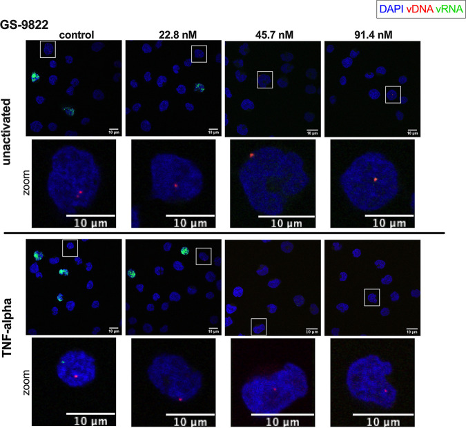
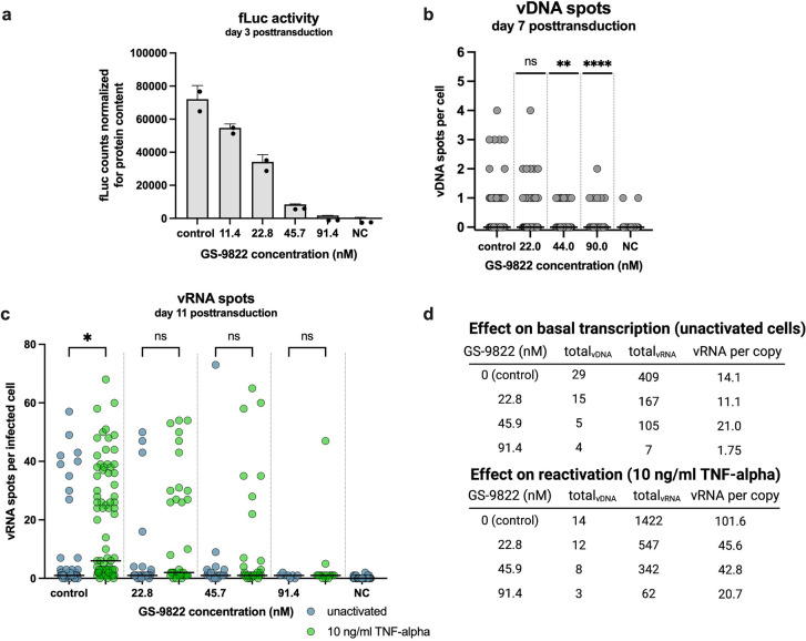
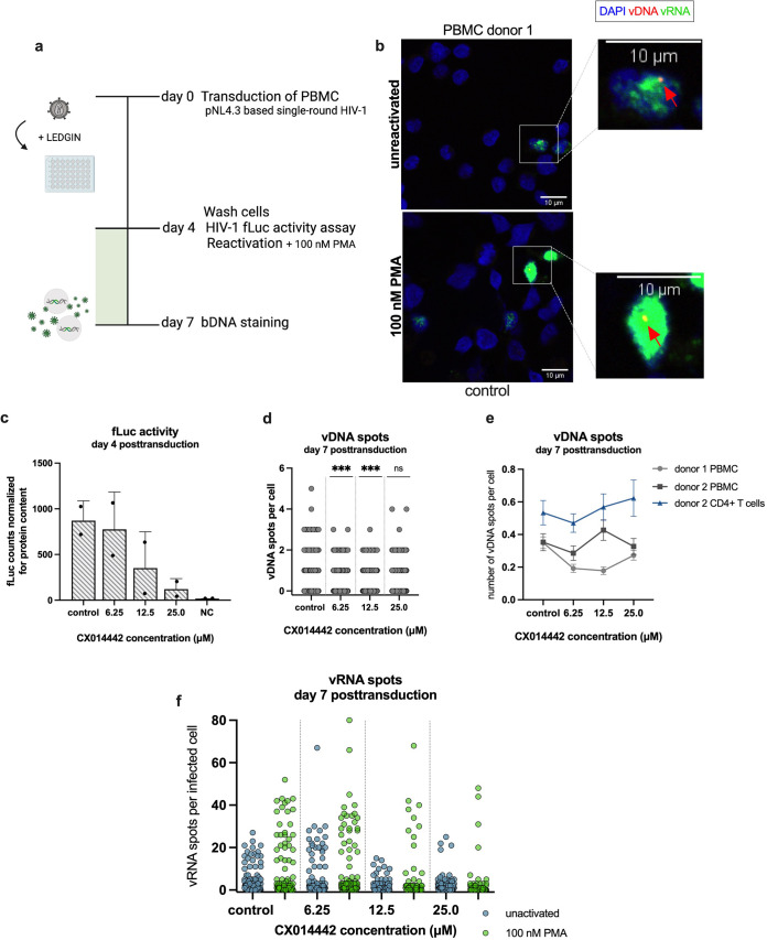
Current antiretroviral treatment fails to cure HIV-1 infection since latent provirus resides in long-lived cellular reservoirs, rebounding whenever therapy is discontinued. The molecular mechanisms underlying HIV-1 latency are complex where the possible link between integration and transcription is poorly understood. HIV-1 integration is targeted toward active chromatin by the direct interaction with a host protein, lens epithelium-derived growth factor (LEDGF/p75). LEDGINs are small-molecule inhibitors of the LEDGF/p75-integrase (IN) interaction that effectively inhibit and retarget HIV-1 integration out of preferred integration sites, resulting in residual provirus that is more latent. Here, we describe a single-cell branched DNA imaging method for simultaneous detection of viral DNA and RNA. We investigated how treatment with LEDGINs affects the location, transcription, and reactivation of HIV-1 in both cell lines and primary cells. This approach demonstrated that LEDGIN-mediated retargeting hampered the baseline transcriptional state and the transcriptional reactivation of the provirus, evidenced by the reduction in viral RNA expression per residual copy. Moreover, treatment of primary cells with LEDGINs induced an enrichment of provirus in deep latency. These results corroborate the impact of integration site selection for the HIV-1 transcriptional state and support block-and-lock functional cure strategies in which the latent reservoir is permanently silenced after retargeting. IMPORTANCE A longstanding question exists on the impact of the HIV-1 integration site on viral gene expression. This unsolved question has significant implications for the search toward an HIV-1 cure, as eradication strategies set up to reactivate and eliminate HIV-1 depend on the site where the provirus is integrated. The main determinant for integration site selection is the interaction of the HIV-1 integrase (IN) and the host chromatin targeting factor, LEDGF/p75. LEDGINs are small-molecule inhibitors of the LEDGF/p75-IN interaction that inhibit and retarget HIV-1 integration out of preferred integration sites. Using both LEDGINs and branched DNA (bDNA) imaging, we now investigated, in much detail, the impact of integration site selection on the three-dimensional location of the provirus, HIV-1 transcription, and reactivation. Our results provide evidence for a "block-and-lock" functional cure strategy that aims to permanently silence HIV-1 by LEDGIN-mediated retargeting to sites that are less susceptible to reactivation after treatment interruption.

Keywords: HIV-1; LEDGF/p75; LEDGINs; integration site selection; single-cell HIV-1 imaging.


Conflict of interest statement

The authors declare no conflict of interest.

Figures

FIG 1
FIG 1
Branched DNA imaging to study HIV-1 latency and reactivation. (a, b) Compound structures of CX014442 (a) and GS-9822 (b). (c) After cell fixation and permeabilization, hybridization with target-specific Z probes allows fluorescence imaging of vDNA (red, as pointed by red arrow) and vRNA (green) with confocal microscopy, allowing us to differentiate between uninfected (vDNA vRNA), actively infected (vDNA+ vRNA+), and latently infected cells (vDNA+ vRNA). Nuclei are visualized with DAPI (blue). Scale bar represents 10 μm. (d) Timeline of the transduction and reactivation experiments. SupT1 cells were transduced with HIV-1 fLuc in the presence of LEDGIN (CX014442; GS-9822) or RAL. Three days posttransduction, the virus and compounds were washed away, and fLuc reporter expression was measured. Ten days posttransduction, the cells were reactivated with 10 ng/mL TNF-α. Twenty-four hours after reactivation, cells were fixed prior to bDNA staining and imaging. (e, f) SupT1 cells were transduced with single-round HIV-1 fLuc in the presence of CX014442 (e) or RAL (f). fLuc activity was measured 3 days posttransduction and normalized for the total protein content. The normalized fLuc activity is shown as mean ± standard deviation from one representative experiment (n = 3), NC, nontransduced negative control.
FIG 2
FIG 2
bDNA imaging of LEDGIN CX014442-treated cells. SupT1 cells were transduced with single-round HIV-1 and reactivated 10 days posttransduction with 10 ng/mL TNF-α. Twenty-four hours after reactivation, the cells were fixed to visualize vDNA (red) and vRNA (green) with bDNA imaging in cells treated with 0 to 50 μM LEDGIN CX014442. Unactivated cells (top) and TNF-α-reactivated cells (bottom) are shown. For better visualization of the vDNA spots, a zoomed image of the cell highlighted by the square box is shown for each condition. Nuclei are stained with DAPI (blue), and scale bars represent 10 μm.
FIG 3
FIG 3
bDNA imaging of RAL-treated cells. SupT1 cells were transduced with single-round HIV-1 and reactivated 10 days posttransduction with 10 ng/mL TNF-α. Twenty-four hours after reactivation, the cells were fixed to visualize vDNA (red) and vRNA (green) with bDNA imaging in cells treated with 0 to 62.3 nM RAL. Unactivated cells (top) and TNF-α-reactivated cells (bottom) are shown. For better visualization of the vDNA spots, a zoomed image of the cell highlighted by the square box is shown for each condition. Nuclei are stained with DAPI (blue), and scale bars represent 10 μm.
FIG 4
FIG 4
LEDGIN CX014442, but not RAL, reduces HIV-1 transcription and reactivation. (a, b) The number of vDNA spots per cell, measured with bDNA imaging 11 days posttransduction after treatment with CX014442 (a) or RAL (b) in SupT1 cells. Pooled data from the unactivated and reactivated cells are shown. The numbers of cells imaged and analyzed per condition are shown in Table S2b and c in the supplemental material. A Kruskal-Wallis test was used to test for statistical significance compared to the control condition: **, P < 0.01; ***, P < 0.001; ****, P < 0.0001. NC, nontransduced negative control. (c, d) The numbers of vRNA spots per infected cell are shown for the unactivated and reactivated cells, measured with bDNA imaging 11 days posttransduction in the presence of CX014442 (c) and RAL (d). The numbers of cells imaged and analyzed per condition are shown in Table S2b and c. The median number of vRNA spots per cell is indicated with a horizontal line. A Kruskal-Wallis test was used to test for statistical significance between the unactivated and reactivated cells of each condition: *, P < 0.05; ***, P < 0.001; ****, P < 0.0001; and ns, nonsignificant. NC, nontransduced negative control (e) Viral RNA expression per residual copy was calculated in the presence of increasing concentrations of CX014442 or RAL by dividing the number of vRNA spots by the number of vDNA spots for the unreactivated cells (top) and reactivated cells (bottom).
FIG 5
FIG 5
bDNA imaging of GS-9822-treated cells. SupT1 cells were transduced with single-round HIV-1 and reactivated 10 days posttransduction with 10 ng/mL TNF-α. Twenty-four hours after reactivation, the cells were fixed to visualize vDNA (red) and vRNA (green) with bDNA imaging in cells treated with increasing concentrations of GS-9822. Unactivated cells (top) and TNF-α-reactivated cells (bottom) are shown. For better visualization of the vDNA spots, a zoomed image of the cell highlighted by the square box is shown for each condition. Nuclei are stained with DAPI (blue), and scale bars represent 10 μm.
FIG 6
FIG 6
LEDGIN GS-9822 reduces HIV-1 transcription and reactivation at nanomolar concentrations. (a) SupT1 cells were transduced with single-round HIV-1 expressing firefly luciferase (fLuc) in the presence of GS-9822. The fLuc activity was measured 3 days posttransduction and normalized for the total protein content. The normalized fLuc activity is shown as mean ± standard deviation from one representative experiment (n = 2), performed in duplicate. NC, nontransduced negative control. (b) The numbers of vDNA spots per cell, measured with bDNA imaging 11 days posttransduction, are shown in the presence of GS-9822. Pooled data from the unactivated and reactivated cells are shown. The numbers of cells that were imaged and analyzed per condition are shown in Table S2d in the supplemental material. A Kruskal-Wallis test was used to test for statistically significant differences compared to the control condition: **, P < 0.01, and ****, P < 0.0001. NC, nontransduced negative control. (c) The numbers of vRNA spots per infected cell are shown for the unactivated and reactivated cells, measured with bDNA imaging 11 days posttransduction in the presence of LEDGIN GS-9822. The median number of vRNA spots per cell is indicated with a horizontal line. A Kruskal-Wallis test was used to test for statistically significant differences between the unactivated and reactivated cells of each condition: *, P < 0.05; ns, nonsignificant. NC, nontransduced negative control. The numbers of cells that were imaged and analyzed per condition are shown in Table S2d. (d) Viral RNA expression per residual copy was calculated in the presence of increasing concentrations of GS-9822 by dividing the number of vRNA spots by the number of vDNA spots for the unreactivated cells (top) and reactivated cells (bottom).
FIG 7
FIG 7
LEDGIN CX014442 treatment in primary cells hampers HIV-1 transcription and reactivation from latency. (a) Peripheral blood mononuclear cells (PBMCs, donor 1) were transduced with single-round HIV-1 expressing firefly luciferase (fLuc) in the presence of LEDGIN CX014442. On day 4 posttransduction, half of the cells were activated with 100 nM PMA, and samples were harvested for bDNA imaging 7 days posttransduction. (b) PBMCs were transduced with single-round HIV-1 and reactivated 4 days posttransduction with 100 nM PMA. On day 7, 72 h after reactivation, the cells were fixed to visualize vDNA (red, as pointed by red arrow) and vRNA (green) with bDNA imaging. Representative images are shown for the control condition. Nuclei are stained with DAPI (blue), and scale bars represent 10 μm. (c) fLuc activity was measured 4 days posttransduction and normalized for the total protein content. The normalized fLuc activity is shown as mean ± standard deviation from one representative experiment (n = 3), performed in duplicate. (d) The number of vDNA spots per cell, measured with bDNA imaging 7 days posttransduction, is shown in the presence of CX014442. Pooled data from the unactivated and reactivated cells are shown. The numbers of cells that were imaged and analyzed per condition are shown in Table S2e in the supplemental material. A Kruskal-Wallis test was used to test for statistically significant differences compared to the control condition: ***, P < 0.001. (e) The average number of vDNA spots per cell is shown as mean ± SEM. (f) The numbers of vRNA spots per infected cell are shown for the unactivated and reactivated cells, measured with bDNA imaging 7 days posttransduction in the presence of CX014442. The numbers of cells that were imaged and analyzed per condition are shown in Table S2e.

Similar articles

Cited by

References

    1. Symons J, Cameron PU, Lewin SR. 2018. HIV integration sites and implications for maintenance of the reservoir. Curr Opin HIV AIDS 13:152–159. doi:10.1097/COH.0000000000000438. - DOI - PMC - PubMed
    1. Jordan A, Defechereux P, Verdin E. 2001. The site of HIV-1 integration in the human genome determines basal transcriptional activity and response to Tat transactivation. EMBO J 20:1726–1738. doi:10.1093/emboj/20.7.1726. - DOI - PMC - PubMed
    1. Lewinski MK, Bisgrove D, Shinn P, Chen H, Hoffmann C, Hannenhalli S, Verdin E, Berry CC, Ecker JR, Bushman FD. 2005. Genome-wide analysis of chromosomal features repressing human immunodeficiency transcription. J Virol 79:6610–6619. doi:10.1128/JVI.79.11.6610-6619.2005. - DOI - PMC - PubMed
    1. Schröder ARW, Shinn P, Chen H, Berry C, Ecker JR, Bushman F. 2002. HIV-1 integration in the human genome favors active genes and local hotspots. Cell 110:521–529. doi:10.1016/S0092-8674(02)00864-4. - DOI - PubMed
    1. Marini B, Kertesz-Farkas A, Ali H, Lucic B, Lisek K, Manganaro L, Pongor S, Luzzati R, Recchia A, Mavilio F, Giacca M, Lusic M. 2015. Nuclear architecture dictates HIV-1 integration site selection. Nature 521:227–231. doi:10.1038/nature14226. - DOI - PubMed

Publication types