. 2022 Nov;32(6):e13097.
doi: 10.1111/bpa.13097. Epub 2022 Jun 13.

Evidence of beta amyloid independent small vessel disease in familial Alzheimer's disease

Affiliations

Evidence of beta amyloid independent small vessel disease in familial Alzheimer's disease

Jessica Lisa Littau et al. Brain Pathol. 2022 Nov.

Abstract

We studied small vessel disease (SVD) pathology in Familial Alzheimer's disease (FAD) subjects carrying the presenilin 1 (PSEN1) p.Glu280Ala mutation in comparison to those with sporadic Alzheimer's disease (SAD) as a positive control for Alzheimer's pathology and Cerebral Autosomal Dominant Arteriopathy with Subcortical Infarcts and Leukoencephalopathy (CADASIL) bearing different NOTCH3 mutations, as positive controls for SVD pathology. Upon magnetic resonance imaging (MRI) in life, some FAD showed mild white matter hyperintensities and no further radiologic evidence of SVD. In post-mortem studies, total SVD pathology in cortical areas and basal ganglia was similar in PSEN1 FAD and CADASIL subjects, except for the feature of arteriosclerosis which was higher in CADASIL subjects than in PSEN1 FAD subjects. Further only a few SAD subjects showed a similar degree of SVD pathology as observed in CADASIL. Furthermore, we found significantly enlarged perivascular spaces in vessels devoid of cerebral amyloid angiopathy in FAD compared with SAD and CADASIL subjects. As expected, there was greater fibrinogen-positive perivascular reactivity in CADASIL but similar reactivity in PSEN1 FAD and SAD groups. Fibrinogen immunoreactivity correlated with onset age in the PSEN1 FAD cases, suggesting increased vascular permeability may contribute to cognitive decline. Additionally, we found reduced perivascular expression of PDGFRβ AQP4 in microvessels with enlarged PVS in PSEN1 FAD cases. We demonstrate that there is Aβ-independent SVD pathology in PSEN1 FAD, that was marginally lower than that in CADASIL subjects although not evident by MRI. These observations suggest presence of covert SVD even in PSEN1, contributing to disease progression. As is the case in SAD, these consequences may be preventable by early recognition and actively controlling vascular disease risk, even in familial forms of dementia.

Keywords: Alzheimer's disease; FAD; cerebral autosomal dominant arteriopathy with subcortical infarcts and leukoencephalopathy; dementia; presenilin; small vessel disease; vascular disease.


Conflict of interest statement

The authors declare no conflict of interest.

Figures
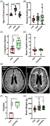
FIGURE 1
FIGURE 1
Characterisation of PSEN1 p.Glu280Ala FAD and CADASIL. An overview of demographical, clinical and imaging data for the Colombian and Newcastle cohort. The FAD and CADASIL cases presented with a significantly lower age of onset in comparison with SAD cases (A). There were no significant differences in disease duration (B) while the rate of cognitive decline was significantly faster in PSEN1 FAD than in CADASIL cases (C). The speed of cognitive decline showed no significant differences (D). Representative MRI images are shown for PSEN1 FAD and CADASIL patients (E). PSEN1 FAD patients presented with significantly lower Fazekas scores compared with CADASIL (F). The brain weight was significantly lower for PSEN1 FAD compared with SAD and CADASIL cases (G).
FIGURE 2
FIGURE 2
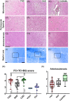
SVD pathology in PSEN1 p.Glu280Ala FAD, SAD and CADASIL. Representative images of vascular pathology are given for PSEN1 FAD, SAD and CADASIL (A–L), scale bar for all panels = 250 μm. The vascular features such as microinfarcts (A–C), perivascular spacing (D–F), arteriosclerosis (G–I) are shown in H&E staining and myelin loss (J–L) is shown in Luxol Fast Blue staining, all features shown in occipital cortex. The average score for frontal (FC) and temporal (TC) cortices and BG (L) was calculated to compare PSEN1 FAD cases with the SAD cohort (Colombian, SAD, n = 10, Newcastle, N‐SAD, n = 17) and different CADASIL mutations (Colombian CADASIL, n = 10, Newcastle CADASIL, n = 12), together with younger (Y‐Ctrl, n = 10) and older healthy (O‐Ctrl, n = 15) controls. Even though PSEN1 FAD tends to show wider variability of vascular pathology, average values are not significantly different to those from pure vascular dementia such as CADASIL. All groups show significant more vascular pathologies than both control groups (p values: **** ≤ 0.0001, *** ≤ 0.001, ** ≤ 0.01, * ≤ 0.05). SAD cases presented with significantly less vascular pathology than the CADASIL cases (p value: ** < 0.01) (M). The arteriosclerosis scores of the evaluated cortices are shown for PSEN1 FAD, SAD and CADASIL (N).
FIGURE 3
FIGURE 3
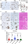
Perivascular spacing in PSEN1 p.Glu280Ala FAD, SAD and CADASIL. Representative images of Aβ pathology are shown for PSEN1 FAD and SAD (A). There are no significant differences in Aβ CAA pathology based on CAA VCING score (B). CAA VCING scores were similar for all vessel types in both groups. Representative images of perivascular spacing for Non CAA and CAA vessels are shown for PSEN1 FAD (n = 21), SAD (n = 10) and CADASIL (n = 10) (C), scale bar all panels = 100 μm. The perivascular space in relation to the diameter of the vessel was calculated to compare perivascular spacing in PSEN1 FAD, SAD and CADASIL. Non CAA FAD vessels showed significant enlargement of the perivascular space compared with all other vessel types measured (D) (p value: **** ≤ 0.0001).
FIGURE 4
FIGURE 4
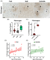
Role of the blood–brain‐barrier in PVS in PSEN1 p.Glu280Ala FAD, SAD and CADASIL. Representative images of leaking (full arrow‐head) and non‐leaking (empty arrow‐head) vessels in PSEN1 FAD (n = 21), SAD (n = 10) and CADASIL (n = 10) are shown (A). PSEN1 FAD and SAD cases presented with significantly less leaking vessels than CADASIL cases (B) (p values: **** ≤ 0.0001, *** ≤ 0.001). Further in PSEN1 FAD significantly less dilated vessels were leaking (C) (p value: **** ≤ 0.0001). The % of fibrinogen leaking vessels is significantly correlated with age of onset in PSEN1 familial Alzheimer's disease but not in CADASIL (D) (p value: 0.014 vs. 0.211, r = 0.526, 0.433).
FIGURE 5
FIGURE 5
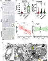
PDGFRβ and the BBB in PSEN1 p.Glu280Ala FAD, SAD and CADASIL. Representative images of PDGFRβ staining are shown for PSEN1 FAD, SAD and CADASIL (A), alongside enlargements of one vessel each, scale bar all panels = 50 μM. The PDGFRβ staining was quantified for PSEN1 FAD (n = 21), SAD (n = 10) and CADASIL (n = 10). The percentage of area covered by PDGFRβ‐positive signal is significantly lower in PSEN1 FAD versus SAD and CADASIL (B) (p values: **** ≤ 0.00001, ** ≤ 0.01). The perivascular PDGFRβ signal was measured for 30 vessels per each group. PSEN1 FAD cases presented with significantly less perivascular PDGFRβ than SAD and CADASIL (C) (p values: **** ≤ 0.0001). The %Area of PDGFRβ is significantly negative correlated with slope of MMSE (r = −0.502, p value: 0.029) while there is no significant correlation observed in CADASIL (r = −0.176, p value: 0.627) (D). Representative EM images are shown for a control (Ctrl) case and a PSEN1 FAD case (F). Thickening of the basement membrane (yellow arrowhead) and a pericyte undergoing apoptosis (yellow arrows) can be observed in FAD (scale bar = 5 μm).
FIGURE 6
FIGURE 6
Influence of perivascular astrocytes in PSEN1 p.Glu280Ala FAD, SAD and CADASIL vessel pathology. Representative images of Fibrinogen+ astrocytes in PS1 FAD, SAD and CADASIL are shown, scale bar all panels = 50 μm (A). Clasmatodendrotic (full arrow‐head) cells can be observed in PSEN1 FAD, SAD and CADASIL as well as star‐shaped astrocytes (empty arrow‐head). Additionally, representative images of AQP4 staining of a dilated and a non‐dilated vessel in PSEN1 FAD (n = 21) are shown. AQP4‐positive astrocyte podocytes (empty arrow‐head) are present for dilated and non‐dilated vessels (B). Signal per area was quantified for 15 dilated and non‐dilated vessels in PSEN1 FAD (n = 21), SAD (n = 10) and CADASIL (n = 10). The signal per area for non‐dilated and dilated vessels in PSEN1 FAD (C) is significantly higher for non‐dilated vessels (p value: * ≤ 0.05).

Similar articles

Cited by

  • Gliovascular alterations in sporadic and familial Alzheimer's disease: APOE3 Christchurch homozygote glioprotection.
    Henao-Restrepo J, López-Murillo C, Valderrama-Carmona P, Orozco-Santa N, Gomez J, Gutiérrez-Vargas J, Moraga R, Toledo J, Littau JL, Härtel S, Arboleda-Velásquez JF, Sepulveda-Falla D, Lopera F, Cardona-Gómez GP, Villegas A, Posada-Duque R. Henao-Restrepo J, et al. Brain Pathol. 2023 Mar;33(2):e13119. doi: 10.1111/bpa.13119. Epub 2022 Sep 21. Brain Pathol. 2023. PMID: 36130084 Free PMC article.
  • Advancements in dementia research, diagnostics, and care in Latin America: Highlights from the 2023 Alzheimer's Association International conference satellite symposium in Mexico City.
    Sosa AL, Brucki SM, Crivelli L, Lopera FJ, Acosta DM, Acosta-Uribe J, Aguilar D, Aguilar-Navarro SG, Allegri RF, Bertolucci PH, Calandri IL, Carrillo MC, Mendez PAC, Cornejo-Olivas M, Custodio N, Damian A, de Souza LC, Duran-Aniotz C, García AM, García-Peña C, Gonzales MM, Grinberg LT, Ibanez AM, Illanes-Manrique MZ, Jack CR Jr, Leon-Salas JM, Llibre-Guerra JJ, Luna-Muñoz J, Matallana D, Miller BL, Naci L, Parra MA, Pericak-Vance M, Piña-Escudero SD, França Resende EP, Ringman JM, Sevlever G, Slachevsky A, Suemoto CK, Valcour V, Villegas-Lanau A, Yassuda MS, Mahinrad S, Sexton C. Sosa AL, et al. Alzheimers Dement. 2024 Jul;20(7):5009-5026. doi: 10.1002/alz.13850. Epub 2024 May 27. Alzheimers Dement. 2024. PMID: 38801124 Free PMC article. Review.
  • Iron accumulation/overload and Alzheimer's disease risk factors in the precuneus region: A comprehensive narrative review.
    Mohammadi S, Ghaderi S, Fatehi F. Mohammadi S, et al. Aging Med (Milton). 2024 Oct 22;7(5):649-667. doi: 10.1002/agm2.12363. eCollection 2024 Oct. Aging Med (Milton). 2024. PMID: 39507230 Free PMC article. Review.
  • The 2022 symposium on dementia and brain aging in low- and middle-income countries: Highlights on research, diagnosis, care, and impact.
    Kalaria R, Maestre G, Mahinrad S, Acosta DM, Akinyemi RO, Alladi S, Allegri RF, Arshad F, Babalola DO, Baiyewu O, Bak TH, Bellaj T, Brodie-Mends DK, Carrillo MC, Celestin KK, Damasceno A, de Silva RK, de Silva R, Djibuti M, Dreyer AJ, Ellajosyula R, Farombi TH, Friedland RP, Garza N, Gbessemehlan A, Georgiou EE, Govia I, Grinberg LT, Guerchet M, Gugssa SA, Gumikiriza-Onoria JL, Hogervorst E, Hornberger M, Ibanez A, Ihara M, Issac TG, Jönsson L, Karanja WM, Lee JH, Leroi I, Livingston G, Manes FF, Mbakile-Mahlanza L, Miller BL, Musyimi CW, Mutiso VN, Nakasujja N, Ndetei DM, Nightingale S, Novotni G, Nyamayaro P, Nyame S, Ogeng'o JA, Ogunniyi A, de Oliveira MO, Okubadejo NU, Orrell M, Paddick SM, Pericak-Vance MA, Pirtosek Z, Potocnik FCV, Raman R, Rizig M, Rosselli M, Salokhiddinov M, Satizabal CL, Sepulveda-Falla D, Seshadri S, Sexton CE, Skoog I, George-Hyslop PHS, Suemoto CK, Thapa P, Udeh-Momoh CT, Valcour V, Vance JM, Varghese M, Vera JH, Walker RW, Zetterberg H, Zewde YZ, Ismail O. Kalaria R, et al. Alzheimers Dement. 2024 Jun;20(6):4290-4314. doi: 10.1002/alz.13836. Epub 2024 May 2. Alzheimers Dement. 2024. PMID: 38696263 Free PMC article. Review.
  • Distinct tau neuropathology and cellular profiles of an APOE3 Christchurch homozygote protected against autosomal dominant Alzheimer's dementia.
    Sepulveda-Falla D, Sanchez JS, Almeida MC, Boassa D, Acosta-Uribe J, Vila-Castelar C, Ramirez-Gomez L, Baena A, Aguillon D, Villalba-Moreno ND, Littau JL, Villegas A, Beach TG, White CL 3rd, Ellisman M, Krasemann S, Glatzel M, Johnson KA, Sperling RA, Reiman EM, Arboleda-Velasquez JF, Kosik KS, Lopera F, Quiroz YT. Sepulveda-Falla D, et al. Acta Neuropathol. 2022 Sep;144(3):589-601. doi: 10.1007/s00401-022-02467-8. Epub 2022 Jul 15. Acta Neuropathol. 2022. PMID: 35838824 Free PMC article.

References

    1. Prince M, Ali GC, Guerchet M, Wu YT, Prina M, Wimo A. World Alzheimer Report 2015: The Global Impact of Dementia. 1st ed. London, UK: Alzheimer's Disease International; 2015.
    1. Scheltens P, de Strooper B, Kivipelto M, Holstege H, Chételat G, Teunissen CE, et al. Alzheimer's disease. Lancet. 2021;397(10284):1577–90. 10.1016/S0140-6736(20)32205-4 - DOI - PMC - PubMed
    1. Sepulveda‐Falla D, Glatzel M, Lopera F. Phenotypic profile of early‐onset familial Alzheimer's disease caused by presenilin‐1 E280A mutation. J Alzheimers Dis. 2012;32(1):1–12. 10.3233/JAD-2012-120907 - DOI - PubMed
    1. Sepulveda‐Falla D, Chavez‐Gutierrez L, Portelius E, Vélez JI, Dujardin S, Barrera‐Ocampo A, et al. A multifactorial model of pathology for age of onset heterogeneity in familial Alzheimer's disease. Acta Neuropathol. 2021;141(2):217–33. 10.1007/s00401-020-02249-0 - DOI - PMC - PubMed
    1. de la Torre JC, Mussivand T. Can disturbed brain microcirculation cause Alzheimer's disease? Neurol Res. 1993;15(3):146–53. 10.1080/01616412.1993.11740127 - DOI - PubMed

Publication types