

Genome-wide characterization of RNA editing highlights roles of high editing events of glutamatergic synapse during mouse retinal development
- PMID: 35685368
- PMCID: PMC9162912
- DOI: 10.1016/j.csbj.2022.05.029
Genome-wide characterization of RNA editing highlights roles of high editing events of glutamatergic synapse during mouse retinal development
Abstract
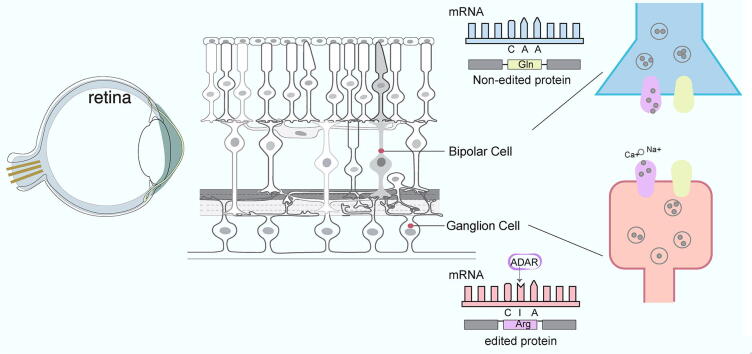
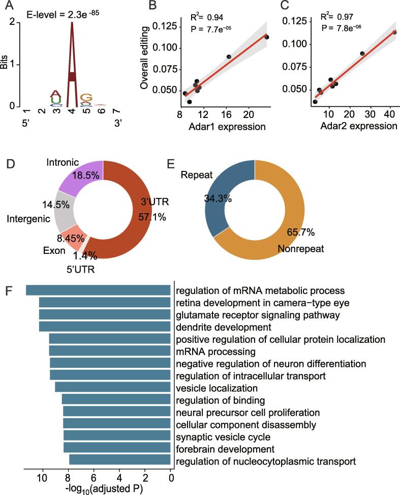
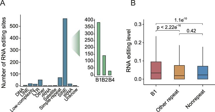
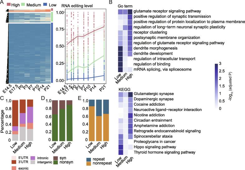
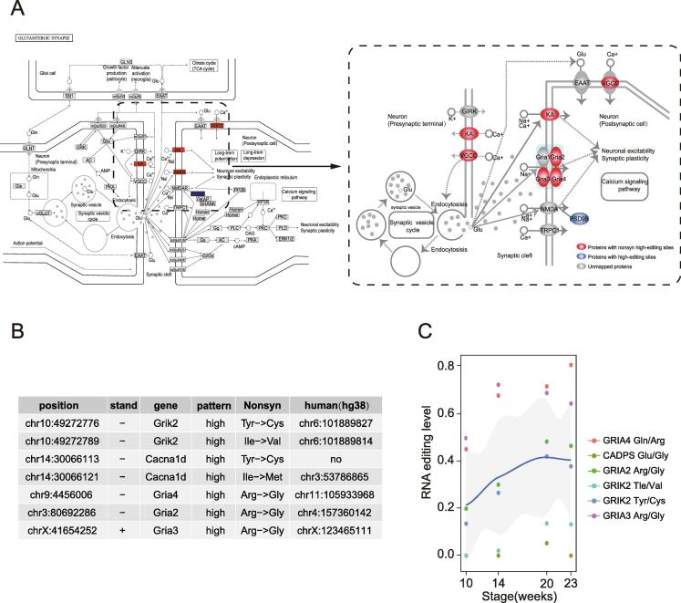
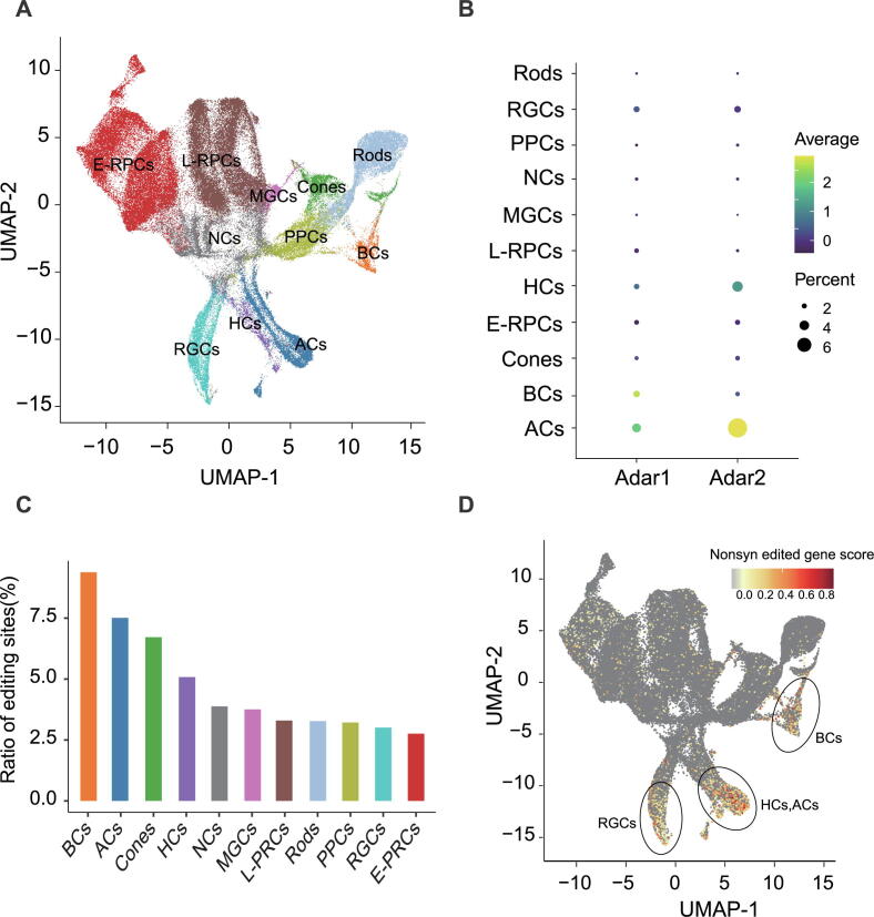
Adenosine-to-inosine (A-to-I) RNA editing leads to functional change of neurotransmitter receptor which is essential for neurotransmission and normal neuronal development. As a highly accessible part of central nervous system, retina has been extensively studied, however, it remains largely unknown how RNA editing regulates its development. Here, a genome-wide screening of high-confidence RNA editing events were performed to decipher the dynamic transcriptome regulation by RNA editing during mouse retinal development. 2000 high-confidence editing sites across eight developmental stages of retina were called. Three unique patterns (RNA-editinghigh pattern, RNA-editingmedium pattern and RNA-editinglow pattern) were identified by clustering these editing sites based on their editing level during retinal development. Editing events from RNA-editinghigh pattern were significantly associated with glutamate receptors and regulated synaptic transmission. Interestingly, most non-synonymous high-editing sites were mapped to ion channel genes of glutamatergic synapse which were associated with neurotransmission by controlling ion channel permeability and affecting exocytosis. Meanwhile, these non-synonymous editing sites were evolutionarily conserved and exhibited a consistently increasing editing levels between mouse and human retinal development. Single-cell RNA-seq data analysis revealed that RNA editing events prefer to occur in two main cell types including bipolar and amacrine cells. Genes with non-synonymous high-editing sites were enriched in both bipolar cells and retina ganglion cells, which may mediate retina ganglion cell differentiation by altering channel ion permeability. Together, our results provide novel insights into mechanism of post-transcriptional regulation during retinal development and help to develop novel RNA editing-guided therapeutic strategies for retinal disorders.
Keywords: Bipolar cells; Non-synonymous; RNA editing; Retinal development; Retinal ganglion cells.
© 2022 The Authors.
Conflict of interest statement
The authors declare that they have no known competing financial interests or personal relationships that could have appeared to influence the work reported in this paper.
Figures
Similar articles
-
The TRPM1 Channel Is Required for Development of the Rod ON Bipolar Cell-AII Amacrine Cell Pathway in the Retinal Circuit.J Neurosci. 2017 Oct 11;37(41):9889-9900. doi: 10.1523/JNEUROSCI.0824-17.2017. Epub 2017 Sep 12. J Neurosci. 2017. PMID: 28899920 Free PMC article.
-
An Evolutionary Landscape of A-to-I RNA Editome across Metazoan Species.Genome Biol Evol. 2018 Feb 1;10(2):521-537. doi: 10.1093/gbe/evx277. Genome Biol Evol. 2018. PMID: 29294013 Free PMC article.
-
Transcriptome-wide identification of A > I RNA editing sites by inosine specific cleavage.RNA. 2013 Feb;19(2):257-70. doi: 10.1261/rna.036202.112. Epub 2012 Dec 21. RNA. 2013. PMID: 23264566 Free PMC article.
-
RNA editing: a molecular mechanism for the fine modulation of neuronal transmission.Acta Neurochir Suppl. 2005;93:53-7. doi: 10.1007/3-211-27577-0_7. Acta Neurochir Suppl. 2005. PMID: 15986727 Review.
-
RNA editing of ion channels and receptors in physiology and neurological disorders.Oxf Open Neurosci. 2022 Jul 11;1:kvac010. doi: 10.1093/oons/kvac010. eCollection 2022. Oxf Open Neurosci. 2022. PMID: 38596706 Free PMC article. Review.
Cited by
-
Temporal landscape and translational regulation of A-to-I RNA editing in mouse retina development.BMC Biol. 2024 May 7;22(1):106. doi: 10.1186/s12915-024-01908-y. BMC Biol. 2024. PMID: 38715001 Free PMC article.
References
-
- Costa Cruz P.H., Kawahara Y. RNA Editing in Neurological and Neurodegenerative Disorders. Methods Mol Biol. 2021;2181:309–330. - PubMed
LinkOut - more resources
Full Text Sources
Research Materials