Loading [MathJax]/jax/output/PreviewHTML/jax.js
Skip to main page content
U.S. flag

An official website of the United States government

Access keys NCBI Homepage MyNCBI Homepage Main Content Main Navigation
. 2022 May 28;12(11):1849.
doi: 10.3390/nano12111849.

Exploring the Effect of Mechanical Anisotropy of Protein Structures in the Unfoldase Mechanism of AAA+ Molecular Machines

Affiliations

Exploring the Effect of Mechanical Anisotropy of Protein Structures in the Unfoldase Mechanism of AAA+ Molecular Machines

Rohith Anand Varikoti et al. Nanomaterials (Basel). .

Abstract

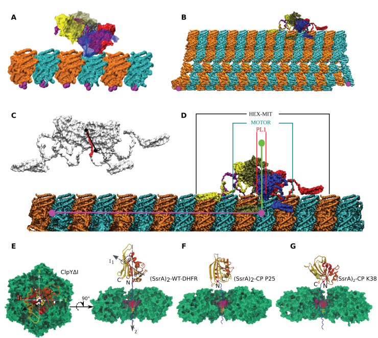
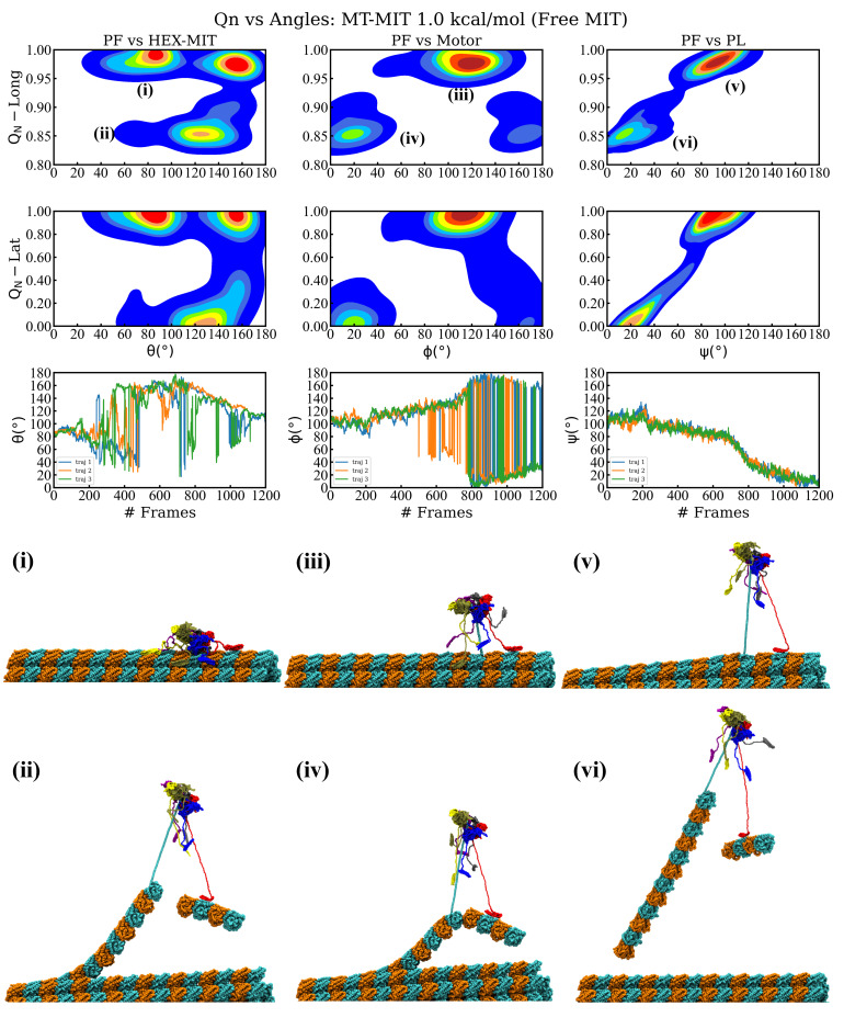
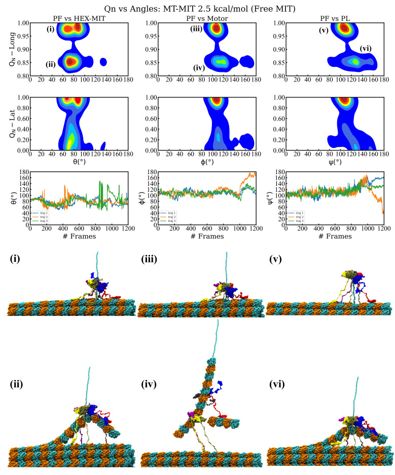
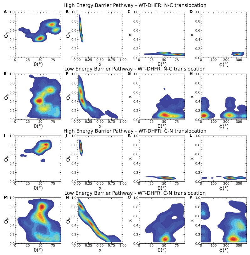
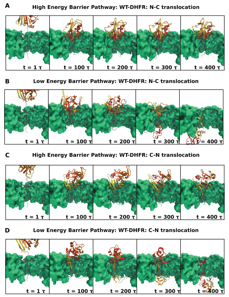
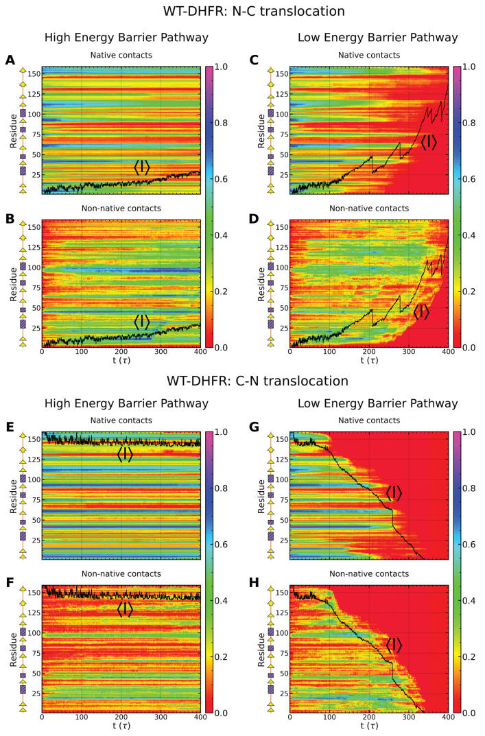
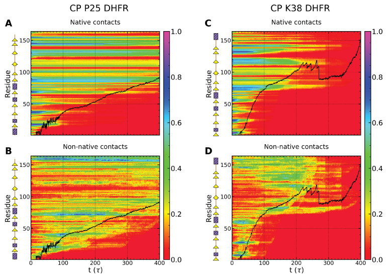
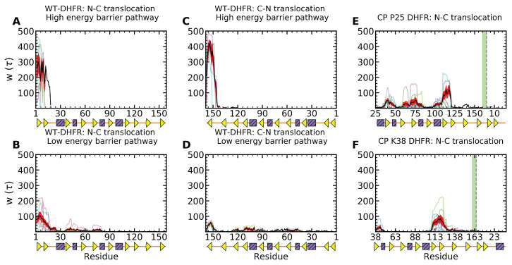
Essential cellular processes of microtubule disassembly and protein degradation, which span lengths from tens of μm to nm, are mediated by specialized molecular machines with similar hexameric structure and function. Our molecular simulations at atomistic and coarse-grained scales show that both the microtubule-severing protein spastin and the caseinolytic protease ClpY, accomplish spectacular unfolding of their diverse substrates, a microtubule lattice and dihydrofolate reductase (DHFR), by taking advantage of mechanical anisotropy in these proteins. Unfolding of wild-type DHFR requires disruption of mechanically strong β-sheet interfaces near each terminal, which yields branched pathways associated with unzipping along soft directions and shearing along strong directions. By contrast, unfolding of circular permutant DHFR variants involves single pathways due to softer mechanical interfaces near terminals, but translocation hindrance can arise from mechanical resistance of partially unfolded intermediates stabilized by β-sheets. For spastin, optimal severing action initiated by pulling on a tubulin subunit is achieved through specific orientation of the machine versus the substrate (microtubule lattice). Moreover, changes in the strength of the interactions between spastin and a microtubule filament, which can be driven by the tubulin code, lead to drastically different outcomes for the integrity of the hexameric structure of the machine.

Keywords: AAA+ superfamily; allostery; microtubule severing; molecular dynamics; molecular machines; protein degradation.


Conflict of interest statement

The authors declare no conflict of interest. The funders had no role in the design of the study; in the collection, analyses, or interpretation of data; in the writing of the manuscript, or in the decision to publish the results.

Figures

Figure 1
Figure 1
Configurations for the spastin and ClpY machines and their substrate proteins (SP) probed in our simulations. (A) Side view of the spastin motor mounted on a MT fragment (α tubulin: orange, β tubulin: cyan, fixed residues: magenta); (B) side view of the entire spastin machine mounted on an 8-dimer long, 13 PF MT lattice; (C) cut-out side view of the HBD (red) bound in the central pore of the spastin hexamer. The largest axis of inertia of the pore loops 1 is indicated by the black line; (D) side view of the spastin machine mounted on a PF showing the various angles calculated in our pulling simulations: between the main axis of the PF (pink) and the principal axis of symmetry of the spastin machine (HEX-MIT), of the spastin motor only (Motor), or of the pore loops 1 (PL1) only (green); (E) Top and side view of the ClpYΔI (green)-SP system with polar angle θ and azimuthal angle ϕ are indicated in the XYZ Cartesian coordinate system. SP is a fusion protein comprising the unfolded (SsrA)2 peptide (blue) and the DHFR domain (color-coded according to secondary structure). ClpY pore loops (red) are also indicated. An external repetitive force F is applied onto SP amino acids transiently located within the ClpY pore region (see Methods). (F,G) Circular permutant variants of DHFR with engineered N- and C-terminals at the (F) P25 and (G) K38 positions are also considered.
Figure 2
Figure 2
Results of the spastin machine acting on an MT filament for the interaction strength between its MIT domains and the MT lattice set to 1.0 kcal/mol. (Top) row plots show the free energy landscape in the plane of the fractional loss of longitudinal native contacts (QN) of the pulled protofilament (PF6) and the angle made by the principal axis of the severing enzyme (HEX-MIT), of the motor, and of the central pore loops (PL), respectively, versus the long axis of the pulled PF (PF). (Middle) row plots show the free energy landscape in the plane of the fractional loss of lateral native contacts of the pulled protofilament and the three angles from the top row panels. The lowest row plots depict the time evolution of the three angles from the upper plots versus the simulation frames. Instantaneous snapshots of the representative structures corresponding to the labeled minima in the free energy plots are shown at the (Bottom).
Figure 3
Figure 3
Results of the spastin machine acting on a MT filament for the interaction strength between its MIT domains and the MT lattice set to 2.5 kcal/mol. (Top) row plots show the free energy landscape in the plane of the fractional loss of longitudinal native contacts (QN) of the pulled protofilament (PF6) and the angle made by the principal axis of the severing enzyme (HEX-MIT), of the motor, and of the central pore loops (PL), respectively, versus the long axis of the pulled PF (PF). (Middle) row plots show the free energy landscape in the plane of the fractional loss of lateral native contacts of the pulled protofilament and the three angles from the top row panels. The lowest row plots depict the time evolution of the three angles from the upper plots versus the simulation frames. Instantaneous snapshots of the representative structures corresponding to the labeled minima in the free energy plots are shown at the (Bottom).
Figure 4
Figure 4
Severing pathways found for the unfoldase severing action of the full spastin machine with free MIT domains on MT8 lattice for all the probed interaction strengths between the MIT domains and the MT lattice. Descriptions and percentage of event occurrences (out of the 21 trajectories) are provided for each main event of a severing mechanism. Colors are used to separate major diverging pathways.
Figure 5
Figure 5
Unfolding of DHFR variants mediated by ClpY. The time evolution of the fraction of native, QN, and non-native, fNN, contacts is shown for wild-type DHFR in (A,B) N-C and (C,D) C-N translocation; (E,F) for CP P25 and (G,H) for CP K38 in N-C translocation. Individual trajectories are indicated by using thin curves. Averages (thick curves, black) and standard errors (red) are also indicated.
Figure 6
Figure 6
Wild-type DHFR substrate orientation at the ClpY pore lumen in unfolding and translocation pathways. Probability density maps of the (A) fraction of native contacts QN vs. polar angle θ and (B) translocation fraction x; (C) translocation fraction vs. polar and azimuthal (ϕ) angles in N-C translocation in the high-energy barrier pathway. (EH) Same as in (AD) in the low-energy barrier pathway. (IP) Same as in (AH) in C-N translocation.
Figure 7
Figure 7
Dynamic orientation of wild-type DHFR at the ClpY pore lumen. The time-dependent orientation of DHFR (color-coded according to secondary structure) near the ClpYΔI (green) pore lumen is shown in translocation in the (A,B) N-C direction in the (A) high- and (B) low-energy barrier pathways and (C,D) C-N direction. Two ClpY protomers are not shown for clarity.
Figure 8
Figure 8
Direction-dependent time evolution of native and non-native content of wild-type DHFR. (AD) The time-dependent fraction of native QN and non-native fNN contacts formed by secondary structural elements of wild-type DHFR during translocation in the N-C direction in the (A,B) high- and (C,D) low-energy barrier pathway. (EH) Same as in (AD) in C-N translocation. The time-dependent average translocation line (black) is also indicated.
Figure 9
Figure 9
Direction-dependent time evolution of native and non-native content of circular permutant variants P25 and K38 of DHFR. (AD) The time-dependent fraction of native QN and non-native fNN contacts formed by secondary structural elements of wild-type DHFR during translocation in the N-C direction in the (A,B) high- and (C,D) low-energy barrier pathway. The time-dependent average translocation line (black) is also indicated.
Figure 10
Figure 10
Translocation hindrance of DHFR variants. The waiting time per residue of the wild-type DHFR is shown in translocation in the (A,B) N-C direction in the (A) high- and (B) low-energy barrier pathways, in the (C,D) C-N direction. (E,F) Same as in (A) for the circular permutant DHFR variants (E) P25 and (F) K38 (the wild-type N- and C-terminal sequence positions are indicated by using dashes and a green line, respectively). Traces for individual trajectories are shown by using thin curves and averaged values by using thick black curves. Standard errors are shown by using red bands.

Similar articles

Cited by

  • Protein Nanomechanics.
    Žoldák G. Žoldák G. Nanomaterials (Basel). 2022 Oct 8;12(19):3524. doi: 10.3390/nano12193524. Nanomaterials (Basel). 2022. PMID: 36234652 Free PMC article.

References

    1. Hanson P.I., Whiteheart S.W. AAA+ Proteins: Have Engine, Will Work. Nat. Rev. Mol. Cell Biol. 2005;6:519–529. doi: 10.1038/nrm1684. - DOI - PubMed
    1. Enemark E.J., Joshua-Tor L. On helicases and other motor proteins. Curr. Opin. Struct. Biol. 2008;18:243–257. doi: 10.1016/j.sbi.2008.01.007. - DOI - PMC - PubMed
    1. Sauer R.T., Baker T.A. AAA+ Proteases: ATP-Fueled Machines of Protein Destruction. Annu. Rev. Biochem. 2011;80:587–612. doi: 10.1146/annurev-biochem-060408-172623. - DOI - PubMed
    1. Sharp D., Ross J. Microtubule Severing Enzymes at the Cutting Edge. J. Cell Sci. 2012;125:2561–2569. doi: 10.1242/jcs.101139. - DOI - PMC - PubMed
    1. Weber-Ban E.U., Reid B.G., Miranker A.D., Horwich A.L. Global unfolding of a substrate protein by the Hsp100 chaperone ClpA. Nature. 1999;401:90–93. doi: 10.1038/43481. - DOI - PubMed

LinkOut - more resources