. 2022 Jun 6;23(11):6349.
doi: 10.3390/ijms23116349.

Impact of N-Terminal Tags on De Novo Vimentin Intermediate Filament Assembly

Affiliations

Impact of N-Terminal Tags on De Novo Vimentin Intermediate Filament Assembly

Saima Usman et al. Int J Mol Sci. .

Abstract

Vimentin, a type III intermediate filament protein, is found in most cells along with microfilaments and microtubules. It has been shown that the head domain folds back to associate with the rod domain and this association is essential for filament assembly. The N-terminally tagged vimentin has been widely used to label the cytoskeleton in live cell imaging. Although there is previous evidence that EGFP tagged vimentin fails to form filaments but is able to integrate into a pre-existing network, no study has systematically investigated or established a molecular basis for this observation. To determine whether a tag would affect de novo filament assembly, we used vimentin fused at the N-terminus with two different sized tags, AcGFP (239 residues, 27 kDa) and 3 × FLAG (22 residues; 2.4 kDa) to assemble into filaments in two vimentin-deficient epithelial cells, MCF-7 and A431. We showed that regardless of tag size, N-terminally tagged vimentin aggregated into globules with a significant proportion co-aligning with β-catenin at cell-cell junctions. However, the tagged vimentin aggregates could form filaments upon adding untagged vimentin at a ratio of 1:1 or when introduced into cells containing pre-existing filaments. The resultant filament network containing a mixture of tagged and untagged vimentin was less stable compared to that formed by only untagged vimentin. The data suggest that placing a tag at the N-terminus may create steric hinderance in case of a large tag (AcGFP) or electrostatic repulsion in case of highly charged tag (3 × FLAG) perhaps inducing a conformational change, which deleteriously affects the association between head and rod domains. Taken together our results shows that a free N-terminus is essential for filament assembly as N-terminally tagged vimentin is not only incapable of forming filaments, but it also destabilises when integrated into a pre-existing network.

Keywords: ectopic protein expression; fusion proteins; immunofluorescence; intermediate filaments; protein domains.


Conflict of interest statement

The authors declare no conflict of interest.

Figures
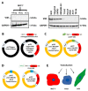
Figure 1
Figure 1
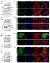
Graphical representation of different constructs used in this study. (A) Vimentin expression analysed in 10 different cancer cell lines by western blotting. A total of 50 µg total protein was loaded for all other cell lines; for MCF-7, 100, 50 and 25 µg protein were loaded onto SDS PAGE gels. GAPDH was used as the loading control. Relevant bands were cropped from different gels and regrouped. Original blots are shown in Supplementary Figure S1. (B) Construct pLPChygro-VIM expressing untagged vimentin (UTV) and pLPChygro its control vector (CV); N and C are the 5′ and 3′ ends of vimentin cDNA, respectively, black color represents the vector backbone. (C) pLPCpuro-NAcGFP-GS10-VIM expressing AcGFP-VIM and pLPCpuro-NAcGFP-GS10 expressing AcGFP its CV. (D) pLPCpuro-NFLAG-GS10-VIM expressing FLAG-VIM and pLPCpuro-NFLAG-GS10 expressing FLAG its CV, the FLAG tag was 3 × FLAG, yellow colour in B and C represents vector backbone. (E): MCF-7, A431 and HFF-1 cell lines (nuclei in blue and the cytoplasm is shown in different colors) were transduced with the constructs described in (BD).
Figure 2
Figure 2
Ectopic expression of vimentin in vimentin-deficient cells. (A) MCF-7 and (B) A431 cells were transduced with the untagged vimentin (UTV) and its control vector (CV) by recombinant retrovirus supernatant. Total protein extract, 10 and 20 µg, from each transduced cell line were loaded onto SDS PAGE gels, transferred onto nitrocellulose and probed with anti-vimentin antibody to confirm the transduction efficiency. GAPDH was used as the loading control. Relevant bands were cropped from different gels and regrouped. Original blots are shown in the Supplementary Figure S2. Immunostaining of (C) MCF-7 and (D) A431 cells transduced with CV and UTV constructs. Untransduced (UT) MCF-7 and A431 cells were also used as controls. Cells were immunostained with anti-vimentin antibody and counter stained with AF-488-labelled goat anti-mouse secondary antibody showing green fluorescence. Nuclei were stained with DAPI in blue and overlapping images are shown as ‘Merge’. Leica DM4000B Epi-fluorescence microscope and DFC350 camera were used for recording images. Immunostaining of (E) MDA-MB-231 and (F) SVFN3 cells expressing endogenous vimentin were also immunostained with anti-vimentin antibody. The vimentin staining in SVFN3 and MDA-MB-231 cells was very similar to that in transduced MCF-7 and A431cells shown in C and D (scale bar = 20 µm).
Figure 3
Figure 3
Ectopic expression of N-terminally tagged vimentin in MCF-7 and A431 cells. Healthy growing cells (A,E) MCF-7 and (B,F) A431 were transduced separately with (A,B) control vector (CV) AcGFP and AcGFP-VIM; (E,F) control vector (CV) FLAG and FLAG-VIM, respectively. Total protein extract, 10 and 20 µg, from each transduced cell line was loaded onto SDS PAGE gels, transferred onto nitrocellulose, and probed with mouse anti-vimentin antibody, or rabbit anti-FLAG antibody to confirm the transduction efficiency. GAPDH was used as the loading control. Molecular weights of the gel bands are given on the right-hand side of the blots. Relevant bands were cropped from different blots and regrouped together. Original blots are shown in Supplementary Figures S3 and S4. (C,G) MCF-7 and (D,H) A431 cells expressing (C,D) AcGFP and AcGFP-VIM and (G,H) FLAG and FLAG-VIM, respectively, were grown on collagen coated glass coverslips and fixed with acetone: methanol (1:1). The cells in (G,H) were incubated with rabbit anti-FLAG antibody and counterstained with AF-488 goat anti-rabbit secondary antibody (green). All coverslips in (C,D,G,H) were incubated with A45-B/B3 (keratins) and counter stained with AF-594 labelled goat anti-mouse secondary antibody (red). Nuclei were stained with DAPI in blue and overlapping of all colours is shown as ‘Merge’. Leica DM4000B Epi-fluorescence microscope was used for visualisation and DFC350 camera was used for image recording (scale bar = 20 µm).
Figure 4
Figure 4
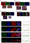
Co-alignment of vimentin globules with β-catenin in MCF-7 and A431 cells at intercellular junction. (A,B) MCF-7 and (C,D) A431 cells stably expressing (A,C) AcGFP-VIM and (B,D) FLAG-VIM were grown on collagen coated glass coverslips. The cells were immunostained with mouse anti-β catenin antibody and AF-594-labelled goat anti-mouse as secondary antibody (red). For cells expressing 3 × FLAG tagged vimentin, cells were stained with rabbit anti-FLAG antibody and AF-488 labelled goat anti-rabbit secondary antibody (green). Nuclei were stained with DAPI in blue and all overlapping images are shown as ‘Merge’. Leica DM4000B Epi-fluorescence microscope was used for visualisation and DFC350 camera was used for recording. Marked areas showing intercellular junctions were magnified for clarity in panels (AC). (E) Images taken by Zeiss 880 laser scanning confocal microscope with Fast Airyscan and Multiphoton (inverted) system. Co-alignment between vimentin aggregates and β catenin is shown by line graph drawn using RGB profiler on maximal single confocal sections. The area used for this analysis is shown by a white line in different panels (scale bar = 20 µm).
Figure 5
Figure 5
Integration of N-terminally tagged vimentin into the pre-existing vimentin filaments. HFF-1 cells expressing AcGFP-VIM, control vector (CV) AcGFP, FLAG-VIM and control vector (CV) FLAG were immunostained with anti-VIM antibody (for endogenous vimentin network) and AF-594-labelled goat anti-mouse as secondary antibody (red). For cells expressing 3 × FLAG constructs, cells were stained with anti-FLAG antibody and AF-488 labelled goat anti-rabbit as secondary antibody (green). Nuclei were stained with DAPI in blue and all overlapping images are shown as ‘Merge’. Leica DM4000B Epi-fluorescence microscope was used for visualisation and DFC350 camera was used for image recording (scale bar = 20 µm). Note that N-terminally tagged vimentin (green colour panels (a,i)) has integrated into the endogenous vimentin (red colour panel (c,k)) filament network in HFF-1 cells which becomes yellow (d,l) in ‘Merge’. No disruption of pre-existing vimentin network was seen in these cells. The western blot analysis showed that the transduced HFF-1 cells had tagged and endogenous vimentin in a ratio of 1:1 (Figure S5).
Figure 6
Figure 6
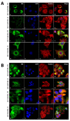
Aggregates of N-terminally tagged vimentin can be reversed by untagged vimentin (UTV). (A) MCF-7 and (B) A431 cells expressing AcGFP-VIM and FLAG-VIM vimentin aggregates were transduced with untagged vimentin (UTV) virus. The control vector (CV) expressed either AcGFP or 3 × FLAG. After 48 h the cells were fixed in acetone:methanol (1:1) and stained with A45-B/B3 for keratins and AF-594-labelled goat anti-mouse secondary antibody (red). For cells expressing FLAG-VIM, the cells were stained with anti-FLAG antibody and AF-488-labelled goat anti-rabbit secondary antibody (green). Nuclei were stained with DAPI in blue and overlapping of all colours is shown as ‘Merge’. Leica DM4000B Epi-fluorescence microscope was used for visualisation and DFC350 camera was used for image recording. (scale bar = 20 µm). Note that vimentin aggregation has been completely reversed by UTV and filaments can be seen in green colour (a,i). The western blot analysis shows a 1:1 ratio between AcGFP-tagged and untagged vimentin (Figure S5).
Figure 7
Figure 7
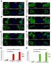
Destabilisation of vimentin network by integration of tagged vimentin. (A) MCF-7 and (B) A431 cells expressing untagged vimentin (UTV) were treated with (+D) or without (−D) 0.1 mM diamide for 10 min and immunostained with anti-vimentin antibody; (C,E) MCF-7 and (D,F) A431 cells expressing (C,D) AcGFP-VIM and (E,F) FLAG-VIM, respectively, were transduced with UTV and treated with (+D) or without (−D) 0.1 mM diamide for 10 min. The AcGFP expressing cells were visualised after mounting whereas the FLAG expressing cells were immunostained with anti-FLAG antibody. Quantification of cells with disrupted vimentin network as a result of diamide treatment in (G) MCF-7 and (H) A431 cells. About 200 cells were visually evaluated for vimentin aggregation under Epi-fluorescence microscope in triplicates for every set. Collapse of filaments by diamide was observed only in 6 ± 0.72% MCF-7 and 3.6 ± 0.91% A431 cells expressing untagged vimentin UTV alone. In UTV + AcGFP-VIM expressing cells, diamide caused filament collapse in 53 ± 1.69% (*** p < 0.001) MCF-7 and 51 ± 0.24% (*** p < 0.001) A431 cells. In UTV + FLAG-VIM expressing cells, diamide caused vimentin aggregation in 60 ± 0.72% (*** p < 0.001) MCF-7 and 63 ± 0.92% (*** p < 0.001) A431 cells, the p values indicate the data are extremely significant. (scale bar = 20 µm).
Figure 8
Figure 8
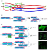
Proposed mechanism of aggregation by N-terminally tagged vimentin. (A) Vimentin tetramer structure in which the rod domains of two dimers (red and blue) associate in a staggered fashion. The head domain (shown in green for one dimer and red for the other dimer) folds back onto the rod domain causing G17 of the head domain to interact with Q137 of coil I (residues highlighted as yellow dots) (modified from [42]). (B) Schematic representation of the two possible ways (I and II) dimers can associate into a tetramer. The N-terminus of the vimentin head domain has a large number (8 out of 26) of serine/threonine residues, phosphorylation of which during mitosis or calyculin A treatment causes repulsion between the head domains leading to disruption of association between head–head and head–rod including G17 and Q137 interaction. This causes conformational alterations leading to collapse of the network. (C) i. Eight tetramers (both types I and II) association to form ULFs which further associate into mature filaments; ii. Tagging the head domain at the N-terminus with AcGFP, which is a larger tag (239 residues; 27 kDa), will therefore interfere with head–head, head–rod and rod–rod interactions (shown by a double arrow) when the head domains fold back, due to steric hindrance, and is likely to have the same effect as induced by phosphorylation of serine/threonine residues located at the N-terminus. This may affect ULF formation or render them incapable of associating into filaments; iii. When vimentin is tagged at the N terminus with 3 × FLAG, a much smaller tag of only 22 residues, aggregates are still formed. This can be explained by the fact that 11 out of 22 residues in 3 × FLAG are aspartic acid making the tag peptide acidic (pI = 3.97), which will create extensive repulsion disrupting not only the head–rod interactions but also inter-rod interactions affecting formation of ULF or making them unable to associate into filaments (redrawn and modified from reference [33]). The proposed mechanism of aggregation by N-terminally tagged vimentin is summarised in Supplementary Video SV1.

Similar articles

Cited by

References

    1. Satelli A., Li S. Vimentin in cancer and its potential as a molecular target for cancer therapy. Cell. Mol. Life Sci. 2011;68:3033–3046. doi: 10.1007/s00018-011-0735-1. - DOI - PMC - PubMed
    1. Pollard T.D., Goldman R.D. Overview of the Cytoskeleton from an Evolutionary Perspective. Cold Spring Harb. Perspect. Biol. 2018;10:a030288. doi: 10.1101/cshperspect.a030288. - DOI - PMC - PubMed
    1. Danielsson F., Peterson M.K., Caldeira Araújo H., Lautenschläger F., Gad A.K. Vimentin Diversity in Health and Disease. Cells. 2018;7:147. doi: 10.3390/cells7100147. - DOI - PMC - PubMed
    1. Nekrasova O.E., Mendez M.G., Chernoivanenko I.S., Tyurin-Kuzmin P.A., Kuczmarski E.R., Gelfand V.I., Goldman R.D., Minin A.A. Vimentin intermediate filaments modulate the motility of mitochondria. Mol. Biol. Cell. 2011;22:2282–2289. doi: 10.1091/mbc.e10-09-0766. - DOI - PMC - PubMed
    1. Schwarz N., Leube R.E. Intermediate Filaments as Organizers of Cellular Space: How They Affect Mitochondrial Structure and Function. Cells. 2016;5:30. doi: 10.3390/cells5030030. - DOI - PMC - PubMed

Grants and funding

This work was supported by Higher Education Commission (HEC), Pakistan for Ph.D. studentship to S.U., Barts Charity for the Dental Centenary Ph.D. studentship to T.K.N.N. and The Rosetrees Trust for financial support (M312-F1 to A.W.).

LinkOut - more resources