High Levels of MFG-E8 Confer a Good Prognosis in Prostate and Renal Cancer Patients
- PMID: 35681775
- PMCID: PMC9179566
- DOI: 10.3390/cancers14112790
High Levels of MFG-E8 Confer a Good Prognosis in Prostate and Renal Cancer Patients
Abstract
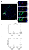
Milk fat globule-epidermal growth factor-8 (MFG-E8) is a glycoprotein secreted by different cell types, including apoptotic cells and activated macrophages. MFG-E8 is highly expressed in a variety of cancers and is classically associated with tumor growth and poor patient prognosis through reprogramming of macrophages into the pro-tumoral/pro-angiogenic M2 phenotype. To date, correlations between levels of MFG-E8 and patient survival in prostate and renal cancers remain unclear. Here, we quantified MFG-E8 and CD68/CD206 expression by immunofluorescence staining in tissue microarrays constructed from renal (n = 190) and prostate (n = 274) cancer patient specimens. Percentages of MFG-E8-positive surface area were assessed in each patient core and Kaplan-Meier analyses were performed accordingly. We found that MFG-E8 was expressed more abundantly in malignant regions of prostate tissue and papillary renal cell carcinoma but was also increased in the normal adjacent regions in clear cell renal carcinoma. In addition, M2 tumor-associated macrophage staining was increased in the normal adjacent tissues compared to the malignant areas in renal cancer patients. Overall, high tissue expression of MFG-E8 was associated with less disease progression and better survival in prostate and renal cancer patients. Our observations provide new insights into tumoral MFG-E8 content and macrophage reprogramming in cancer.
Keywords: M2 macrophage; MFG-E8; prostate cancer; renal cancer.
Conflict of interest statement
The authors declare no conflict of interest.
Figures


Similar articles
-
Polarization of prostate cancer-associated macrophages is induced by milk fat globule-EGF factor 8 (MFG-E8)-mediated efferocytosis.J Biol Chem. 2014 Aug 29;289(35):24560-72. doi: 10.1074/jbc.M114.571620. Epub 2014 Jul 8. J Biol Chem. 2014. PMID: 25006249 Free PMC article.
-
MFG-E8 overexpression is associated with poor prognosis in breast cancer patients.Pathol Res Pract. 2019 Mar;215(3):490-498. doi: 10.1016/j.prp.2018.12.036. Epub 2018 Dec 31. Pathol Res Pract. 2019. PMID: 30612778
-
Milk fat globule epidermal growth factor-8 limits tissue damage through inflammasome modulation during renal injury.J Leukoc Biol. 2016 Nov;100(5):1135-1146. doi: 10.1189/jlb.3A0515-213RR. Epub 2016 Jun 3. J Leukoc Biol. 2016. PMID: 27260955
-
Role of milk fat globule-epidermal growth factor 8 in osteoimmunology.Bonekey Rep. 2016 Jul 20;5:820. doi: 10.1038/bonekey.2016.52. eCollection 2016. Bonekey Rep. 2016. PMID: 27579162 Free PMC article. Review.
-
Identification of MFG-E8 as a novel therapeutic target for diseases.Expert Opin Ther Targets. 2013 Nov;17(11):1275-85. doi: 10.1517/14728222.2013.829455. Epub 2013 Aug 23. Expert Opin Ther Targets. 2013. PMID: 23972256 Review.
Cited by
-
MFG-E8 promotes M2 polarization of macrophages and is associated with poor prognosis in patients with gastric cancer.Heliyon. 2023 Dec 18;10(1):e23917. doi: 10.1016/j.heliyon.2023.e23917. eCollection 2024 Jan 15. Heliyon. 2023. PMID: 38192793 Free PMC article.
-
CircSMAD2 accelerates endometrial cancer cell proliferation and metastasis by regulating the miR-1277-5p/MFGE8 axis.J Gynecol Oncol. 2023 Mar;34(2):e19. doi: 10.3802/jgo.2023.34.e19. Epub 2023 Jan 2. J Gynecol Oncol. 2023. PMID: 36659830 Free PMC article.
References
Grants and funding
LinkOut - more resources
Full Text Sources
Miscellaneous

