Review
. 2022 Aug:287:106821.
doi: 10.1016/j.bpc.2022.106821. Epub 2022 Apr 29.

Protein folding in vitro and in the cell: From a solitary journey to a team effort

Affiliations
Review

Protein folding in vitro and in the cell: From a solitary journey to a team effort

Miranda F Mecha et al. Biophys Chem. 2022 Aug.

Abstract

Correct protein folding is essential for the health and function of living organisms. Yet, it is not well understood how unfolded proteins reach their native state and avoid aggregation, especially within the cellular milieu. Some proteins, especially small, single-domain and apparent two-state folders, successfully attain their native state upon dilution from denaturant. Yet, many more proteins undergo misfolding and aggregation during this process, in a concentration-dependent fashion. Once formed, native and aggregated states are often kinetically trapped relative to each other. Hence, the early stages of protein life are absolutely critical for proper kinetic channeling to the folded state and for long-term solubility and function. This review summarizes current knowledge on protein folding/aggregation mechanisms in buffered solution and within the bacterial cell, highlighting early stages. Remarkably, teamwork between nascent chain, ribosome, trigger factor and Hsp70 molecular chaperones enables all proteins to overcome aggregation propensities and reach a long-lived bioactive state.

Keywords: Aggregation; Chaperones; Cotranslational folding; Energy landscapes; Kinetic trapping; Ribosome.


Figures

Figure 1.
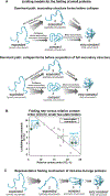
Figure 1.. Overview of protein folding mechanisms upon dilution from denaturant or upon recovery from temperature jumps.
Some small (50–60 residues) proteins fold via (A) a mechanism dominated by secondary structure formation before chain collapse, or via (B) chain collapse preceding the formation of most secondary structure. (C) Plot of folding rate as a function of relative contact order (CO) for small (60–110 residues) two-state folding proteins. The graph is reprinted with permission from Figure 1A of J. Mol. Biol., 277, Plaxco, K. W.; Simons, K. T.; Baker, D., 985–994, Copyright (1998) [35]. (D) Larger proteins (> 60 residues) fold via more complex folding mechanisms, often including folding intermediates.
Figure 2.
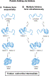
Figure 2.. Foldons and protein folding mechanisms.
Scheme illustrating how proteins may fold via native-like intermediates denoted as foldons, which are generated either (A) sequentially or (B) in parallel.
Figure 3.
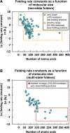
Figure 3.. Effect of size and folding mechanism on protein folding rates.
(A) Plot illustrating the dependence of protein folding rate constant (kf) on the number of residues for two-state folding proteins. Small (<50 residues) two-state proteins fold quickly with ln(kf) > 9.4 (green box). Many larger two-state folders fold more slowly (orange box). (B) Dependence of protein folding rate constant (kf) on the number of residues for multi-state folding proteins. Large (>200 residues) multi-state proteins have the slowest folding rates, with ln(kf) <−2.5 (red box). A list of the proteins and references for the data in this plot is available as Supplementary Information Table S1.
Figure 4.
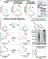
Figure 4.. Kinetic trapping of E. coli proteome relative to insoluble aggregates.
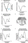
(A-C) Representative standard-state Gibbs free energy landscapes for (A) non-aggregating proteins, (B) proteins that have kinetically-trapped native and unfolded states relative to aggregated states, and (C) proteins that have kinetically-trapped native states relative to unfolded states or folding intermediates. (D) Variations in the population of proteins described in panels A, B and C, respectively, after heating and cooling. (E) SDS-Page analysis of soluble E. coli proteome upon heating for 20 hrs at 70 °C followed by slow cooling to room temperature. Sample centrifugation generated a supernatant (S) and an insoluble pellet (I), shown separately in the gel [92]. (F) Fraction of insoluble, aggregated E. coli proteome generated by procedure described above as a function of total protein concentration. The solid line is meant to guide the eye. Error bars denote the standard error for three independent experiments [92]. Panels E and F are adapted with permission from Varela, A. E.; Lang, J. F.; Wu, Y.; Dalphin, M. D.; Stangl, A. J.; Okuno, Y.; Cavagnero, S. J. Phys. Chem. B 2018, 122, 7682–7698. Copyright (2018) American Chemical Society.
Figure 5.
Figure 5.. Representative protein folding energy landscapes.
(A) Folding funnel proposed by Wolynes and coworkers, showing how protein conformational entropy decreases in concert with effective potential energy, as a protein folds to its native state [104, 116]. (B) Helmholtz free energy landscape for proteins that do not have a free-energy transition state for folding. (C) Helmholtz free energy landscape for proteins that have a free-energy transition state for folding. In panels A-C, the native state has 100% native contacts (Q = 1), and the unfolded state has Q = 0. (D) Multidimensional energy landscape. The vertical axis represents the potential energy of any given protein conformation plus the free energy of solvation [113]. Figure 1D is reprinted with permission from John Wiley and Sons [41] from figure 37C in Protein Science 4, Dill, K. A.; Bromberg, S.; Yue, K.; Chan, H. S.; Ftebig, K. M.; Yee, D. P.; Thomas, P. D. Principles of Protein Folding — a Perspective from Simple Exact Models. 4, 561–602. Copyright (1995). (E) Standard-state Gibbs free energy landscape of a protein that cannot form aggregates at a given temperature, pressure and solution conditions. (F) Gibbs free energy landscape for a protein that can form aggregates at a given temperature, pressure and solution conditions.
Figure 6.
Figure 6.. Funnel landscapes of multi-domain proteins.
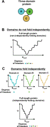
Some multi-domain proteins show independent folding of domains, and their folding can be described by combining the independent folding funnels for each domain [76]. (A) Cartoon of three-domain protein. (B) Folding funnel for multidomain protein with non-independently folding domains. The total number of degrees of freedom of the full-length protein is equal to the product of the degrees of freedom of each individual domain [76]. (C) Folding funnels for individual domains and combined folding funnel for multi-domain protein whose domains fold independently. In this case, the total number of degrees of freedom for the full-length protein is equal to the sum of the degrees of freedom for each individual domain [76].
Figure 7.
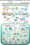
Figure 7.. Pictorial representation of major pathways leading to protein native-structure formation in prokaryotes.
This scheme applies to gram-negative bacteria, e.g., E. coli. Abbreviations: SRP = signal recognition particle, REMPs = redox enzyme maturation protein, TatABC = twin-arginine translocation, TF = trigger factor.
Figure 8.
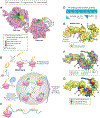
Figure 8.. Protein folding in the presence of the trigger factor (TF) chaperone.
(A) Crystal structure of the RBD of TF bound to the 50s unit of the ribosome from eubacterium Deinococcus radiodurans. PDB: 2AAR [223]. (B) TF cycle. Note that TF is in rapid equilibrium with the ribosome. (C) Multiple TFs can be associated with the same nascent chain during translation or with the client protein in solution. (D) Nascent chain binding site for TF. (E) Crosslinking sites used to track the progression of the nascent chain as it travels throughout the TF [220]. PDB: 2MLX. (F) E. coli TF amino acids (aa) highlighted according to type. Note that the TF binding sites for PhoA, shown in the next panel, are all either nonpolar or polar. PDB: 2MLX. (G) E. coli TF associated with the 220–310 fragment of the PhoA client protein. PDB: 2MLX (Abbreviations: NC = nascent chain, TF = trigger factor, residues = amino acids).
Figure 9.
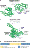
Figure 9.. Key structural features required for the interactions of a client protein with the Hsp70 chaperone system.
(A) Structure of ADP-bound (or nucleotide-free) Hsp70 chaperone (DnaK from E. coli). PDB ID: 2KHO. (B) Structure of ATP-bound DnaK chaperone. PDB ID: 4B9Q. (C) Client-protein binding motif for interaction with the E. coli Hsp70 chaperone DnaK, defined according to [284, 333]. Note that the positively charged residues flanking the central nonpolar core are progressively less important, as the sequence separation from the core increases.
Figure 10.
Figure 10.. Scheme illustrating the major steps of the E. coli Hsp70 (a.k.a. DnaK) chaperone cycle.
Hsp70 cooperates with co-chaperones DnaJ and GrpE through an ATP-dependent cycle to promote the folding of client proteins.
Figure 11.
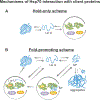
Figure 11.. Simplified schemes illustrating chaperone-assisted protein folding.
The diagrams in this figure are consistent with experimental results achieved with distinct classes of client proteins. (A) Hold-only model consistent with both computational and experimental results on non-aggregation-prone proteins bearing one Hsp70 binding site [307, 315]. (B) Fold-promoting models consistent with experimental results obtained with aggregation-prone client proteins bearing multiple chaperone binding sites per molecule. For instance, firefly luciferase (fluc) populate their native states more quickly and avoid generating aggregates in the presence of the Hsp70 chaperone system [259]. According to this fold-promoting model, the Hsp70 chaperone system catalyzes the conversion of misfolded monomers (M*) to the native state and, in so doing, increases the yields and observed rates of native-structure formation.
Figure 12.
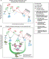
Figure 12.. Summary of protein folding standard-state Gibbs free energy landscapes.
(A) Protein folding in vitro upon dilution from denaturant, and (B) ribosome and chaperone-assisted protein folding within the bacterial cell.

Similar articles

Cited by

References

    1. Theillet F-X, Binolfi A, Frembgen-Kesner T, Hingorani K, Sarkar M, Kyne C, Li C, Crowley PB, Gierasch L, Pielak GJ, Elcock AH, Gershenson A, Selenko P, Physicochemical properties of cells and their effects on intrinsically disordered proteins (IDPs), Chem. Rev. 114 (2014) 6661–6714. - PMC - PubMed
    1. Yamaguchi H, Miyazaki M, Refolding techniques for recovering biologically active recombinant proteins from inclusion bodies, Biomolecules 4 (2014) 235–251. - PMC - PubMed
    1. Betts SD, King J, Cold rescue of the thermolabile tailspike intermediate at the junction between productive folding and off-pathway aggregation, Protein Sci. 7 (1998) 1516–1523. - PMC - PubMed
    1. Speed MA, Wang DI, King J, Specific aggregation of partially folded polypeptide chains: The molecular basis of inclusion body composition, Nat. Biotechnol. 14 (1996) 1283–1287. - PubMed
    1. Tycko R, Amyloid polymorphism: structural basis and neurobiological relevance, Neuron 86 (2015) 632–645. - PMC - PubMed

Publication types

Substances

LinkOut - more resources