

BifA Triggers Phosphorylation of Ezrin to Benefit Streptococcus equi subsp. zooepidemicus Survival from Neutrophils Killing
- PMID: 35625669
- PMCID: PMC9138245
- DOI: 10.3390/biomedicines10050932
BifA Triggers Phosphorylation of Ezrin to Benefit Streptococcus equi subsp. zooepidemicus Survival from Neutrophils Killing
Abstract
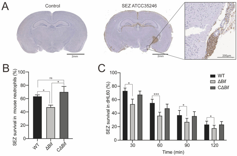
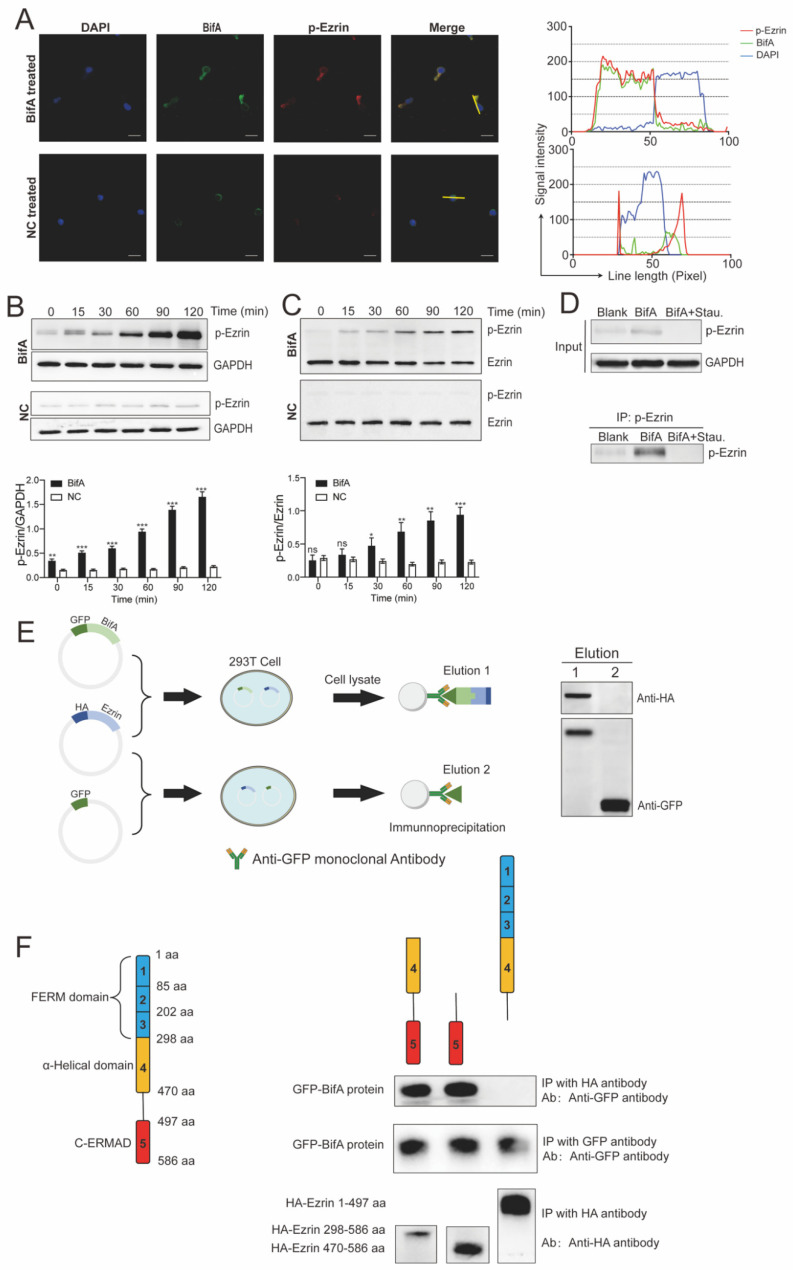
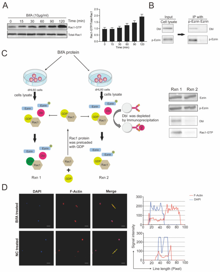
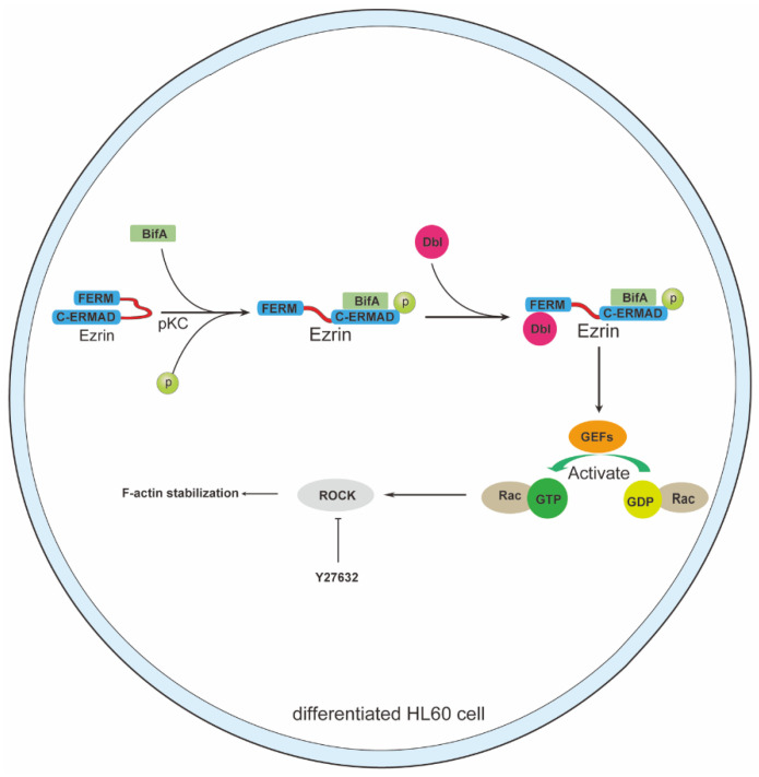
Streptococcus equi subsp. zooepidemicus (SEZ) ATCC35246 can invade the brain and cause severe neutrophils infiltration in brain tissue. This microorganism can survive and reproduce to an extremely high CFU burden (108-109/organ) under stressful neutrophils infiltration circumstances. The aim of this research is to explore the mechanism of the SEZ hypervirulent strain with its specific bifA gene which avoids being eliminated by neutrophils in the brain. We isolated the primary mouse neutrophils to treat SEZ WT and bifA gene defective (ΔBif) strains. The ΔBif strain had a weakened function of defending against neutrophils killing in vitro. The interaction between BifA and ezrin proteins in neutrophils were identified by co-IP and immunoblot. In neutrophils, the BifA interacts with ezrin and triggers the phosphorylation of ezrin at its Thr567 site in a PKC-dependent manner, then the excessive elevation of phosphorylated-ezrin recruits Dbl and activates Rac1. Since the Rac1 is closely relevant to several critical cellular functions, its abnormal activation will lead to neutrophils dysfunction and benefit to SEZ survival from neutrophils killing. Our findings reveal a novel consequence of BifA and ERM family protein (for ezrin, radixin, moesin) interaction, which happens between BifA and ezrin in neutrophils and contributes to SEZ survival in the brain. BifA should be considered as a potential target for drug development to prevent SEZ infection.
Keywords: Streptococcus equi subsp. zooepidemicus; bifA; ezrin; neutrophils.
Conflict of interest statement
The authors declare no conflict of interest.
Figures
Similar articles
-
A streptococcal Fic domain-containing protein disrupts blood-brain barrier integrity by activating moesin in endothelial cells.PLoS Pathog. 2019 May 9;15(5):e1007737. doi: 10.1371/journal.ppat.1007737. eCollection 2019 May. PLoS Pathog. 2019. PMID: 31071198 Free PMC article.
-
Neutrophil accumulation raises defence against Streptococcus equi ssp. zooepidemicus in the absence of Gasdermin D.Int Immunopharmacol. 2024 Apr 20;131:111891. doi: 10.1016/j.intimp.2024.111891. Epub 2024 Mar 17. Int Immunopharmacol. 2024. PMID: 38498953
-
Differences in the Accessory Genomes and Methylomes of Strains of Streptococcus equi subsp. equi and of Streptococcus equi subsp. zooepidemicus Obtained from the Respiratory Tract of Horses from Texas.Microbiol Spectr. 2022 Feb 23;10(1):e0076421. doi: 10.1128/spectrum.00764-21. Epub 2022 Jan 12. Microbiol Spectr. 2022. PMID: 35019696 Free PMC article.
-
Protection Efficacy of Monoclonal Antibodies Targeting Different Regions of Specific SzM Protein from Swine-Isolated Streptococcus equi ssp. zooepidemicus Strains.Microbiol Spectr. 2022 Dec 21;10(6):e0174222. doi: 10.1128/spectrum.01742-22. Epub 2022 Oct 18. Microbiol Spectr. 2022. PMID: 36255327 Free PMC article.
-
Perspectives for Ezrin and Radixin in Astrocytes: Kinases, Functions and Pathology.Int J Mol Sci. 2019 Aug 2;20(15):3776. doi: 10.3390/ijms20153776. Int J Mol Sci. 2019. PMID: 31382374 Free PMC article. Review.
Cited by
-
Endogenous Type I-C CRISPR-Cas system of Streptococcus equi subsp. zooepidemicus promotes biofilm formation and pathogenicity.Front Microbiol. 2024 May 22;15:1417993. doi: 10.3389/fmicb.2024.1417993. eCollection 2024. Front Microbiol. 2024. PMID: 38841053 Free PMC article.
References
-
- Feng Z.G., Hu J.S. Outbreak of swine streptococcosis in Sichuan province and identification of pathogen. Anim. Husb. Vet. Med. Lett. 1977;2:7–12.
-
- Chen X., Resende-De-Macedo N., Sitthicharoenchai P., Sahin O., Burrough E., Clavijo M., Derscheid R., Schwartz K., Lantz K., Robbe-Austerman S., et al. Genetic characterization of Streptococcus equi subspecies zooepidemicus associated with high swine mortality in the United States. Transbound Emerg. Dis. 2020;67:2797–2808. doi: 10.1111/tbed.13645. - DOI - PubMed
-
- Ma Z., Geng J., Yi L., Xu B., Jia R., Li Y., Meng Q., Fan H., Hu S. Insight into the specific virulence related genes and toxin-antitoxin virulent pathogenicity islands in swine streptococcosis pathogen Streptococcus equi ssp. zooepidemicus strain ATCC35246. BMC Genom. 2013;14:377. doi: 10.1186/1471-2164-14-377. - DOI - PMC - PubMed
Grants and funding
LinkOut - more resources
Full Text Sources
Research Materials
