Rho GTPase-activating protein 10 (ARHGAP10/GRAF2) is a novel autoantibody target in patients with autoimmune encephalitis
- PMID: 35624318
- PMCID: PMC9468106
- DOI: 10.1007/s00415-022-11178-9
Rho GTPase-activating protein 10 (ARHGAP10/GRAF2) is a novel autoantibody target in patients with autoimmune encephalitis
Abstract
Background: In 2010, we described a novel immunoglobulin G (IgG) autoantibody (termed anti-Ca after the index case) targeting Rho GTPase-activating protein 26 (ARHGAP26, also termed GTPase regulator associated with focal adhesion kinase [GRAF], or oligophrenin-like protein 1 [OPHN1L]) in autoimmune cerebellar ataxia (ACA). Later, ARHGAP26-IgG/anti-Ca was reported in patients with limbic encephalitis/cognitive decline or peripheral neuropathy. In several of the reported cases, the syndrome was associated with cancer. ARHGAP10/GRAF2, which is expressed throughout the central nervous system, shares significant sequence homology with ARHGAP26/GRAF. Mutations in the ARHGAP10 gene have been linked to cognitive and psychiatric symptoms and schizophrenia.
Objective: To assess whether ARHGAP26-IgG/anti-Ca co-reacts with ARHGAP10.
Methods: Serological testing for ARHGAP10/GRAF2 autoantibodies by recombinant cell-based assays and isotype and IgG subclass analyses.
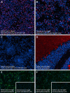
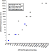
Results: 26/31 serum samples (84%) from 9/12 (75%) ARHGAP26-IgG/anti-Ca-positive patients and 4/6 ARHGAP26-IgG/anti-Ca-positive CSF samples from four patients were positive also for ARHGAP10-IgG. ARHGAP10-IgG (termed anti-Ca2) remained detectable in the long-term (up to 109 months) and belonged mainly to the complement-activating IgG1 subclass. Median ARHGAP26-IgG/anti-Ca and median ARHGAP10-IgG/anti-Ca2 serum titres were 1:3200 and 1:1000, respectively, with extraordinarily high titres in some samples (ARHGAP26-IgG/anti-Ca: up to 1:1000,000; ARHGAP10-IgG: up to 1:32,000). ARHGAP26/anti-Ca serum titres exceeded those of ARHGAP10-IgG in all samples but one. A subset of patients was positive also for ARHGAP10-IgM and ARHGAP10-IgA. CSF/serum ratios and antibody index calculation suggested intrathecal production of ARHGAP26-IgG/anti-Ca and anti-ARHGAP10. Of 101 control samples, 100 were completely negative for ARHGAP10-IgG; a single control sample bound weakly (1:10) to the ARHGAP10-transfected cells.
Conclusions: We demonstrate that a substantial proportion of patients with ARHGAP26-IgG/anti-Ca-positive autoimmune encephalitis co-react with ARHGAP10. Further studies on the clinical and diagnostic implications of ARHGAP10-IgG/anti-Ca2 seropositivity in patients with autoimmune encephalitis are warranted.
Keywords: Anti-Ca; Anti-Ca2; Antibodies; Antigen; Autoantibodies; Autoimmune encephalitis; Cerebellar ataxia; Cognitive decline; GRAF2; GTPase regulator associated with focal adhesion kinase (GRAF); Immunoglobulin G (IgG); Limbic encephalitis; Medusa head ataxia; Oligophrenin-like protein 1 (OPHN1L); Polyneuropathy; Rho GTPase-activating protein 10 (ARHGAP10); Rho GTPase-activating protein 26 (ARHGAP26).
© 2022. The Author(s).
Conflict of interest statement
S.J., B.W., J.H. and J.U.R. report no conflicts of interest. L.K. and S.B. are employees of Euroimmun AG, Lübeck, Germany.
Figures


Similar articles
-
Rho GTPase-activating protein 17 (ARHGAP17) as additional autoimmune target in ARHGAP26-IgG/anti-Ca autoantibody-associated autoimmune encephalitis.J Neurol. 2023 Mar;270(3):1776-1780. doi: 10.1007/s00415-022-11417-z. Epub 2022 Nov 4. J Neurol. 2023. PMID: 36333454 Free PMC article. No abstract available.
-
Two new cases of anti-Ca (anti-ARHGAP26/GRAF) autoantibody-associated cerebellar ataxia.J Neuroinflammation. 2013 Jan 15;10:7. doi: 10.1186/1742-2094-10-7. J Neuroinflammation. 2013. PMID: 23320754 Free PMC article.
-
Anti-Ca/anti-ARHGAP26 antibodies associated with cerebellar atrophy and cognitive decline.J Neuroimmunol. 2014 Feb 15;267(1-2):102-4. doi: 10.1016/j.jneuroim.2013.10.010. Epub 2013 Nov 6. J Neuroimmunol. 2014. PMID: 24439423
-
Inositol 1,4,5-trisphosphate receptor type 1 autoantibody (ITPR1-IgG/anti-Sj)-associated autoimmune cerebellar ataxia, encephalitis and peripheral neuropathy: review of the literature.J Neuroinflammation. 2022 Jul 30;19(1):196. doi: 10.1186/s12974-022-02545-4. J Neuroinflammation. 2022. PMID: 35907972 Free PMC article. Review.
-
'Medusa head ataxia': the expanding spectrum of Purkinje cell antibodies in autoimmune cerebellar ataxia. Part 2: Anti-PKC-gamma, anti-GluR-delta2, anti-Ca/ARHGAP26 and anti-VGCC.J Neuroinflammation. 2015 Sep 17;12:167. doi: 10.1186/s12974-015-0357-x. J Neuroinflammation. 2015. PMID: 26377184 Free PMC article. Review.
Cited by
-
Case report: Anti-ARHGAP26 autoantibodies in atypical dementia with Lewy bodies.Front Dement. 2023 Aug 3;2:1227823. doi: 10.3389/frdem.2023.1227823. eCollection 2023. Front Dement. 2023. PMID: 39081998 Free PMC article.
-
Genetic Rare Variants Affecting Multiple Pathways in Japanese Patients with Palindromic Rheumatism.Kobe J Med Sci. 2024 Apr 30;70(1):E26-E38. doi: 10.24546/0100489391. Kobe J Med Sci. 2024. PMID: 38719338 Free PMC article.
-
Rho GTPase-activating protein 17 (ARHGAP17) as additional autoimmune target in ARHGAP26-IgG/anti-Ca autoantibody-associated autoimmune encephalitis.J Neurol. 2023 Mar;270(3):1776-1780. doi: 10.1007/s00415-022-11417-z. Epub 2022 Nov 4. J Neurol. 2023. PMID: 36333454 Free PMC article. No abstract available.
References
-
- Geis C, Weishaupt A, Hallermann S, Grunewald B, Wessig C, Wultsch T, Reif A, Byts N, Beck M, Jablonka S, Boettger MK, Uceyler N, Fouquet W, Gerlach M, Meinck HM, Siren AL, Sigrist SJ, Toyka KV, Heckmann M, Sommer C. Stiff person syndrome-associated autoantibodies to amphiphysin mediate reduced GABAergic inhibition. Brain. 2010;133:3166–3180. doi: 10.1093/brain/awq253. - DOI - PubMed
MeSH terms
Substances
Supplementary concepts
LinkOut - more resources
Full Text Sources
Other Literature Sources
Medical
Research Materials
Miscellaneous

