

Endocardial Regulation of Cardiac Development
- PMID: 35621833
- PMCID: PMC9144171
- DOI: 10.3390/jcdd9050122
Endocardial Regulation of Cardiac Development
Abstract
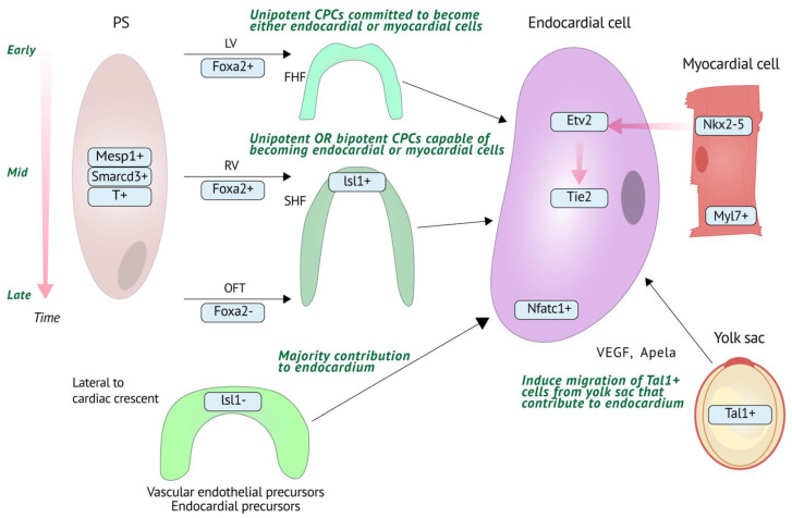
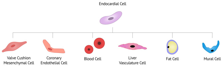
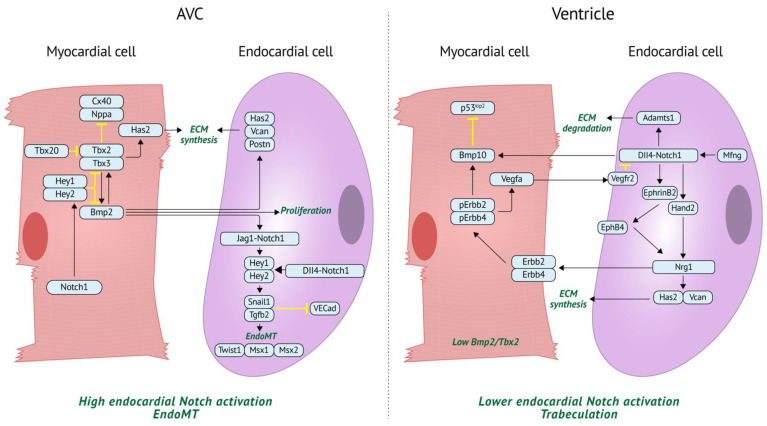
The endocardium is a specialized form of endothelium that lines the inner side of the heart chambers and plays a crucial role in cardiac development. While comparatively less studied than other cardiac cell types, much progress has been made in understanding the regulation of and by the endocardium over the past two decades. In this review, we will summarize what is currently known regarding endocardial origin and development, the relationship between endocardium and other cardiac cell types, and the various lineages that endocardial cells derive from and contribute to. These processes are driven by key molecular mechanisms such as Notch and BMP signaling. These pathways in particular have been well studied, but other signaling pathways and mechanical cues also play important roles. Finally, we will touch on the contribution of stem cell modeling in combination with single cell sequencing and its potential translational impact for congenital heart defects such as bicuspid aortic valves and hypoplastic left heart syndrome. The detailed understanding of cellular and molecular processes in the endocardium will be vital to further develop representative stem cell-derived models for disease modeling and regenerative medicine in the future.
Keywords: BMP; Notch; endocardium; heart development; heart disease.
Conflict of interest statement
The authors declare no conflict of interest.
Figures
Similar articles
-
Intrinsic Endocardial Defects Contribute to Hypoplastic Left Heart Syndrome.Cell Stem Cell. 2020 Oct 1;27(4):574-589.e8. doi: 10.1016/j.stem.2020.07.015. Epub 2020 Aug 17. Cell Stem Cell. 2020. PMID: 32810435 Free PMC article.
-
Development of the endocardium.Pediatr Cardiol. 2010 Apr;31(3):391-9. doi: 10.1007/s00246-010-9642-8. Epub 2010 Feb 5. Pediatr Cardiol. 2010. PMID: 20135106 Free PMC article. Review.
-
Endocardial Notch Signaling in Cardiac Development and Disease.Circ Res. 2016 Jan 8;118(1):e1-e18. doi: 10.1161/CIRCRESAHA.115.305350. Epub 2015 Dec 3. Circ Res. 2016. PMID: 26635389 Review.
-
Endocardial Cell Plasticity in Cardiac Development, Diseases and Regeneration.Circ Res. 2018 Mar 2;122(5):774-789. doi: 10.1161/CIRCRESAHA.117.312136. Circ Res. 2018. PMID: 29496799 Review.
-
Cooperative Response to Endocardial Notch Reveals Interaction With Hippo Pathway.Circ Res. 2023 Dec 8;133(12):1022-1039. doi: 10.1161/CIRCRESAHA.123.323474. Epub 2023 Nov 14. Circ Res. 2023. PMID: 37961886 Free PMC article.
Cited by
-
Ion Channels in the Development and Remodeling of the Aortic Valve.Int J Mol Sci. 2023 Mar 20;24(6):5860. doi: 10.3390/ijms24065860. Int J Mol Sci. 2023. PMID: 36982932 Free PMC article. Review.
-
Transient Notch Activation Converts Pluripotent Stem Cell-Derived Cardiomyocytes Towards a Purkinje Fiber Fate.bioRxiv [Preprint]. 2024 Sep 28:2024.09.22.614353. doi: 10.1101/2024.09.22.614353. bioRxiv. 2024. PMID: 39386729 Free PMC article. Preprint.
-
Rapid Progression of Heterotopic Ossification in Severe Variant of Fibrodysplasia Ossificans Progressiva with p.Arg258Gly in ACVR1: A Case Report and Review of Clinical Phenotypes.Case Rep Genet. 2022 Aug 25;2022:5021758. doi: 10.1155/2022/5021758. eCollection 2022. Case Rep Genet. 2022. PMID: 36060212 Free PMC article.
-
Automated endocardial cushion segmentation and cellularization quantification in developing hearts using optical coherence tomography.Biomed Opt Express. 2022 Oct 4;13(11):5599-5615. doi: 10.1364/BOE.467629. eCollection 2022 Nov 1. Biomed Opt Express. 2022. PMID: 36733755 Free PMC article.
-
Isolation and Characterization of Porcine Endocardial Endothelial Cells.Tissue Eng Part C Methods. 2023 Aug;29(8):371-380. doi: 10.1089/ten.TEC.2023.0009. Epub 2023 Jul 7. Tissue Eng Part C Methods. 2023. PMID: 37310900 Free PMC article.
References
Publication types
Grants and funding
LinkOut - more resources
Full Text Sources
Miscellaneous
