Molecular and Immune Profiling of Syngeneic Mouse Models Predict Response to Immune Checkpoint Inhibitors in Gastric Cancer
- PMID: 35609622
- PMCID: PMC9873335
- DOI: 10.4143/crt.2022.094
Molecular and Immune Profiling of Syngeneic Mouse Models Predict Response to Immune Checkpoint Inhibitors in Gastric Cancer
Abstract
Purpose: Appropriate preclinical mouse models are needed to evaluate the response to immunotherapeutic agents. Immunocompetent mouse models have rarely been reported for gastric cancer. Thus, we investigated immunophenotypes and responses to immune checkpoint inhibitor (ICI) in immunocompetent mouse models using various murine gastric cancer cell lines.
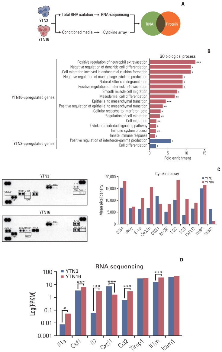
Materials and methods: We constructed subcutaneous syngeneic tumors with murine gastric cancer cell lines, YTN3 and YTN16, in C57BL/6J mice. Mice were intraperitoneally treated with IgG isotype control or an anti-programmed death-ligand 1 (PD-L1) neutralizing antibody. We used immunohistochemistry to evaluate the tumor-infiltrating immune cells of formalin-fixed paraffin-embedded mouse tumor tissues. We compared the protein and RNA expression between YTN3 and YTN16 cell lines using a mouse cytokine array and RNA sequencing.
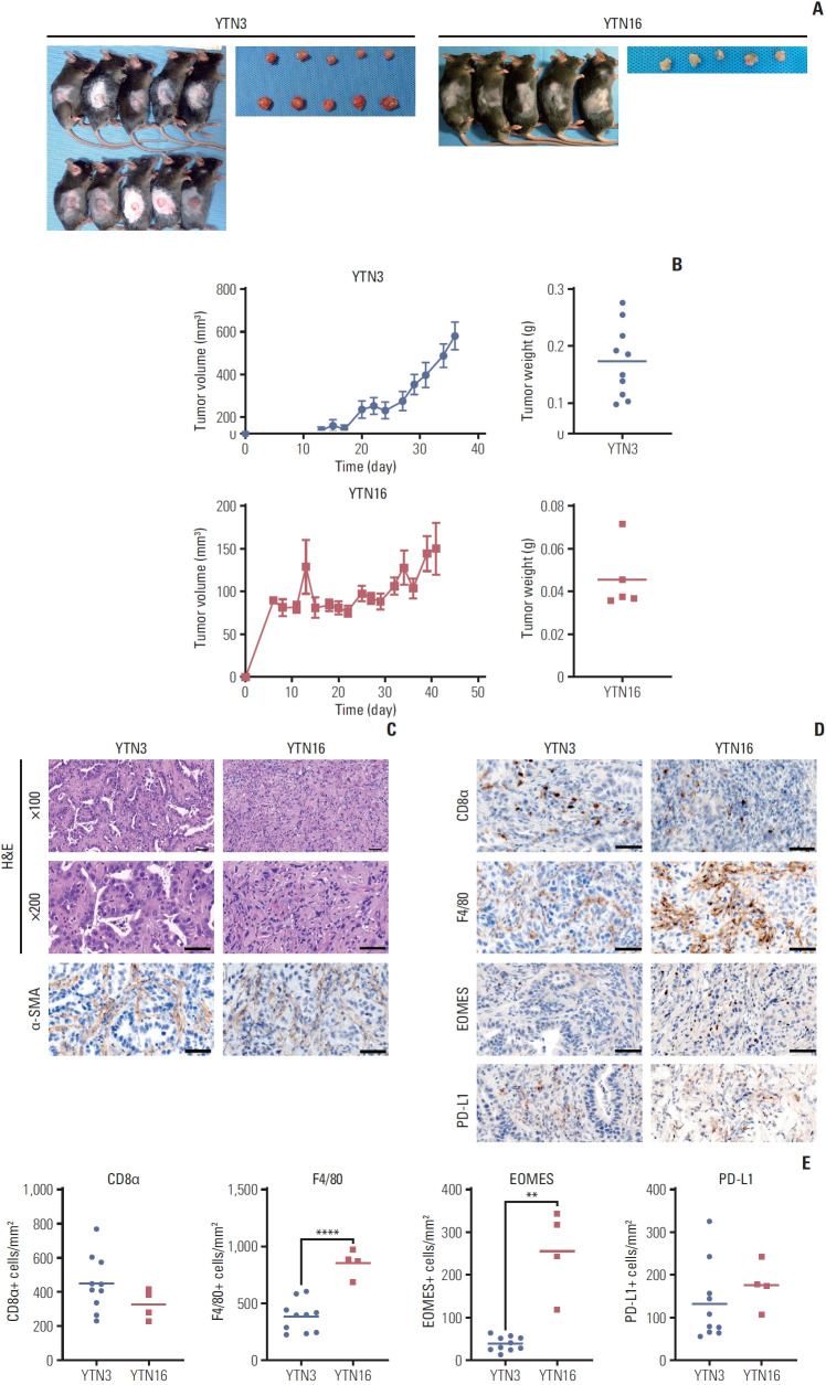
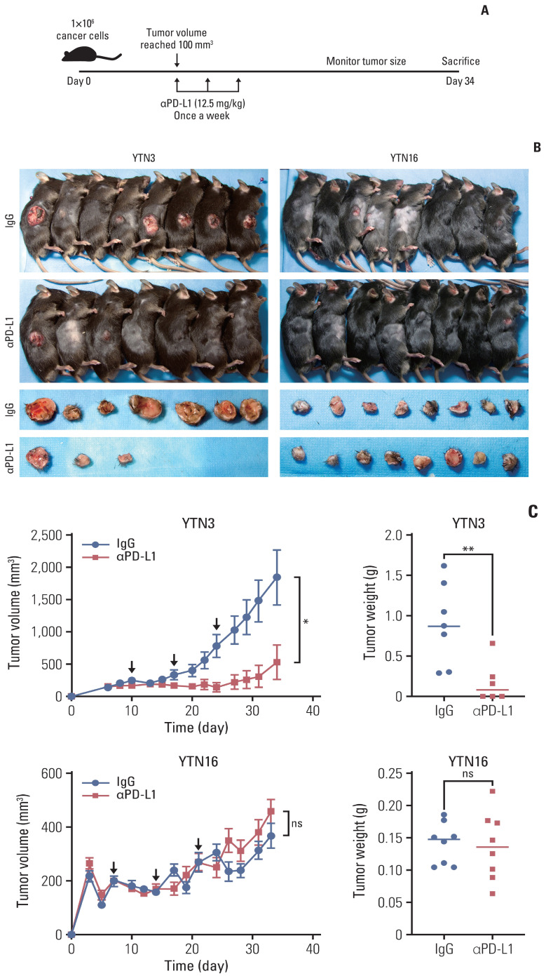
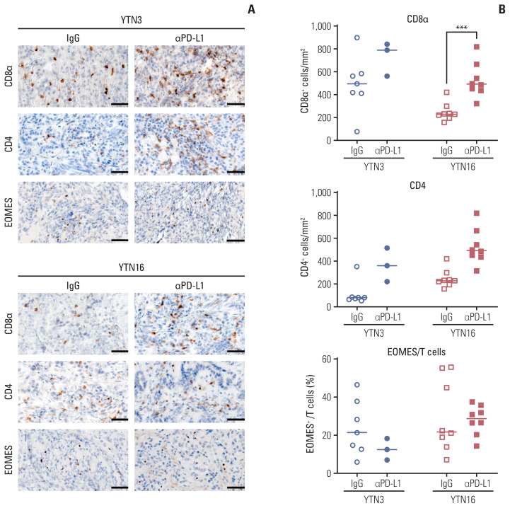
Results: The mouse tumors revealed distinct histological and molecular characteristics. YTN16 cells showed upregulation of genes and proteins related to immunosuppression, such as Ccl2 (CCL2) and Csf1 (M-CSF). Macrophages and exhausted T cells were more enriched in YTN16 tumors than in YTN3 tumors. Several YTN3 tumors were completely regressed by the PD-L1 inhibitor, whereas YTN16 tumors were unaffected. Although treatment with a PD-L1 inhibitor increased infiltration of T cells in both the tumors, the proportion of exhausted immune cells did not decrease in the non-responder group.
Conclusion: We confirmed the histological and molecular features of cancer cells with various responses to ICI. Our models can be used in preclinical research on ICI resistance mechanisms to enhance clinical efficacy.
Keywords: Immune checkpoint inhibitors; Macrophages; Stomach neoplasms; Syngeneic mouse; Tumor immunity.
Figures
Similar articles
-
Targeting GAS6/AXL signaling improves the response to immunotherapy by restoring the anti-immunogenic tumor microenvironment in gastric cancer.Life Sci. 2023 Dec 15;335:122230. doi: 10.1016/j.lfs.2023.122230. Epub 2023 Nov 11. Life Sci. 2023. PMID: 37952835
-
Deep immunophenotyping at the single-cell level identifies a combination of anti-IL-17 and checkpoint blockade as an effective treatment in a preclinical model of data-guided personalized immunotherapy.J Immunother Cancer. 2020 Oct;8(2):e001358. doi: 10.1136/jitc-2020-001358. J Immunother Cancer. 2020. PMID: 33093158 Free PMC article.
-
High baseline tumor burden-associated macrophages promote an immunosuppressive microenvironment and reduce the efficacy of immune checkpoint inhibitors through the IGFBP2-STAT3-PD-L1 pathway.Cancer Commun (Lond). 2023 May;43(5):562-581. doi: 10.1002/cac2.12420. Epub 2023 Apr 8. Cancer Commun (Lond). 2023. PMID: 37031362 Free PMC article.
-
Current status of immune checkpoint inhibitors for gastric cancer.Gastric Cancer. 2020 Jul;23(4):565-578. doi: 10.1007/s10120-020-01090-4. Epub 2020 May 28. Gastric Cancer. 2020. PMID: 32468420 Review.
-
Low Programmed Death-Ligand 1-Expressing Subgroup Outcomes of First-Line Immune Checkpoint Inhibitors in Gastric or Esophageal Adenocarcinoma.J Clin Oncol. 2022 Feb 1;40(4):392-402. doi: 10.1200/JCO.21.01862. Epub 2021 Dec 3. J Clin Oncol. 2022. PMID: 34860570 Review.
Cited by
-
Safety and Efficacy of Neoadjuvant Chemoimmunotherapy in Gastric Cancer Patients with a PD-L1 Positive Status: A Case Report.Curr Issues Mol Biol. 2023 Sep 19;45(9):7642-7649. doi: 10.3390/cimb45090481. Curr Issues Mol Biol. 2023. PMID: 37754265 Free PMC article.
-
Modeling human gastric cancers in immunocompetent mice.Cancer Biol Med. 2024 Jun 28;21(7):553-70. doi: 10.20892/j.issn.2095-3941.2024.0124. Cancer Biol Med. 2024. PMID: 38940675 Free PMC article. Review.
-
Tubulointerstitial nephritis antigen-like 1 from cancer-associated fibroblasts contribute to the progression of diffuse-type gastric cancers through the interaction with integrin β1.J Transl Med. 2024 Feb 14;22(1):154. doi: 10.1186/s12967-024-04963-9. J Transl Med. 2024. PMID: 38355577 Free PMC article.
References
-
- Sung H, Ferlay J, Siegel RL, Laversanne M, Soerjomataram I, Jemal A, et al. Global cancer statistics 2020: GLOBOCAN estimates of incidence and mortality worldwide for 36 cancers in 185 countries. CA Cancer J Clin. 2021;71:209–49. - PubMed
-
- Kang YK, Boku N, Satoh T, Ryu MH, Chao Y, Kato K, et al. Nivolumab in patients with advanced gastric or gastro-oesophageal junction cancer refractory to, or intolerant of, at least two previous chemotherapy regimens (ONO-4538-12, ATTRACTION-2): a randomised, double-blind, placebo-controlled, phase 3 trial. Lancet. 2017;390:2461–71. - PubMed
-
- Wu T, Dai Y. Tumor microenvironment and therapeutic response. Cancer Lett. 2017;387:61–8. - PubMed
MeSH terms
Substances
LinkOut - more resources
Full Text Sources
Medical
Research Materials
Miscellaneous